Review
. 1993 Jun;17(6):1127-52.

Cell migration and chimerism after whole-organ transplantation: the basis of graft acceptance

Affiliations
Review

Cell migration and chimerism after whole-organ transplantation: the basis of graft acceptance

T E Starzl et al. Hepatology. 1993 Jun.

Abstract

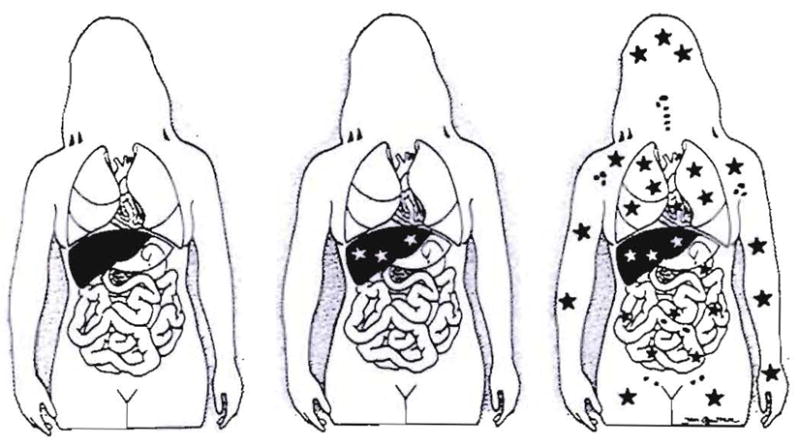
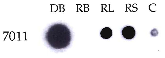
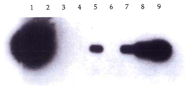
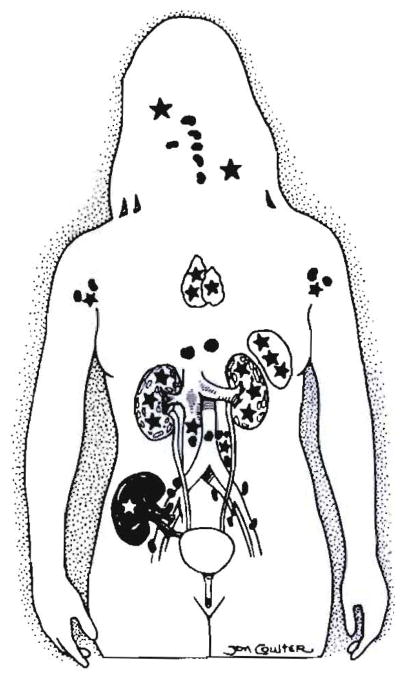
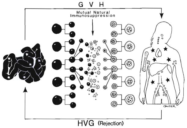
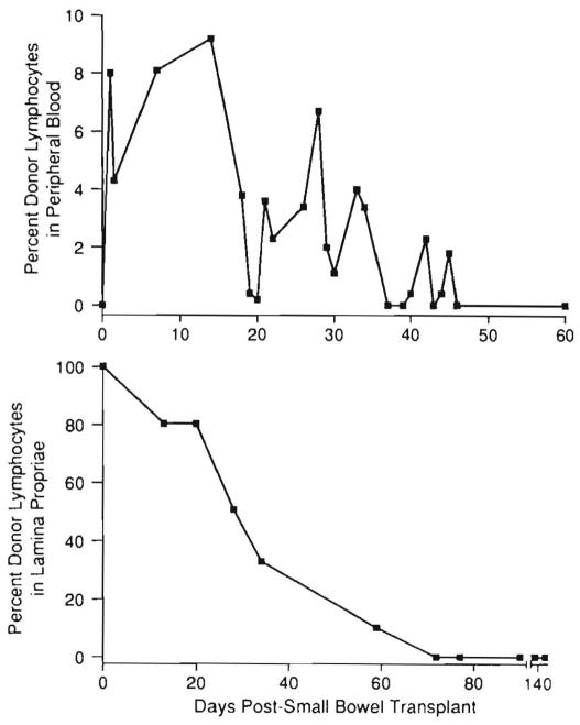
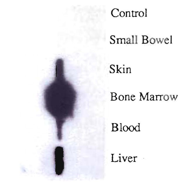
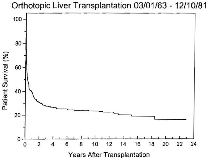
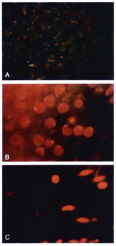
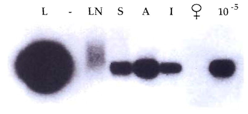
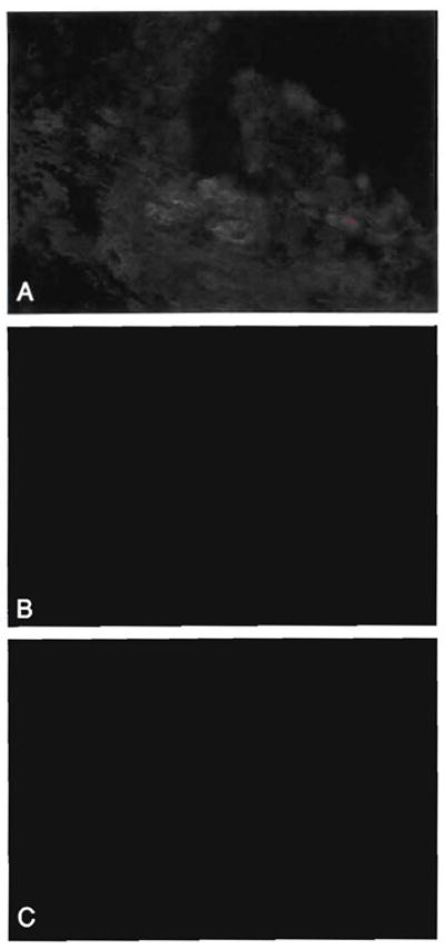
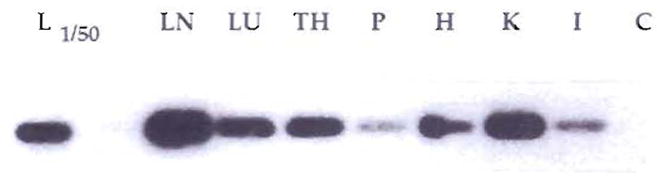
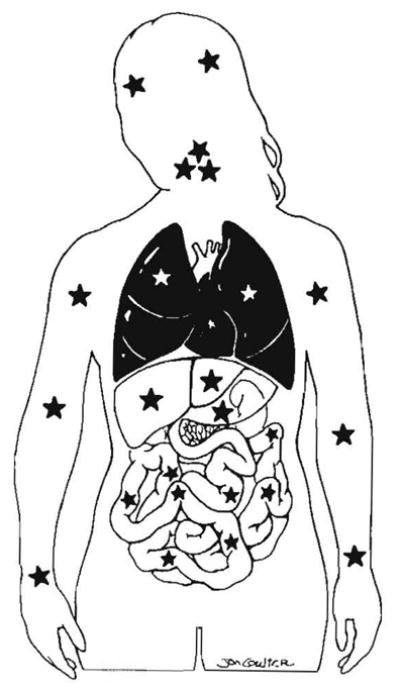
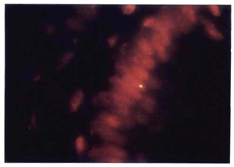
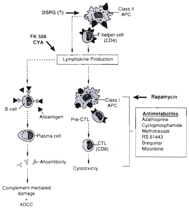
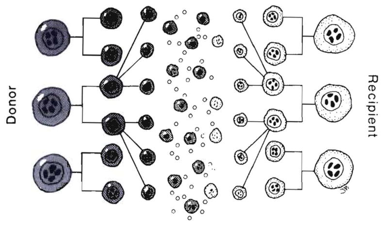
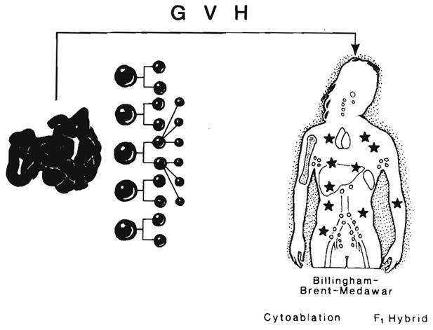
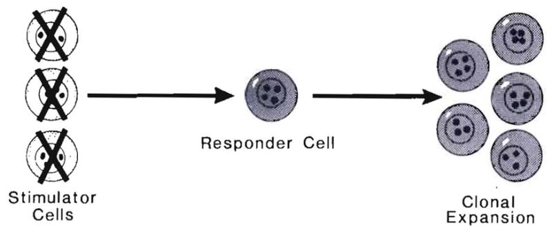
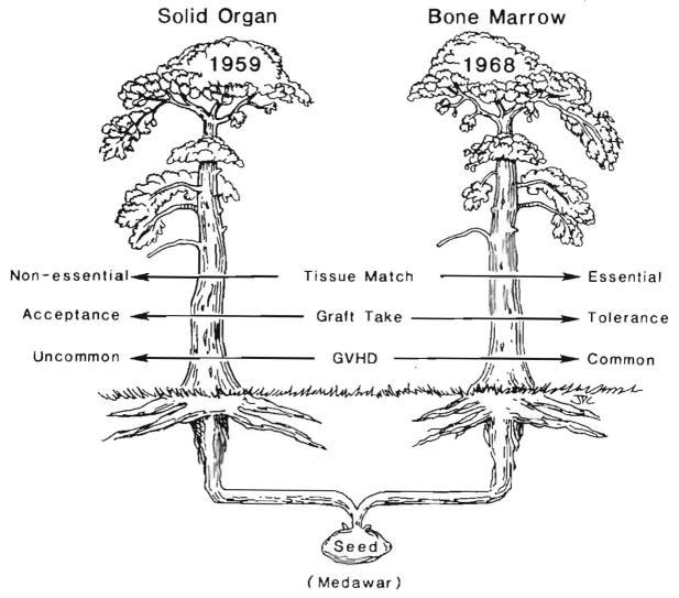
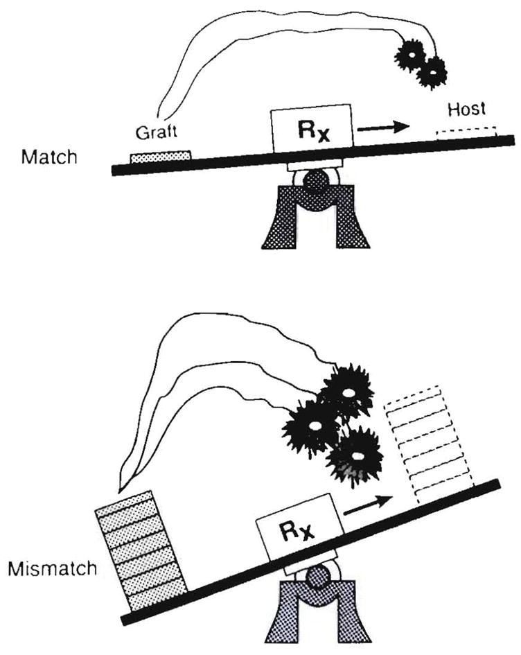
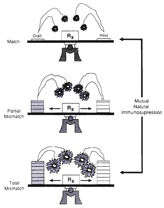
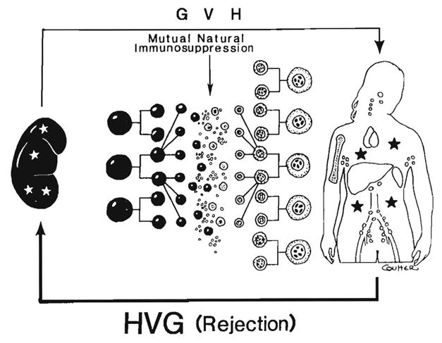
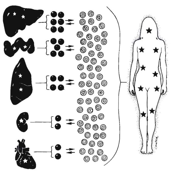
Improvements in the prevention or control of rejection of the kidney and liver have been largely interchangeable (1, 2) and then applicable, with very little modification, to thoracic and other organs. However, the mechanism by which anti rejection treatment permits any of these grafts to be “accepted” has been an immunological enigma (3, 4). We have proposed recently that the exchange of migratory leukocytes between the transplant and the recipient with consequent long-term cellular chimerism in both is the basis for acceptance of all whole-organ allografts and xenografts (5). Although such chimerism was demonstrated only a few months ago, the observations have increased our insight into transplantation immunology and have encouraged the development of alternative therapeutic strategies (6).


Figures

Fig. 1
Fig. 1
Steps in understanding liver transplantation: left– historical view; middle – realization in 1969 that the liver graft became a genetic composite (chimera); right–proof in 1992 of systemic chimerism. Stars represent cell exchange between graft and host.
Fig. 2
Fig. 2
Repopulation of the lamina propria of human small intestinal grafts, demonstrated by HLA allele phenotyping. Monoclonal antibodies directed at Bw loci were used to differentiate donor (Bw6) from recipient (Bw4) cells. (A) Backtable graft biopsy specimen showed no recipient cells as expected. (Immunoperoxidase staining for Bw4 [left] and Bw6 [right]; original magnification × 250.) (B) Biopsy specimen 54 days after transplantation. The recipient cells have repopulated the lamina propria, but the epithelium and endothelium remained of donor origin. Ommunoperoxidase staining with DAB [brown] for Bw4 [left] and Bw6 [right); original magnification × 250.)
Fig. 3
Fig. 3
Transfer of positive skin test results from kidney donors to recipients in cases at the University of Colorado from 1962 to 1964 (32). Although inexplicable at the time, these observations reflected adoptive transfer after cell migration, repopulation and chimerism.
Fig. 4
Fig. 4
Patient LD51. Immunostained biopsy samples 29 yr after transplantation of (A) an allograft kidney (original magnification × 250) and (B) an inguinal lymph node (original magnification × 100) stained for HLA B7,40–an antigen present in the donor but not the recipient; note the sparse and evenly scattered donor cells (rust colored) in the lymph node. (C) As a control the lymph node was also stained with anti-A2,28–present in neither the donor nor the recipient (original magnification × 100).
Fig. 5
Fig. 5
Chimerism demonstrated by HLA molecular typing in a kidney recipient, LD5l. The donor’s DR2 allele was specifically amplified by PCR from DNA extracted from the recipient’s blood (RB), lymph node (RL) and skin (RS). The amplified product was dot blotted and hybridized with 7011, a DR2-specific oligonucleotide probe. DNA from the donor’s blood (DB) was also amplified. The negative control (C) of the PCR amplification performed in the absence of DNA is also shown.
Fig. 6
Fig. 6
PCR demonstration of chimerism in a female recipient (LD52) of a kidney from her father. The DNA of the SRY region of the Y chromosome was PCR amplified, Southern blotted and hybridized with a homologous radioactive probe. DNA from biopsy specimens of the patient’s kidney (1), blood (3), lymph node (4) and skin (5) were tested. As controls, DNA from an unrelated female (6) and serial dilutions of male DNA in female DNA 10−4 (9), 10−5 (8) and 10−6 (7) were also analyzed. No sample was present in lane 2.
Fig. 7
Fig. 7
Systemic and graft chimerism in all five tested patients treated who had borne continuously functioning allografts for 27 to 29 yr. The stars represent the migrated cells. In these studies only the kidney, skin, lymph nodes and blood were sampled, but the more ubiquitous chimerism was inferred from the more extensive studies in recipients of livers and other organs.
Fig. 8
Fig. 8
Current understanding of the graft and systemic chimerism that occurs after intestinal transplantation. The automatic production of mixed allogenic chimerism after intestinal transplantation, providing the preexisting immunological apparatus, is not iatrogenically damaged on either donor or recipient side (see text). Evolution of this concept permitted successful clinical intestinal transplantation trials (108). The stars represent the cell exchange between the graft and host.
Fig. 9
Fig. 9
Appearance of donor mononuclear cells in the blood of an intestinal human transplant recipient (upper) and the demonstration of progressive replacement of interstitial donor leukocytes from the graft during the same general time (lower). (Data from Iwaki et al. [13].)
Fig. 10
Fig. 10
(A) An endomyocardial biopsy specimen obtained in 1989, at the time of liver transplantation, revealed diffuse amylopectin deposits within and between cells. (B) A repeat endomyocardial biopsy sample taken in 1992 revealed only traces of extracellular deposits. (Periodic acid–Schiff-D; original magnification ×100.)
Fig. 11
Fig. 11
(A) Hepatic hilar lymph node removed in April 1990 at the time of liver replacement from a patient with Gaucher’s disease and (B) an inguinal lymph node obtained in June 1992. Note the marked decrease in the number of Gaucher’s cells, which were difficult to find in the latter specimen. (Periodic acid–Schiff; original magnification ×250.)
Fig. 12
Fig. 12
Allograft liver and inguinal lymph node biopsy specimens obtained 26 mo after the liver replacement from the recipient with Gaucher’s disease. These were stained with antibodies to DR1 (donor) and DR3 (control). (A) Note the presence of DR1-positive donor cells (green) in the portal tract of the liver (original magnification × 400). (B) Absence of staining in the control (antibodies to DR3; original magnification × 400). (C) Rare DR1-positive (donor) cells were also seen in the inguinal lymph node (original magnification × 250). (D) The lymph node control (antibodies to DR3) was negative (original magnification × 250). (All were stained with FITC indirect immunofluorescence.)
Fig. 13
Fig. 13
Detection of chimerism by molecular HLA class II typing in various tissues after liver transplantation in a patient with type 1 Gaucher’s disease. Southern blot analysis of DR1-specific amplification of the DNA extracted from small bowel, skin, bone marrow, blood and liver. The denaturated DNA present on the nylon membrane was hybridized to a radioactively labeled DR1 (donor) specific oligonucleotide probe (7001). In the case of the liver, only 1/100 of the amplification product was used. Note negative control of the PCR amplification.
Fig. 14
Fig. 14
Life survival curve of the first 210 patients in our Colorado-Pittsburgh series. The survival is actual to 11 yr and actuarial thereafter. Fifty of the original 210 patients survived for more than 10 yr; 43 are still alive after 11 to 23 yr. The other 7 died after 11.1 to 18.4 yr.
Fig. 15
Fig. 15
Studies of patient OT64, 19.5 yrs after her liver replacement from a male donor. Fluorescent in situ hybridization with the DYZ1 probe for the Y chromosome was used to differentiate male from female cells. (A) The allograft liver was used as a positive control. About 60% of hepatocytes and a few sinusoidal cells retained the donor genotype; the Y chromosome in the negative hepatocytes was likely excluded from the 2 μm–thick sectioning plane. The yellow cytoplasmic material is autofluorescence (original magnification × 250). (B) Oil immersion microscopy was used to illustrate the variety of signals obtained in the liver, which showed a beaded to reticular pattern, often located at the periphery of the nucleus (original magnification × 1,000). (C) Skin biopsy specimen: spindle-shaped stromal cells, with a similar signal for the Y chromosome, were sparsely distributed (original magnification × 1,000).
Fig. 16
Fig. 16
Detection of chimerism by PCR in a female recipient 12 yr after transplantation of a liver from a male donor. The samples were obtained at the time of successful retransplantation, necessitated by HCV infection of the primary graft. The DNA of the SRY region of the Y chromosome was PCR amplified, Southern blotted and hybridized with a homologous radioactive probe. Liver (L), lymph node (LN), skin (S), aorta (A) and intestine (I) of the patient were tested. DNA from an unrelated female (♀) and male DNA diluted 1: 100,000 in the female DNA (10−6) were also amplified as negative and positive controls.
Fig. 17
Fig. 17
Studies of patient OT46, 21 yr after liver transplantation. An HLA-A1 (donor) antibody was used to detect donor cells in the allograft liver and in an excised inguinal lymph node. Immunofluorescence staining was used. (A) Note the staining of bile duct and endothelial cells of a mildly inflamed portal tract, whereas the inflammation is not stained. Faint sinusoidal A1 positivity can also be seen (antibodies to A1; original magnification × 400). (B) Rare A1-positive cells were also seen in the inguinal lymph node (antibodies to A1; original magnification × 400). (C) Substitution of mouse IgM for the primary antibody in the same node was negative (mouse IgM; original magnification × 400).
Fig. 18
Fig. 18
Systemic chimerism by PCR of multiple autopsy tissues of patient 77, 18.4 yr after liver transplantation; death was caused by HBV. DNA was extracted from liver (L), lymph node (LN), lung (LU), thymus (TH), pancreas (P), heart (H), kidney (K) and intestine (I) and was PCR amplified with DR4 (donor)–specific primers. A Southern blot of the amplified DNA was hybridized with a DR4 allele–specific oligonucleotide probe. In the case of liver, only 1/50 of the amplification product was used. C is the negative control of the PCR amplification performed in the absence of DNA.
Fig. 19
Fig. 19
Lung and heart graft acceptance is also by the mechanism of cell migration and repopulation (see text). Stars represent chimeric donor cells.
Fig. 20
Fig. 20
Fluorescent in situ hybridization for Y chromosome in a colon biopsy specimen of a woman who was given a lung allograft from a male donor (original magnification × 1,000). PCR analysis of the same specimen was also positive for male DNA.
Fig. 21
Fig. 21
Sites of action of immunosuppressive drugs. All can induce a state of donor-specific nonreactivity if they permit cell migration and repopulation.
Fig. 22
Fig. 22
The mutual leukocyte engagement and interaction after whole-organ transplantation of cell migration. This is in essence a two-way in vivo MLR.
Fig. 23
Fig. 23
Absence of the two-way cell interaction with the removal (cytoablation) or nonreactivity (Billingham-Brent-Medawar and F1 hybrid models) of one population. These conditions are the in vivo equivalents of the one-way MLR shown in Figure 24. They are conducive to the development of GVHD. Stars represent chimeric donor cells.
Fig. 24
Fig. 24
One-way MLR in which only one cell population is reactive. This is the in vitro analog of the in vivo models shown in Figure 23.
Fig. 25
Fig. 25
The division of transplantation into two separate disciplines by divergent therapeutic dogmas that created one-way vs. two-way in vivo MLR analogs. The policies used in bone marrow transplantation inhibited or precluded bidirectional cell migration, whereas this phenomenon was the fundamental basis for graft acceptance with the policies of whole-organ transplanters.
Fig. 26
Fig. 26
Explanation for uncontrollability of GVHD with major histocompatibility complex-mismatched donors when mutual cell engagement is prevented, thereby eliminating the element of mutual natural immunosuppression that is shown in Figure 27.
Fig. 27
Fig. 27
The reciprocal alteration of alloreactivity by coexisting immunocyte populations (mixed allogeneic chimerism). The cardinal requirement for GVHD resistance is to preserve both populations contributing to the reaction. The factor of mutual natural immunosuppression also explains the nondiscrimination of HLA matching in predicting the outcome after whole-organ transplantation (see text).
Fig. 28
Fig. 28
The David (kidney) and Goliath (recipient) conditions of kidney transplantation in which the graft brings a limited number of leukocytes to the mutual cell engagement. Nevertheless, the migratory donor leukocytes were demonstrated in all of the patients with continuously surviving allografts who underwent biopsies 27 to 29 yr after transplantation. Stars represent chimeric donor cells.
Fig. 29
Fig. 29
Skin biopsy specimen of a female recipient of a liver and pancreatic islets obtained from a male donor. The clinical and histopathological diagnosis was GVHD. This was easily controlled with increased immunosuppression. (A) The skin was stained for donor (Bw4) antigen, showing infiltrating cells of donor specificity. These cells also stained positive for the Y probe (not shown). (B) Stain for recipient (A3) antigen in same specimen, showing normal distribution of recipient cells (original magnification × 200).
Fig. 30
Fig. 30
Infiltrative donor cells in the (A) epidermis and (B) adnexa in a patient with clinical GVHD 34 days after combined liver and bone marrow transplantation (antibodies to B55; original magnification × 200). (C) The GVHD was progressive and at 6 wk stored, autologous (recipient) bone marrow was infused. At 14 wk this wedge biopsy specimen of the liver showed that the biliary epithelium and endothelium were of donor origin (antibodies to B55; original magnification × 250). (D) Biliary epithelium and endothelium were negative for recipient antigens. A few positive infiltrative recipient cells were noted (antibodies to A2; original magnification × 250). (E) Scattered donor cells were detected in the lamina propria of a small bowel biopsy sample, indicating that microchimerism persisted after the GVHD was eliminated (antibodies to Bw4; original magnification × 400).
Fig. 31
Fig. 31
Studies in a female recipient of a male donor’s liver, who developed GVHD and a B-cell lymphoma in the first 3 postoperative mo. Note the Y chromosomes in a cervical lymph node biopsy sample containing an Epstein-Barr virus–positive posttransplant lymphoproliferative disorder (fluorescent in situ hybridization for Y chromosome; original magnification × 400). Double labeling for the Y probe and lymphocyte phenotypic markers revealed that at least one third of the donor cells were activated T cells (UCHL-1 +, not shown).
Fig. 32
Fig. 32
The explanation for the variable ability under immuno-suppression to induce acceptance and ultimately tolerance of different organs. We postulate that the dendritic leukocyte is the single most important, although not the only, tolerogenic cell. The tissue content of these potentially migratory cells is liver > intestine > lung > kidney and heart. Stars represent chimeric donor cells in the recipient.
Fig. 33
Fig. 33
Hamster cells in the interstitium of the recipient heart 100 days after hamster-to-rat xenotransplantation (hamster antibody immunofluorescence; original magnification × 400). The periarterial lymphatic sheath of the spleen also contained these cells.
Fig. 34
Fig. 34
PCR amplification of a baboon-specific DNA from recipient tissues. Lane LI is the PCR product from the baboon liver and contains only 1% of the PCR reaction so that the other lanes won’t be overwhelmed (HE, heart; LU, lung; KI, kidney; LN1 and LN2, lymph nodes 1 and 2; and HU, human blood used as a negative control).

Comment in

References

    1. Starzl TE. Experience in renal transplantation. Philadelphia: WB Saunders Co; 1964. pp. 1–383.
    1. Starzl TE, Putnam CW. Experience in hepatic transplantation. Philadelphia: WB Saunders Co; 1969. pp. 1–553.
    1. Murase N, Kim DG, Todo S, Cramer DV, Fung JJ, Starzl TE. Suppression of allograft rejection with FK 506 I: prolonged cardiac and liver survival in rats following short course therapy. Transplantation. 1990;50:186–189. - PMC - PubMed
    1. Murase N, Kim DG, Todo S, Cramer DV, Fung JJ, Starzl TE. FK 506 suppression of heart and liver allograft rejection II: the induction of graft acceptance in rat. Transplantation. 1990;50:739–744. - PMC - PubMed
    1. Starzl TE, Demetris AJ, Murase N, Ildstad S, Ricordi C, Trucco M. Cell migration, chimerism, and graft acceptance. Lancet. 1992;339:1579–1582. - PMC - PubMed

Publication types