

Review
doi: 10.1016/s0091-679x(08)61390-4.
Laser killing of cells in Caenorhabditis elegans
Affiliations
- PMID: 8531727
- PMCID: PMC4442485
- DOI: 10.1016/s0091-679x(08)61390-4
Item in Clipboard
Review
Laser killing of cells in Caenorhabditis elegans
Methods Cell Biol.
1995.
No abstract available
Figures
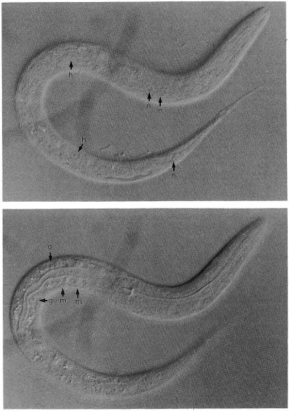
Appearance of different cell types. L1 animal viewed by Nomarski optics under a 100× Neuflour objective (Zeiss). h, hypodermal nucleus; n, neuronal nucleus; g, gut nucleus; m, muscle nucleus.
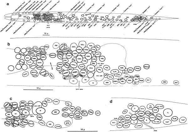
Position of nuclei in L1 larvae. (a) Positions of nuclei in L1 larvae (left lateral view). (b) Neuronal nuclei in the head (left lateral view). (c) Neuronal nuclei in the head (ventral view). (d) Neuronal nuclei in the tail (left lateral view). Anterior is to the left. In a, b, and d, only the left laternl nuclei and the medial nuclei arc shown. Most right lateral nuclei occupy positions similar to those of their homologs on the left side; the exceptions arc found most on the ventral side (see c). The thickness of the nuclear outline is inversely related to the depth of the nucleus within the worm (e.g., in b. lateral nuclei have thick outlines and medial nuclei have thin outlines). Reprinted, with permission, from Sulston et al. (1983).
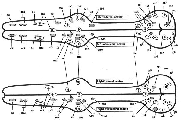
Positions of nuclei in the pharynx. Courtesy of Ron Ellis.
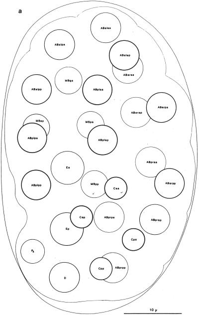
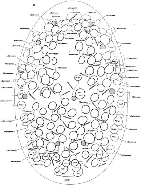
Embryonic nuclei. (a) Twenty-eight-cell embryo 100 minutes, left dorsal aspect. (b) Embryo, 260 minutes, dorsal aspect, superficial nuclei. (c) Embryo, 270 minutes, ventral aspect, superficial nuclei. Anterior is at top. The thickness of the nuclear outline is inversely related to the depth of the nucleus within the worm. Reprinted, with permission, from Sulston et al. (1983). For detailed descriptions of embryonic and postembryonic cell divisions, see Sulston and Horvitz (1977), Kimble and Hirsh (1979), and Sulston et al. (1983).

Embryonic nuclei. (a) Twenty-eight-cell embryo 100 minutes, left dorsal aspect. (b) Embryo, 260 minutes, dorsal aspect, superficial nuclei. (c) Embryo, 270 minutes, ventral aspect, superficial nuclei. Anterior is at top. The thickness of the nuclear outline is inversely related to the depth of the nucleus within the worm. Reprinted, with permission, from Sulston et al. (1983). For detailed descriptions of embryonic and postembryonic cell divisions, see Sulston and Horvitz (1977), Kimble and Hirsh (1979), and Sulston et al. (1983).
Embryonic nuclei. (a) Twenty-eight-cell embryo 100 minutes, left dorsal aspect. (b) Embryo, 260 minutes, dorsal aspect, superficial nuclei. (c) Embryo, 270 minutes, ventral aspect, superficial nuclei. Anterior is at top. The thickness of the nuclear outline is inversely related to the depth of the nucleus within the worm. Reprinted, with permission, from Sulston et al. (1983). For detailed descriptions of embryonic and postembryonic cell divisions, see Sulston and Horvitz (1977), Kimble and Hirsh (1979), and Sulston et al. (1983).
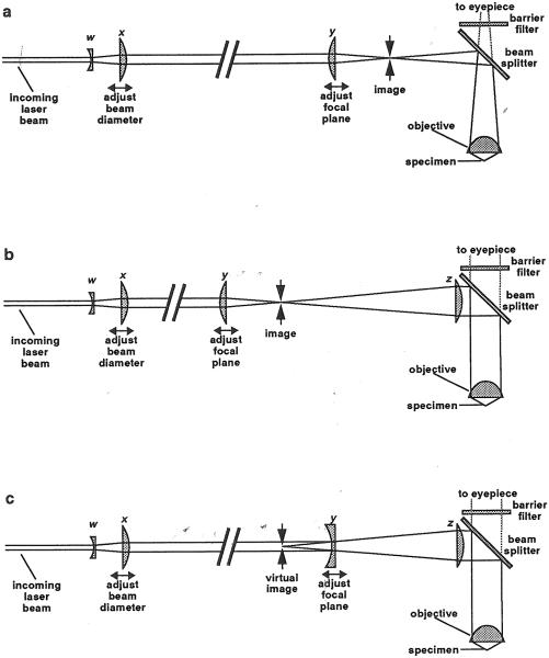
Optics for laser ablation. (a) Optics for use with a microscope objective that forms an image 160 mm away. Lenses w and x form a Galilean telescope. Beam diameter can be changed by moving lens x. Lens y focuses the beam to a point in the image of the specimen, so that it will also focus to a point within the specimen itself. The distance between x and y is larger in proportion to other distances than shown. This is indicated by a break in the beam. The beam splitter reflects blue laser light into the specimen while passing longer-wavelength light to the eyepiece so that the worm can be seen. The barrier filter prevents stray laser reflections from reaching the eyepiece. (b) Optics for use with a microscope objective that forms an image at infinity. This arrangement is identical except for the addition of a new lens z. With this type of objective, all the rays from a point in the specimen come out parallel, so that there is no image plane. Lens z form an image in which the laser beam can be focused. Lens z may be an additional lens you insert, or it may be part of the microscope. It may be more complex than a single lens. (c) A variation, useful if there is not enough space for the arrangement shown in b. Lens y is now concave instead of convex, and causes the laser light to diverge to the right of the lens as if it were emanating from a point (the virtual image) to the left.
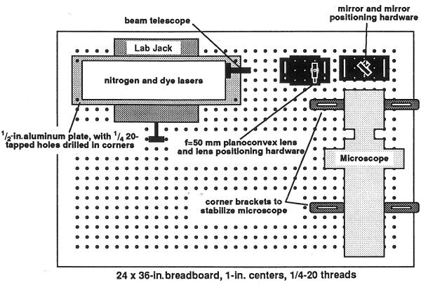
Mechanical arrangement of the laser, microscope, and coupling optics.
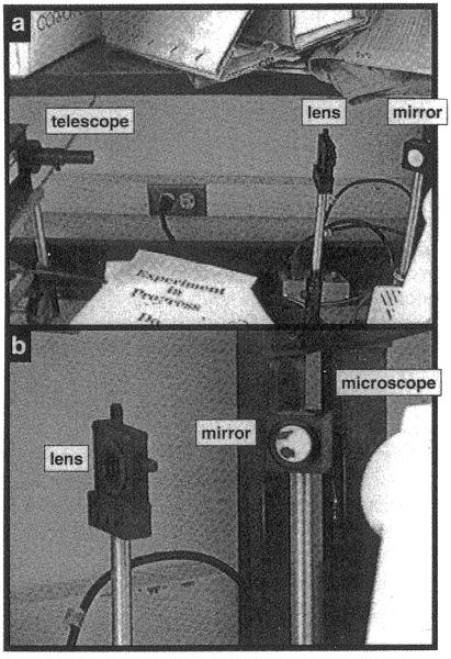
Photographs of the optical elements. (a) The telescope, mounted on the laser, corresponds to lenses w and x in Fig. 5. The lens corresponds to lens y of Fig. 5. Lens z of Fig. 5b is inside the microscope. The mirror (not shown in Fig. 5) is between lenses y and z. The lens, mirror, and laser are mounted on steel posts. The lens and mirror mounts allow their positions to be adjusted. (b) Closeup showing the lens, mirror, and back of the microscope.
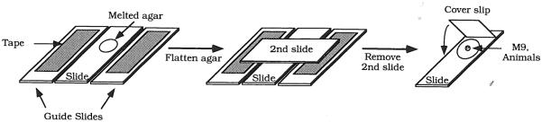
Slide preparation. Melted agar is placed on the surface of the slide. A second slide is used to flatten the agar into a thin pad. The thickness of the agar pad is set using two guide slides to which a piece of tape has been attached. After liquid and worms have been placed on the slide, a coverglass is placed on the slide.
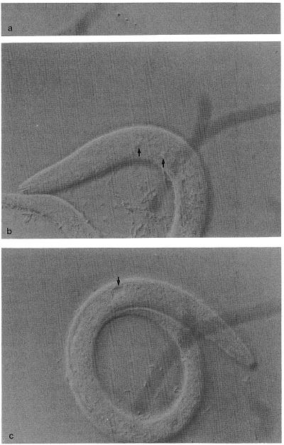
Laser damage. (a) Damage to the glass coverslip, used to locate the laser spot. (b) Damaged nuclei following laser operations (arrows). (c) A ruptured basement membrane (note the fluid-filled cavity).
References
-
- Ashkin A, Dziedzic J, Yamane T. Optical trapping and manipulation of single cells using infrared laser beams. Nature. 1987;330:769–772. - PubMed
-
- Austin J, Kimble J. glp-1 is required in the germ line for regulation of the decision between mitosis and meiosis in C. elegans. Cell. 1987;51:589–599. - PubMed
-
- Avery L, Horvitz HR. Pharyngeal pumping continues after laser killing of the pharyngeal nervous system of C. elegans. Neuron. 1989;3:473–485. - PubMed
-
- Bargmann CI, Horvitz HR. Chemosensory neurons with overlapping functions direct chemotaxis to multiple chemicals in C. elegans. Neuron. 1991a;7:729–742. - PubMed
Publication types
MeSH terms
Grants and funding
LinkOut - more resources
Full Text Sources