Review
. 1995:48:251-69.

Electrophysiological methods

Affiliations
Review

Electrophysiological methods

L Avery et al. Methods Cell Biol. 1995.
No abstract available


Figures

Fig. 1
Fig. 1
Equivalent circuit of the pharynx.
Fig. 2
Fig. 2
Electropharyngeogram electrodes and minirig. (a) Electrode and bath during recording. The bath is about 15 mm in diameter. (b) A worm with its head trapped in the pipet for recording. The worm is slightly more than 1 mm long. (c) Complete rig with dissecting microscope, micromanipulator, microelectrode amplifier, Faraday cage, and oscilloscope. (d) Closeup of the microscope and micromanipulator.
Fig. 3
Fig. 3
Pharynx dissection. The worm is cut transversely through the body just behind the pharynx with a surgical knife. (The knife blade is actually much larger relative to the worm than pictured.) The tail section, consisting of everything posterior to the pharynx, is discarded. The smaller head section contains the pharynx. The muscles underlying the head cuticle contract, exposing most of the pharynx.
Fig. 4
Fig. 4
Typical normal electropharyngeogram.
Fig. 5
Fig. 5
Equivalent circuit of the electropharyngeogram recording arrangement. This figure shows the recording configuration in voltage-clamp mode, where currents are recorded with a low-impedance current-following amplifier diagrammed internally as a short-circuit. In current-clamp recording the current follower is replaced with a high-impedance voltage amplifier, which acts roughly as an open circuit.
Fig. 6
Fig. 6
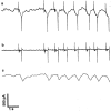
Dissection of the electropharyngeogram into low- and high-frequency components. (a) A typical electropharyngeogram. (b) High-frequency component of a, obtained by high-pass filtering with a time constant of 10 milliseconds. This component is caused mostly by electrical events in pharyngeal muscle. (c) Low-frequency component of a, obtained by low-pass filtering with a time constant of 100 milliseconds. This component comes from many sources, such as drift in the electronic components and probably motions of the worm and the pharynx:
Fig. 7
Fig. 7
Schematic drawing of a simple recording chamber. A rectangular hole is cut in a plexiglass plate. A coverslip is glued (epoxy cement) over the hole on the underside of the plate. Dimensions are in centimeters.
Fig. 8
Fig. 8
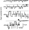
Single-channel currents recorded from a neuron in the cell-attached patch confiiuration. Downward deflections represent inward current. The numbers at the left are pipet potential with respect to the bath (in mV) and the dotted lines indicate zero current. The pipet contained the intracellular solution in Table I. Open probability increased when the patch was hyperpolarized by making the pipet potential more positive. At 80 mV, prolonged closures sometimes occurred revealing an exponential (rather than stepwise) relaxation of the current (arrowhead). This suggests that current flowing through a single ion channel is sufficient to alter the membrane potential of the neuron.

Similar articles

Cited by

References

    1. Albertson DG, Thomson JN. The pharynx of Caenorhabditis elegans. Phil. Trans. R. Soc. Lond. 1976;B275:299–325. - PubMed
    1. Avery L, Horvitz HR. Effects of starvation and neuroactive drugs on feeding in Caenorhabditis elegans. J. Exp. Zool. 1990;253:263–270. - PubMed
    1. Avery L. The genetics of feeding in Caenorhabditis elegans. Genetics. 1993;133:897–917. - PMC - PubMed
    1. Brown KT, Flaming DG. Advanced Micropipette Techniques for Cell Physiology. New York; Wiley: 1986.
    1. Byerly L, Masuda MO. Voltage-clamp analysis of the potassium current that produces a negative-going action potential in Ascaris muscle. J. Physiol. 1979;288:263–284. - PMC - PubMed

MeSH terms

LinkOut - more resources