. 1996 Jan 5;271(5245):64-7.
doi: 10.1126/science.271.5245.64.

Role of mutant CFTR in hypersusceptibility of cystic fibrosis patients to lung infections

Affiliations

Role of mutant CFTR in hypersusceptibility of cystic fibrosis patients to lung infections

G B Pier et al. Science. .

Abstract

Cystic fibrosis (CF) patients are hypersusceptible to chronic Pseudomonas aeruginosa lung infections. Cultured human airway epithelial cells expressing the delta F508 allele of the cystic fibrosis transmembrane conductance regulator (CFTR) were defective in uptake of P. aeruginosa compared with cells expressing the wild-type allele. Pseudomonas aeruginosa lipopolysaccharide (LPS)-core oligosaccharide was identified as the bacterial ligand for epithelial cell ingestion; exogenous oligosaccharide inhibited bacterial ingestion in a neonatal mouse model, resulting in increased amounts of bacteria in the lungs. CFTR may contribute to a host-defense mechanism that is important for clearance of P. aeruginosa from the respiratory tract.


Figures

Fig. 1
Fig. 1
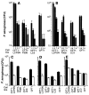
Internalization of P. aeruginosa by 105 transformed airway epithelial cells (2). Cells were grown for 72 hours and bacteria were then allowed to invade the cells for 3 or 4 hours. Bars indicate the mean of the determinations and error bars the SD. Cell lines are as follows: 1, CFT1-LCFSN; 2, CFT1; 3, CFT1-LC3; and 4, CFT1-ΔF508. (A) Cells were grown at 37°C, a temperature that inhibits membrane expression of ΔF508 CFTR (5) and the assay was carried out at 37°C. Multiple comparisons for all three bacterial strains were significant (P < 0.001, ANOVA); the CFT1-LCFSN line was significantly different from any other cell line (P < 0.01, Fisher's PLSD statistic) for all three bacterial strains. (B) Cells were grown at 26°C and internalization was assessed at 26°C. (C) Cells were grown at 26°C and internalization was assessed at 37°C. When cells were grown at 26°C to promote surface expression of ΔF508 CFTR (5), there were no significant (P > 0.2, ANOVA) differences in bacterial uptake among the cell lines for any P. aeruginosa strain tested.
Fig. 2
Fig. 2
Role of the complete outer-core region of P. aeruginosa LPS in internalization by airway epithelial cells (2). Bars indicate the mean of the determinations and error bars the SD. (A and B) Assays with bacterial strains of defined LPS phenotype (9); 1, PAC1 R, wild-type, smooth; 2, PAC557, complete core; 3, PAC1R algC::tet, incomplete core; 4, PAC605, incomplete core; 5, PAO1, wild-type, smooth; 6, AK44, complete core; 7, PAO1 algC::tet, incomplete core; and 8, AK1012, incomplete core. All eight strains were internalized by the CFT1-LCFSN cell line significantly better than by the three cell lines expressing mutant ΔF508 CFTR (P < 0.001, ANOVA; P < 0.05 for all pair-wise comparisons, Fisher PLSD). Bacterial strains with a smooth or complete-core LPS were internalized by all of the cell lines significantly better than strains expressing an incomplete core (P < 0.01, ANOVA; P < 0.05 for all pair-wise comparisons, Fisher PLSD). (C to E) Increased internalization by airway epithelial cells of recombinant LPS-smooth P. aeruginosa strains carrying cloned rfb genes (solid bars) compared with parental clinical isolates expressing an incomplete LPS structure and carrying only the DNA cloning vector (open bars) (10). (C) Strain 2192; (D) strain 332; (E) strain 9125. Ingestion of all of the LPS-smooth strains by all of the cell lines was significantly better than ingestion of the corresponding LPS-incomplete isolate (P ≤ 0.002, unpaired t test), except for strain 9125 by the CFT1 cells. There was also significantly greater (P < 0.001, ANOVA) uptake of all of the LPS-smooth bacteria by the CFT1-LCFSN cells compared with the other three cell lines expressing ΔF508 CFTR.
Fig. 3
Fig. 3
Effect of intact LPS (A) and an oligosaccharide with a lipid A–free core (B) on internalization of P. aeruginosa into CFT1-LCFSN cells. Each point is the mean of three to seven replicates; error bars are SD. Pseudomonas aeruginosa strains: (□), PAO1; (Δ), 149; and (Ο), 324. Solid symbols in (B) represent the mean CFU of bacteria internalized in the presence of intact LPS (100 μg/ml) from strain PAC1R algC::tet, which has an incomplete-core oligosaccharide. Amounts of internalization < 104 CFU differed significantly (P < 0.01, ANOVA and Fisher PLSD for pair-wise comparisons) from amounts of internalization in the presence of incomplete-core LPS (100 μg/ml).
Fig. 4
Fig. 4
Effect of complete-core oligosaccharide on P. aeruginosa infection in neonatal mouse lungs. Each point indicates the median bacterial CFU for 8 to 10 lungs obtained from each group of mice and the bars indicate the upper and lower quartiles. Inhibitor delivered with inoculum: (●), none; (□), complete-core oligosaccharide; and (■), incomplete-core oligosaccharide. Groups of 7-day-old neonatal BALB/c mice were infected intranasally with ~ 108 CFU of strain PAO1 (14) alone or mixed with either complete-core oligosaccharide (50 μg) or incomplete-core oligosaccharide (50 μg) (11). After 1, 24, or 48 hours, four to five mice were killed and their lungs were removed, weighed, and dispersed into single-cell suspensions. The total CFU of bacteria present in each lung was determined from a sample treated with Triton X-100 (0.5%) to release intracellular bacteria. The remaining cells were treated with gentamicin (300 μg/ml) for 60 min to kill extracellular P. aeruginosa, then the cells were centrifuged, washed twice in tissue-culture medium, and resuspended in 200 μl of 0.5% Triton X-100 to release intracellular bacteria. These suspensions were diluted and plated. Differences among groups were analyzed by nonparametric statistics: P < 0.0001, Kruskall-Wallis nonparametric ANOVA; P < 0.001, Dunn procedure for individual pair-wise differences between the groups at 1 and 24 hours. Also, at 1 hour the group receiving the incomplete-core oligosaccharide had a reduced amount (P = 0.05, Dunn procedure) of intracellular bacteria compared with the group receiving nothing with the inoculum. At 48 hours, the group treated initially with complete-core inhibitor had significantly more bacteria in their lungs (P = 0.003, Kruskall-Wallis; P < 0.05, Dunn procedure for all pair-wise comparisons) than did the other groups.

References

    1. Aronson M, Medalia O, Amichay D, Nativ O. Infect. Immun. 1988;56:1615. - PMC - PubMed
    2. Dalal E, Medalia O, Harari O, Aronson M. ibid. 1994;62:5505. - PMC - PubMed
    3. Orikasa S, Hinman F., Jr. Invest. Urol. 1977;15:185. - PubMed
    1. Cells were released from monolayers in tissue culture flasks by 5-min incubation with a trypsin-versene mixture (BioWhitaker, Walkersville, MD) and washed, counted, and seeded into 96-well tissue culture plates at a concentration of 105 cells per well in supplemented F-12 medium (3). Plates were incubated at either 26° or 37°C in 5% CO2. Fresh cultures of P. aeruginosa grown overnight at 37°C on a tryptic soy agar plate were suspended in supplemented F-12 medium to prepare the bacterial inoculum; then ~106 colony-forming units (CFU; range from 6 × 10⁵ to 2.3 × 10⁶ CFU) were added per well of 105 epithelial cells. Bacteria were allowed to be ingested by the epithelial cells for 3 or 4 hours at either 26° or 37°C, after which nonadherent bacteria were removed by washing. The remainder of the assay and controls were as described [S. M. J. Fleiszig, T. S. Zaidi, G. B. Pier, Infect. Immun. 63, 4072 (1995)]. Three to nine replicates were obtained per point and were analyzed by analysis of variance (ANOVA) and the Fisher PLSD statistic to determine pair-wise differences.

    1. Olsen JC, et al. Hum. Gene Ther. 1992;3:253. - PubMed
    2. Sarkadi B, et al. J. Biol. Chem. 1992;267:2087. - PubMed
    1. Pier GB, DesJardins D, Aguilar T, Barnard M, Speert DP. J. Clin. Microbiol. 1986;24:189. - PMC - PubMed
    1. Denning GM, et al. Nature. 1992;358:761. - PubMed
    2. Denning GM, Ostedgaard LS, Welsh MJ. J. Cell Biol. 1992;118:551. - PMC - PubMed
    3. Denning GM, Ostedgaard LS, Cheng SH, Smith AE, Welsh MJ. J. Clin. Invest. 1992;89:339. - PMC - PubMed

Publication types

MeSH terms

Substances