. 1995 Oct 1;21(2):145-52.
doi: 10.1002/cyto.990210206.

Novel flow-cytometric method for separating cell types in differentiated F9 embryoid bodies

Affiliations

Novel flow-cytometric method for separating cell types in differentiated F9 embryoid bodies

C A Burdsal et al. Cytometry. .

Abstract

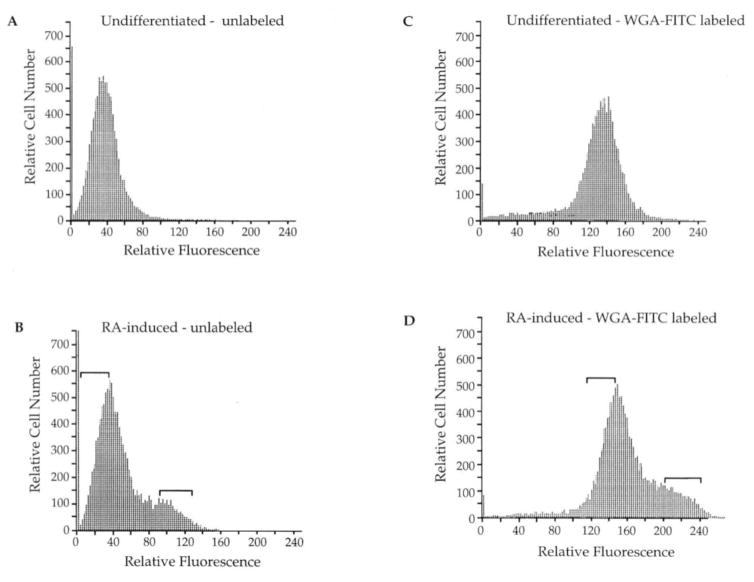
The differentiation of F9 teratocarcinoma cells mimics the formation of a mouse embryonic tissue, the primitive endoderm. In vitro, small aggregates of F9 cells, termed embryoid bodies, differentiate in response to retinoic acid and develop a surface epithelium that is characterized by the production of alpha-fetoprotein. In the present study, cellular autofluorescence profiles obtained by fluorescence-activated embryoid bodies were composed of a single type of cell. In contrast, retinoic acid-induced embryoid bodies were composed of two cell types: a major population displaying autofluorescence levels similar to those of cells from undifferentiated embryoid bodies and a second population displaying higher autofluorescence. RNA analyses demonstrated that the transcription of alpha-fetoprotein was associated only with the more highly autofluorescent population, indicating that flow cytometry provides a novel mechanism for the separation of undifferentiated cells from differentiated endoderm cells in F9 embryoid bodies.


Figures

Fig 1
Fig 1
Morphology of undifferentiated and RA-induced embryoid bodies. A: Bright-field photograph of undifferentiated F9 embryoid bodies. B: Bright-field photograph of RA-induced embryoid bodies. In C and D embryoid bodies were embedded in plastic, sectioned, and stained with hematoxylin and eosin. C: Undifferentiated embryoid bodies D: RA-induced embryoid body. Size bar for A and B shown in 9. Size bar for C and D shown in D. Bar = 40 μm.
Fig. 2
Fig. 2
Immunocytochemical detection of AFP expression. Embryoid bodies were fixed in acidified ethanol, embedded in paraffin, and stained with a polyclonal antibody specific for AFP. A: Undifferentiated F9 embryoid body. B: RA-induced embryoid body. Bar = 20 μm.
Fig. 3
Fig. 3
Fluorescence profiles of unlabeled and WGA-FITC-labeled embryoid bodies. Cellular fluorescence measurements are presented on a single-parameter logarithmic display. A: Autofluorescence profile of cells from unlabeled, undifferentiated F9 embryoid bodies. B: Autofluorescence profile of cells from unlabeled, RA-induced embryoid bodies. C: Fluorescence profile of cells from WGA-FITC-labeled, undifferentiated embryoid bodies. D: Fluorescence profile of two cell populations from WGA-FITC-labeled, RA-induced embryoid bodies. These profiles were taken from a single collection; however, the same profiles were reproducible over all collection periods. The small signal in the left corner of histograms represents debris resulting from trypsinization of the samples. Brackets indicate sorting windows for the collection of cells.
Fig. 4
Fig. 4
Confirmation of sorting parameters. The two cell populations collected from WGA-FITC-labeled, RA-induced embryoid bodies were re-analyzed to assess the purity provided by sorting parameters. A: Fluorescence profile of cell populations of WGA-FITC-labeled, RA-induced embryoid bodies before separation and collection. B: Fluorescence profile of the same populations after collected cells were recombined in equal numbers.
Fig. 5
Fig. 5
RNA analysis of AFP and TPI expression. Total RNA was prepared from fetal liver, adult liver, intact embryoid bodies, and sorted cell populations. RNAs were fractionated by agarose gel electrophoresis, blotted onto nitrocellulose, and hybridized to 32P-labeled AFP cDNA (upper panel) or 32P-labeled TPI cDNA (lower panel). Lane 1: 15 μg of fetal liver RNA; lane 2: 1:4 dilution of fetal liver RNA; lane 3: 1:16 dilution of fetal liver RNA; lane 4: RNA from intact undifferentiated F9 embryoid bodies; lane 5: RNA from intact RA-induced embryoid bodies; lane 6: RNA from the low autofluorescent sorted population of unlabeled, RA- induced embryoid bodies; lane 7: RNA from the high autofluorescent sorted population of unlabeled RA-induced embryoid bodies; lane 8: RNA from the low fluorescent sorted population of WGA-FITC-labeled, RA-induced embryoid bodies; lane 9: RNA from the high fluorescent sorted population of WGA-FITC-labeled, RA-induced embryoid bodies; lane 10: empty lane; lane 11: adult liver RNA.

References

    1. Adamson EA, Grover A. The production and maintenance of a functioning epithelial layer from embryonal carcinoma cells. In: Silver LM, Martin GR, Strickland S, editors. Teratocarcinoma Stem Cells. Cold Spring Harbor Press; Cold Spring Harbor, NY: 1983. pp. 69–81.
    1. Aubin JE. Autofluorescence of viable cultured mammalian cells. J Histochem Cytochem. 1979;27:36–43. - PubMed
    1. Becker S, Casanova J, Grabel LB. Localization of endoderm-specific mRNAs in differentiating F9 embryoid bodies. Mech Devel. 1992;37:3–12. - PubMed
    1. Benson RC, Meyer RA, Zaruba ME, McKhann GM. Cellular autofluorescence—is it due to flavins? J Histochem Cytochem. 1979;27:44–48. - PubMed
    1. Bernstine EG, Hooper ML, Grandchamp S, Ephrussi B. Alkaline phosphatase activity in mouse teratoma. Proc Natl Acad Sci USA. 1973;70:3899–3903. - PMC - PubMed

Publication types

LinkOut - more resources