. 1995 Nov;121(11):3583-92.
doi: 10.1242/dev.121.11.3583.

Neurofibromin-deficient fibroblasts fail to form perineurium in vitro

Affiliations

Neurofibromin-deficient fibroblasts fail to form perineurium in vitro

T Rosenbaum et al. Development. 1995 Nov.

Abstract

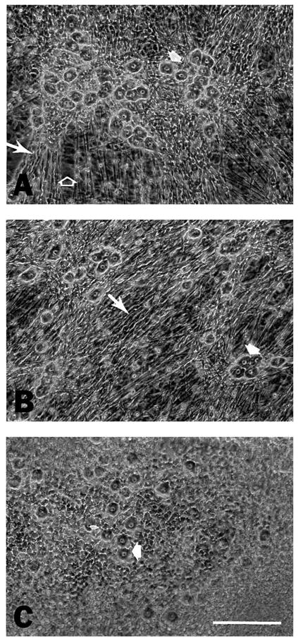
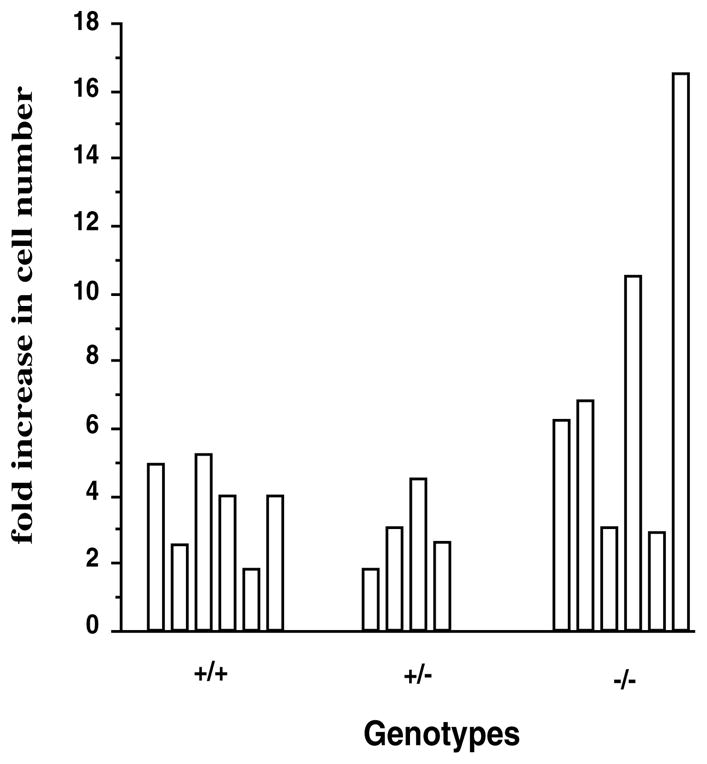
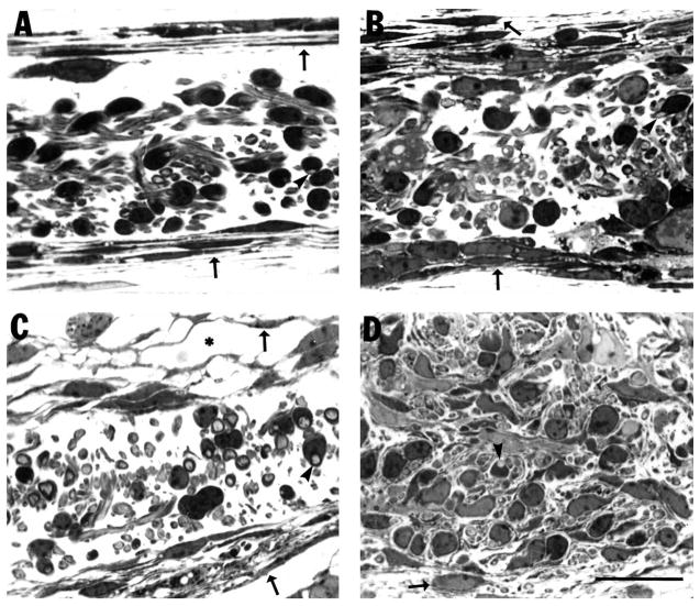
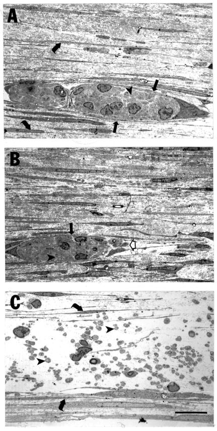
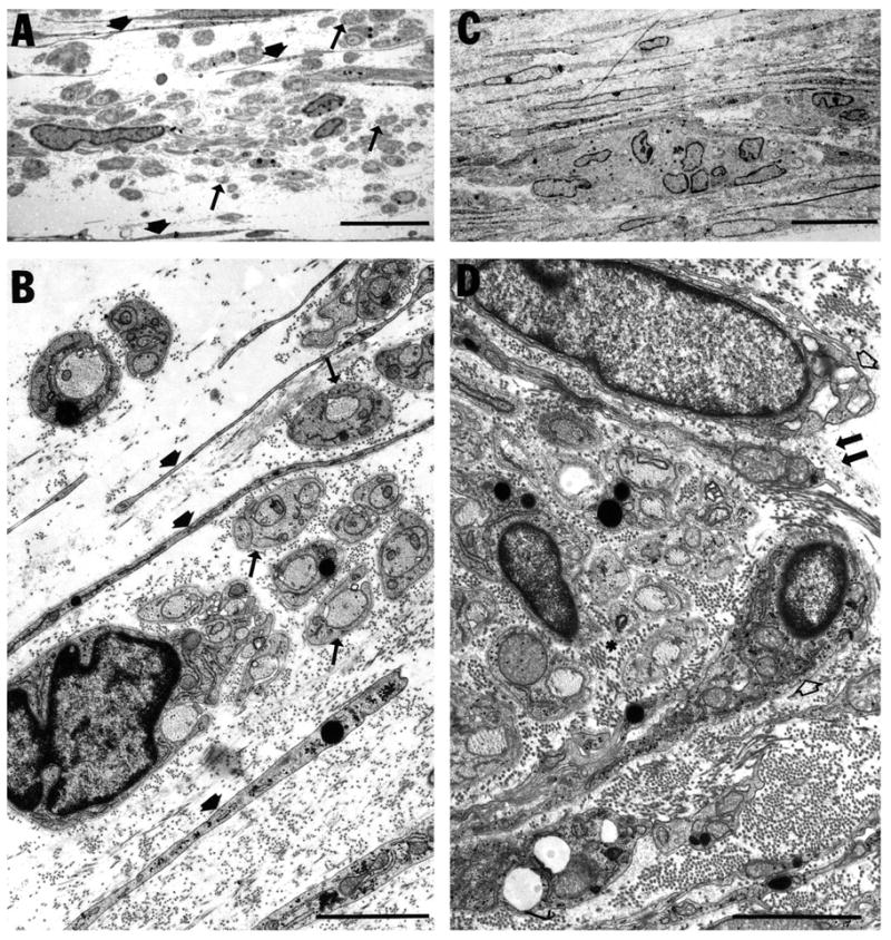
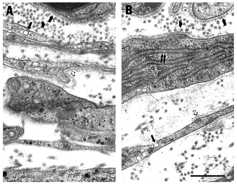
To identify cell type(s) that might contribute to nerve sheath tumors (neurofibromas) in patients with neurofibromatosis type 1, we generated cell cultures containing neurons. Schwann cells and fibroblasts from transgenic mouse embryos in which the type 1 neurofibromatosis gene was disrupted by homologous recombination (Brannan et al. (1994) Genes Development, 8,1019-1029). Normal fascicle formation by perineurial cells failed to occur in the absence of neurofibromin. Fascicles were reduced in number and showed abnormal morphology when normal neurons and Schwann cells were cultured up to 37 days with fibroblasts lacking neurofibromin. Proliferation was increased in a majority of fibroblast cell strains analyzed from embryos lacking neurofibromin. These observations suggest that mutations in the neurofibromatosis type I gene affect fibroblast behavior that might contribute to neurofibroma formation in patients with neurofibromatosis type 1.


Figures

Fig. 1
Fig. 1
Morphology of 9 day old organotypic cell cultures from Nf1 mice by phase-contrast microscopy. Cell suspensions containing neurons, Schwann cell precursors and fibroblasts were obtained from embryonic day 12.5 mice and plated onto collagen-coated 35 mm dishes. Neurons were identified as round phase-dark cells with prominent nuclei and nucleoli (white arrowheads) that extended processes. Schwann cells were defined based on their attachment to neuronal processes (arrows), fibroblasts formed a cellular background layer (open arrowheads). While neuronal processes and accompanying Schwann cells were easily identified in wild-type (A) and heterozygous (B) cultures, in many homozygous null cultures (C) Schwann cell-covered axons appeared more obscured. Bar, 160 μm.
Fig. 2
Fig. 2
Increased growth rate in neurofibromin-deficient fibroblasts. Fresh fibroblasts derived from single E12.5 mouse embryos were plated onto 24-well or 6-well plates in DMEM with 10% FBS at a density of 2500 or 5000 cells/cm2, respectively. Every second day after plating, cell number was determined by counting duplicate samples in a hemocytometer. After 8 days in vitro the fold increase in cell number/cm2 was calculated for each fibroblast culture.
Fig. 3
Fig. 3
Failure of organization in cultures lacking neurofibromin. Cell cultures containing neurons, Schwann cells and fibroblasts were obtained from wild-type, Nf1 (+/−) or Nf1 (−/−) mouse embryos, fixed after 18 days in vitro and analyzed in semi-thin plastic sections. Wild-type cultures showed a high level of organization with axon-Schwann cell units sandwiched between multiple layers of elongated fibroblasts (A). In most cultures lacking neurofibromin, fibroblasts were loosely arranged in two multilayers sandwiching axon-Schwann cell units (C). In some Nf1 (−/−) cultures, this organizational pattern was completely disrupted (D) and fibroblasts were intermingled with axon-Schwann cell units. Cultures from Nf1 (+/−) animals exhibited an intermediate phenotype (B). Arrows point to layers of fibroblasts above and below axon-Schwann cell units, arrowheads to axons associated with Schwann cells. In C, * denotes spaces between adjacent fibroblasts layers. Bar, 30 μm.
Fig. 4
Fig. 4
Fibroblasts lacking neurofibromin fail to fasciculate axons and Schwann cells. Fibroblasts derived from E 12.5 Nf1 mouse embryos were added to cultures of normal rat neurons fully populated with rat Schwann cells. 30 days after addition of fibroblasts, cultures were fixed and analyzed in semithin plastic sections. In cultures containing wild-type fibroblasts (A), axon-Schwann cell units (arrowhead) were organized into compact fascicles and surrounded by flattened fibroblasts (arrows). Fascicles were further sandwiched between compact multilayers of fibroblasts (curved arrow). In most cultures containing fibroblasts without neurofibromin (C), fascicle-formation did not occur. Axon-Schwann cell units (arrowhead) were loosely arranged between two multilayers of fibroblasts (curved arrow). Cultures containing Nf1 (+/−) fibroblasts (B) appeared well organized but often showed incomplete perineurium-formation (open arrow). Bar, 15 μm.
Fig. 5
Fig. 5
Abnormal fascicles in representative cultures containing Nf1 (−/−) fibroblasts. Most cultures containing fibroblasts without neurofibromin (prepared as described in Fig. 4) did not show any fascicle-formation at all (A,B). Axon-Schwann cell units (arrows) remained distant from each other and were intermingled with fibroblasts (short filled arrows). Some regions, however, showed a high level of organization with apparently normal fascicle-formation (C). Examination at higher magnification (D) revealed that ensheathment of axon-Schwann cell bundles by perineurial fibroblasts (open arrow) was in fact incomplete (double arrow) and endoneurial space was increased (asterisk). Bars: A,C, 10 μm; B,D, 2 μm.
Fig. 6
Fig. 6
Perineurial cells differentiate in the absence of neurofibromin. Cultures were prepared as described in Fig. 4. Both wild-type (A) and Nf1 (−/−) (B) mouse fibroblasts exhibit characteristics of perineurial cell differentiation. Cells become elongated, are covered with patchy basal lamina (open arrows) on both sides and contain caveolae (arrows). Collagen fibers of similar diameter are present in both cultures (short filled arrows). Basal lamina also covered Schwann cells in cultures (curved arrows). In neurofibromin-deficient cultures, however, relatively undifferentiated fibroblasts (with distended rER) occurred adjacent to axon-Schwann cell units (double arrow). Bar, 0.25 μm.

Similar articles

Cited by

References

    1. Barbacid M. ras genes. Annu Rev Biochem. 1987;56:779–827. - PubMed
    1. Baron P, Kreider B. Axons induce differentiation of neurofibroma Schwann-like cells. Acta Neuropathol Berl. 1991;81:491–495. - PubMed
    1. Basu TN, Gutmann DH, Fletcher JA, Glover TW, Collins FS, Downward J. Aberrant regulation of ras proteins in malignant tumour cells from type 1 neurofibromatosis patients. Nature. 1992;356:713–715. - PubMed
    1. Bolande RP. The neurocristopathies: a unifying concept of disease arising in neural crest maldevelopment. Hum Pathol. 1974;5:409–429. - PubMed
    1. Bottenstein J, Sato GH. Growth of a neuroblastoma cell line in serum free supplemented medium. Proc Natn Acad Sci USA. 1979;76:514–517. - PMC - PubMed

Publication types

MeSH terms