. 1996 Apr 4;380(6573):451-3.
doi: 10.1038/380451a0.

Direct observation of single kinesin molecules moving along microtubules

Affiliations

Direct observation of single kinesin molecules moving along microtubules

R D Vale et al. Nature. .

Abstract

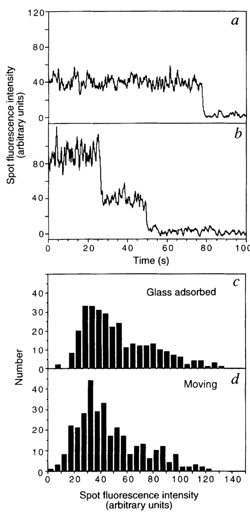
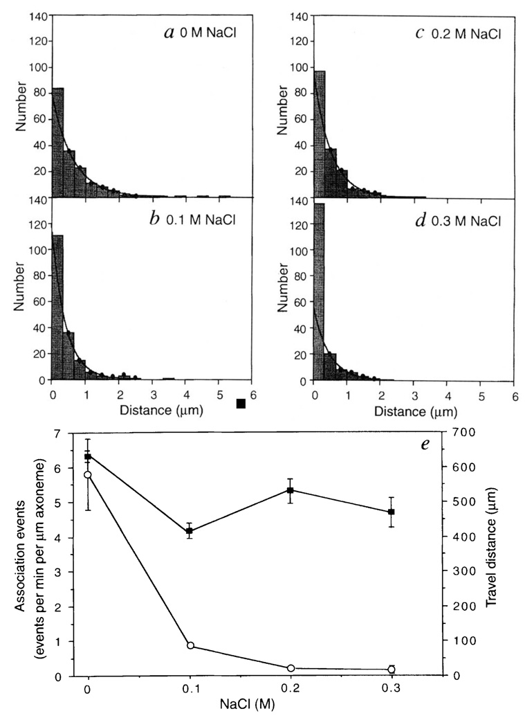
Kinesin is a two-headed motor protein that powers organelle transport along microtubules. Many ATP molecules are hydrolysed by kinesin for each diffusional encounter with the microtubule. Here we report the development of a new assay in which the processive movement of individual fluorescently labelled kinesin molecules along a microtubule can be visualized directly; this observation is achieved by low-background total internal reflection fluorescence microscopy in the absence of attachment of the motor to a cargo (for example, an organelle or bead). The average distance travelled after a binding encounter with a microtubule is 600 nm, which reflects a approximately 1% probability of detachment per mechanical cycle. Surprisingly, processive movement could still be observed at salt concentrations as high as 0.3 M NaCl. Truncated kinesin molecules having only a single motor domain do not show detectable processive movement, which is consistent with a model in which kinesin's two force-generating heads operate by a hand-over-hand mechanism.


Figures

FIG. 1
FIG. 1
Movement of a single fluorescently labelled kinesin molecule along an axoneme. a, Kinesin (uhK560cys) labelled at the C-terminal cysteine residue with Cy3 fluorescent dye was combined with Cy5-labelled axonemes in the presence of ATP. The specimen was illuminated by the evanescent field resulting from total internal reflection of a laser beam at the boundary between the fused silica slide and the aqueous solution. b, Cy5-axoneme. c, uhK560-Cy3. The caret (^) marks a stationary fluorescent spot, and the arrows track a moving fluorescence spot (time in seconds). Other spots visible in individual panels are ones that associated briefly (< 2 s) with the axoneme. Bar, 5 µm. METHODS. uhK560 with an additional C-terminal peptide containing a reactive cysteine (PSIVHRKCF) was expressed in Escherichia coli and purified by phosphocellulose followed by mono-Q chromatography. uhK560-Cy3 (concentration determined from absorbance at 280 nm, A280; ref. 22) was labelled to a stoichiometry of 0.1 to 0.5 dyes per polypeptide chain as described. The uhK560-Cy3 preparation was centrifuged at 100,000 r.p.m. for 10 min in a table-top ultracentrifuge to remove any aggregates; additional microtubule-affinity purification was sometimes performed to improve motility. Analysis of low concentrations of uhK560-Cy3 by sucrose gradient velocity sedimentation (5 S) and Superose-6 gel filtration chromatography indicates that it is dimeric. The concentrations used in the sucrose gradient (5 nM) suggest that uhK560-Cy3 remains dimerized under the conditions of the assay. The microtubule-stimulated ATPase properties of uhK560 labelled with Cy3 at a 0.43 molar ratio of dye to polypeptide (kcat = 22.2 ± 1.0 s−1/site; K0.5,MT = 1.1 ± 0.2 µM) and at 0.96 stoichiometry (kcat = 19.2 ± 0.2 s−1/site; K0.5,MT = 1.6 ± 0.06 µM) were similar to that of the unlabelled protein (kcat = 25.8 ± 0.8 s−1/site; K0.5,MT = 0.7 ± 0.08 µM) (measured in the motility buffer: 12 mM PIPES, pH 6.8, 2 mM MgCl2, 1 mM EGTA). Cy5-labelled axonemes were combined with 0.5–1.5 nM uhK560-Cy3 in motility buffer containing 1 mM ATP, 7.5 mg ml−1 BSA, 0.5% mercaptoethanol and an oxygen scavenger system at 25 °C. BSA inhibited nonspecific adsorption of kinesin but did not prevent axonemes from attaching to the slide. Details of low-background total internal reflection fluorescence microscopy are described elsewhere. In these experiments, 5.8 ± 1.0 association events per min per µm axoneme (n = 8 axonemes; 5-min observation for each axoneme) were observed when the uhK560-Cy3 concentration was 0.6 nM, whereas only 0.05 ± 0.01 events per min were scored on glass without an axoneme. Kinesin spots moving on an axoneme disappeared at 0.42 ± 0.03 s−1, which was significantly faster than the rate of photobleaching of glass-attached kinesin (0.02 ± 0.0005 s−1).
FIG. 2
FIG. 2
Photobleaching properties and fluorescence intensities of fluorescently labelled kinesin. uhK560-Cy3 was adsorbed onto a fused-silica slide, and the intensities of individual fluorescent spots plotted against time. Typical examples of one-step (a) or two-step (b) photobleaching events are shown, reflecting one or two Cy3 dye molecules attached to kinesin, respectively. The distribution of the fluorescence intensities of isolated uhK560-Cy3 spots adsorbed onto the slide (c) or moving along the axoneme (d) are comparable, which suggests that moving fluorescent spots are single molecules and not higher-order aggregates. Intensities were measured using a rolling 8-frame average to reduce fluctuations, summing intensities from a 7 × 7 pixel window, and subtracting the background intensity from an adjacent region on the slide. The intensities of two dye molecules (b) and three dye molecules (not shown) are in the linear range of the camera, as photobleaching caused approximately equal decreases in intensity.
FIG. 3
FIG. 3
Effects of ionic strength on the travel distance and association frequencies of uhK560-Cy3. The distances travelled by single uhK560-Cy3 molecules in motility buffer containing 0 M (a), 0.1 M (b), 0.2 M (c) or 0.3 M NaCl (d) are shown. The dots indicate the bins that were used for exponential fitting. In e, the travel distances (filled squares) in ad are compared with that of uhK560-Cy3 association events with the axoneme (open circles). Mean and standard deviations of association events were scored from 5–12 different axonemes (5–40 associations per axoneme). The velocities of uhK560–Cy3 that travelled for > 0.5 µm were 300 ± 60, 380 ± 120, 310 ± 80 and 250 ± 60 nm s−1 at 0, 0.1, 0.2 and 0.3 M NaCl, respectively (n = 60, 41, 52 and 20). METHODS. Measurements of solitary uhK560-Cy3 molecules were made by tracking fluorescent spots that were positioned over the axoneme for at least 5 video frames. Static associations of > 3 s were rare, and were excluded as they probably represented nonspecific associations. Although travel distances of < 0.3 µm could not be measured accurately, all measurable associations, including those with < 0.3 µm translocations, are included in these histograms. As the first bin may contain nonspecific associations with the axoneme, it was excluded from the two-parameter exponential fits. The means determined from the decay constants (630, 415, 520 and 470 nm at 0, 0.1, 0.2, 0.3 M NaCl, respectively) were in general agreement with the distribution averages (620, 430, 500 and 300 nm).

Similar articles

Cited by

References

    1. Bloom G, Endow S. Motor Proteins 1: Kinesin. London: Academic; 1994. - PubMed
    1. Hackney DD. Nature. 1995;377:448–450. - PubMed
    1. Gilbert SP, Webb MR, Brune M, Johnson KA. Nature. 1995;373:671–676. - PMC - PubMed
    1. Funatsu T, Harada Y, Tokunaga M, Saito K, Yanagida T. Nature. 1995;374:555–559. - PubMed
    1. Yang JT, Laymon RA, Goldstein LS. Cell. 1989;56:879–889. - PubMed

Publication types