. 1996 Apr 11;380(6574):550-5.
doi: 10.1038/380550a0.

Crystal structure of the kinesin motor domain reveals a structural similarity to myosin

Affiliations

Crystal structure of the kinesin motor domain reveals a structural similarity to myosin

F J Kull et al. Nature. .

Abstract

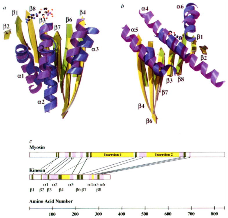
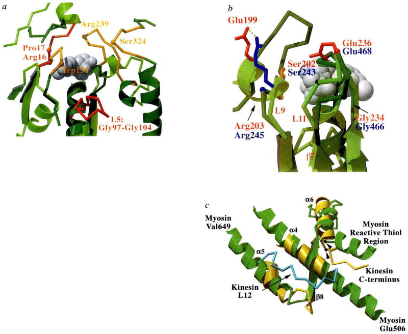
Kinesin is the founding member of a superfamily of microtubule based motor proteins that perform force-generating tasks such as organelle transport and chromosome segregation. It has two identical approximately 960-amino-acid chains containing an amino-terminal globular motor domain, a central alpha-helical region that enables dimer formation through a coiled-coil, and a carboxy-terminal tail domain that binds light chains and possibly an organelle receptor. The kinesin motor domain of approximately 340 amino acids, which can produce movement in vitro, is much smaller than that of myosin (approximately 850 amino acids) and dynein (1,000 amino acids), and is the smallest known molecular motor. Here, we report the crystal structure of the human kinesin motor domain with bound ADP determined to 1.8-A resolution by X-ray crystallography. The motor consists primarily of a single alpha/beta arrowhead-shaped domain with dimensions of 70 x 45 x 45 A. Unexpectedly, it has a striking structural similarity to the core of the catalytic domain of the actin-based motor myosin. Although kinesin and myosin have virtually no amino-acid sequence++ identity, and exhibit distinct enzymatic and motile properties, our results suggest that these two classes of mechanochemical enzymes evolved from a common ancestor and share a similar force-generating strategy.


Figures

FIG. 1
FIG. 1
Structure of the human kinesin motor domain. a, Stereo view showing electron density in the MgADP binding area from the final SIGMAA weighted 1.8-Å 2FoFc map. The final refined structure is shown. The red spheres indicate water molecules and the purple sphere indicates the magnesium ion. Map contours are a 1σ. b, MOLSCRIPT rendition of the kinesin structure, in which β-strands are shown in green, α-helices in purple, and loops in cyan. The ADP is shown as a ball-and-stick figure. The small lobe containing a three-stranded β-sheet is seen in the upper left of the molecule. Two adjacent strands of the core β-sheet, β4 (amino acids 126–138) and β6 (206–216), are unusually long and run the entire length of the molecule; one of the edge strands (β5; 141–144 and 171–173) contains an insertion of 24 amino acids that forms an extended loop with two short β-strands that are isolated from the main β-sheet. Several of the α-helices also exhibit unusual features. Most unusual is α2 (91–122), which contains a 9-amino-acid insertion (97–104) flanked by two conserved glycines. This ‘hairpin’ loop extends perpendicular to the helix, but leaves the overall helical path of α2 unperturbed. Helices α4 (257–269) and α5 (281–292) are notable in that they are oriented at 45° to the β-sheet, unlike the other four helices that are in a parallel orientation. These helices are the only ones linked directly by a short loop rather than by an intervening β-strand. Although loop L11 is shown, it is disordered at the tip (238–254) and is not part of the present model.
FIG. 2
FIG. 2
Comparison of overlapping secondary structure in kinesin and myosin. The overlap was achieved by aligning nine α-carbon atoms (kinesin amino acids 84–92 and myosin 178–186) in the P-loops of the two structures. The similarity between kinesins and myosins was first noted when the ncd tertiary structure was aligned with other proteins in the coordinate data bank (L. Holm, personal communication). a, Front view of the molecule, showing overlapping β-sheets and the three front α-helices. Secondary structure elements of kinesin are indicated. Myosin residue numbers refer to chicken skeletal muscle myosin. Shown are: β-strands (kinesin (k) yellow; myosin (m), green); left to right, β2, k50–k52, m116–119; β1, k9–k15, m122–m126; β8, k295–k302, m668–m675; β3, k79–k84, m173–m177; β7, k222–k231, m457–m463; β6, k206–k216, m247–m255; β4, k126–k138, m263–m268. Helices (kinesin, blue; myosin, purple); left to right, α1, k58–k74, m155–m169; α2, k91–k122, m186–m199; α3, k176–k189, m220–m231. b, Rear view of the molecule, showing overlapping β-sheets and the three rear α-helices. β-strands, same as in a, but rotated by 180°; α-helices, left to right, α5, k281–k292, m649–m665; α4, k257–k269, m475–m506; α6, k306–k320, m690–m697. c, Placement of corresponding structural elements in the linear sequence of kinesin and myosin. Helices and strands are indicated in purple and green, respectively. Insertions in the core motor domains are shown in yellow. In myosin, these insertions contain elements that interact with actin. Lines connect overlapping structural elements; broken lines indicate structural elements that are in a different relative location in the primary sequence; the asterisk indicates the position of the P-loops.
FIG. 3
FIG. 3
Nucleotide environment of kinesin and comparison of functional regions with myosin, a, Loops surrounding the entrance to the nucleotide-binding pocket are shown in this view of kinesin (light green with red loops) overlaid with myosin (dark green with orange loops). The bound ADP is shown as grey spheres, and highlighted residues and loops are discussed in the text. b, The back of the nucleotide-binding pocket with kinesin in light green and myosin in dark green. Residues that may be involved in γ-phosphate sensing are indicated (kinesin residues labelled in red, myosin in blue). Side chains are coloured red for Glu, blue for Arg, orange for Ser, and grey for Gly. Myosin residues are indicated by their α-carbon positions. The bound ADP is shown as grey spheres. This view has been rotated by approximately 180° relative to that in a. c, Structures potentially involved in transducing conformational changes from the nucleotide and filament binding site to a mechanical amplifier in kinesin (yellow) and myosin (green). Secondary structure elements of kinesin are indicated. Orientation is similar to that in Fig. 2b. The kinesin loop (L12; amino acids 272–280) that may be involved in microtubule binding is shown in light blue. The 142-amino-acid actin-binding domain of myosin is not shown, but is located between myosin residues Glu 506 and Val 649. Myosin’s reactive thiol region that may bend or melt during myosin’s ATPase cycle and the C terminus of the current kinesin model are indicated. The position of glycine residues that may be involved as pivot points for conformational movements are shown for myosin Gly 699 and kinesin Gly 319 (arrowheads).

Comment in

References

    1. Bloom G, Endow S. Motor Proteins 1: Kinesin. Academic; London: 1994. - PubMed
    1. Goldstein LS. A Rev Genet. 1993;27:319–351. - PubMed
    1. Yang JT, Saxton WM, Stewart RJ, Raff EC, Goldstein LS. Science. 1990;249:42–47. - PubMed
    1. Hackney DD. Proc natn Acad Sci USA. 1988;85:6314–6318. - PMC - PubMed
    1. Gilbert SP, Webb MR, Brune M, Johnson KA. Nature. 1995;373:671–676. - PMC - PubMed

Publication types

Associated data