. 1996 May 1;16(9):3045-55.
doi: 10.1523/JNEUROSCI.16-09-03045.1996.

Immunological instability of persistent adenovirus vectors in the brain: peripheral exposure to vector leads to renewed inflammation, reduced gene expression, and demyelination

Affiliations

Immunological instability of persistent adenovirus vectors in the brain: peripheral exposure to vector leads to renewed inflammation, reduced gene expression, and demyelination

A P Byrnes et al. J Neurosci. .

Abstract

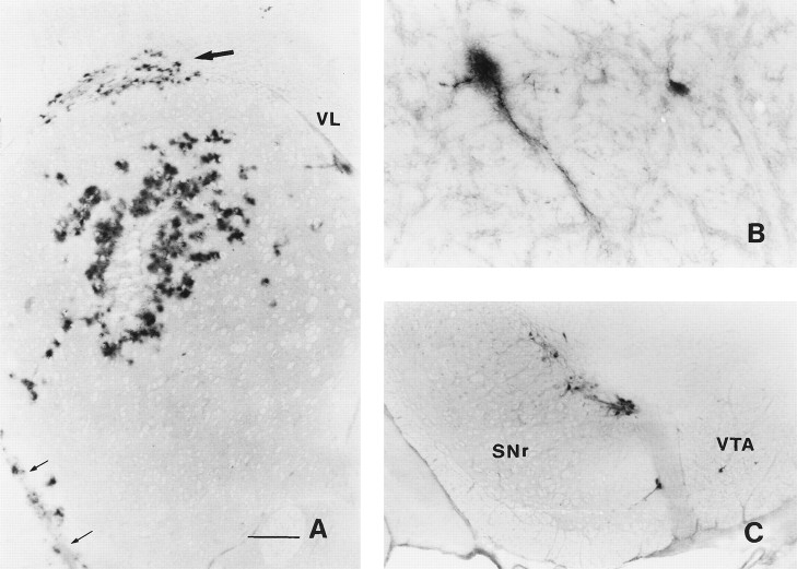
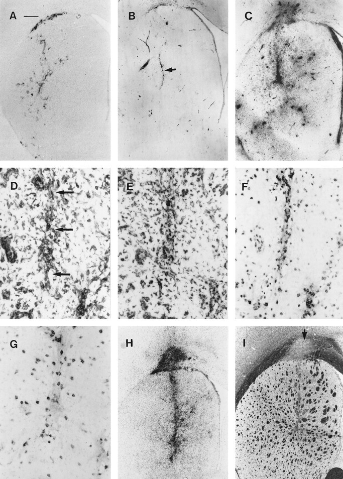
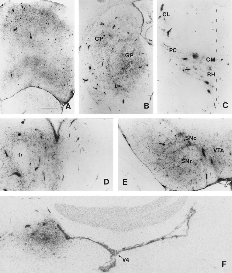
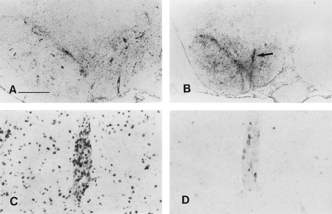
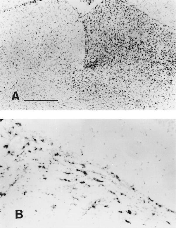
Nonreplicating adenovirus vectors are being developed as vehicles for the delivery of therapeutic genes in vivo. Whereas in many organs an antiviral T cell response eliminates the vector and damages local tissue, when adenovirus vectors are injected into the brain the subsequent immune attack can be ineffective, allowing the vector to persist. In the present study, E1-deleted human adenovirus vectors were injected into the caudate nucleus of rats. Two months later, expression of protein from the vector was still evident and little inflammation was seen. A subcutaneous injection of adenovirus vector at this time, however, led within 2 weeks to severe mononuclear inflammation and microglial activation in the caudate. This caused local demyelination and a decrease in detectable protein expression from the vector. Interestingly, intense microglial activation and numerous lymphocytes and monocytes were also seen in brain areas containing neurons capable of retrogradely transporting the adenovirus vector from the caudate. Control experiments established that this inflammation in distant brain areas was not a nonspecific consequence of degeneration. These experiments demonstrate that although adenovirus vectors can persist in the brain without causing chronic inflammation, they remain the potential target of a damaging cell-mediated immune response brought about by a subsequent peripheral exposure to vector. The finding of lymphocytes in brain areas that project to the caudate further shows that viral antigens that are retrogradely transported by neurons can also be the target of a T cell attack.


Figures

Fig. 1.
Fig. 1.
β-Galactosidase expression 3 d after an injection of AdRL into the right caudate nucleus. A, Numerous β-galactosidase-positive cells were seen surrounding the injection site in the caudate. In addition, positive cells were found in the corpus callosum (large arrow) and along blood vessels. The anterior striate artery is indicated by small arrows. VL, Lateral ventricle. B, Two β-galactosidase-positive cells, one of which is clearly a neuron, are shown in the ipsilateral entopeduncular nucleus (medial globus pallidus). C, Many positive neurons were seen in the ipsilateral substantia nigra, mostly in the pars compacta.SNr, Substantia nigra pars reticulata; VTA, ventral tegmental area. Scale bars, A, 500 μm;B, 85 μm; C, 350 μm.
Fig. 2.
Fig. 2.
Reaction in the brain after peripheral exposure to AdRL. A, Sixty days after injection of AdRL into the caudate, many β-galactosidase-expressing cells were still seen in the caudate and corpus callosum. B, Little MHC I expression was seen at 60 d in the caudate. The injection site is marked by anarrow. MHC I is also seen on vascular endothelium, the ventricular ependyma, and some microglia. C, After a peripheral injection of AdRL into rats that had previously received AdRL in the caudate, MHC I expression was greatly upregulated in the caudate, shown here 3 weeks after the peripheral injection. This was accompanied by many perivascular cuffs containing MHC I-positive leukocytes. D, Expression of MHC II was seen on infiltrating leukocytes in the injection tract (arrows), in nearby perivascular cuffs, and on microglia. Panels D–G are taken from an animal killed 2 weeks after the peripheral AdRL injection. E, ED1-positive macrophages were seen in perivascular cuffs and in the parenchyma, as well as ED1-positive cells with microglial morphology. F, Many T cells expressing the αβ T cell receptor were seen in the cuffs and in the parenchyma.G, Numerous OX-62-positive cells were also found. These were most likely dendritic cells. H, Expression of OX-42, an antigen expressed on macrophages and activated microglia, is shown 4 weeks after a peripheral injection of AdRL. I, In a nearby section from the same animal, staining with luxol fast blue reveals severe demyelination in areas of inflammation. An arrowindicates demyelination in the corpus callosum. Scale bar: A–C, H, I, 420 μm; D–G, 53 μm.
Fig. 3.
Fig. 3.
MHC I expression in afferents to the caudate.A, Contralateral frontal cerebral cortex (mainly secondary motor areas) at 4 weeks after peripheral exposure to AdRL. Note the numerous MHC I-positive perivascular cuffs. B, Ipsilateral globus pallidus (GP) and caudal caudate putamen (CP) at 3 weeks. C, Ipsilateral thalamus at 4 weeks; inflammation is seen in the rostral intralaminar nuclei. The midline is indicated. RH, Rhomboid nucleus; CM, central median; PC, paracentral; CL, central lateral. D, Ipsilateral thalamus: parafascicular nucleus at 3 weeks. Note lack of inflammation on the contralateral side. fr, Fasciculus retroflexus. E, Ipsilateral substantia nigra at 3 weeks. SNc, Pars compacta;SNr, pars reticulata; VTA, ventral tegmental area. F, Ipsilateral locus coeruleus at 4 weeks. No inflammation is seen in the contralateral locus coeruleus.V4, Fourth ventricle. Scale bar: A, B, 1 mm;CE, 500 μm; F, 390 μm.
Fig. 4.
Fig. 4.
Inflammation in the ipsilateral substantia nigra.A, MHC II expression at 4 weeks after peripheral exposure to AdRL. B, OX-42 expression at 3 weeks. No inflammation is seen on the contralateral side of the brain. The blood vessel indicated with an arrow is shown stained with other monoclonal antibodies in C and D. C, T cells that express the αβ T cell receptor. Note their presence both in the perivascular cuff and in the parenchyma of the brain. D, A nearby section stained with OX-39, which detects the high-affinity IL-2 receptor, which is present on activated T cells, indicates that most of the T cells are not activated. Staining with OX-40 found similar small numbers of cells (not shown). Scale bar: A, 500 μm;B, 1 mm; C, D, 125 μm.
Fig. 5.
Fig. 5.
Microglial activation caused by Wallerian degeneration of the optic nerve. A, OX-42 expression in the superior colliculus was upregulated 1 week after sectioning the optic nerve. No excess expression is seen on the other side of the brain, which is innervated by the uncut optic nerve. B, ED1 upregulation in the optic tract, shown at 2 weeks after optic nerve transection. Scale bar: A, = 500 μm; B, 125 μm.

References

    1. Akli S, Caillaud C, Vigne E, Stratford-Perricaudet LD, Poenaru L, Perricaudet M, Kahn A, Pechanski MR. Transfer of a foreign gene into the brain using adenovirus vectors. Nature Genet. 1993;3:224–228. - PubMed
    1. Backes MG, Lund RD, Lagenaur CF, Kunz HW, Gill TJ. Cellular events associated with peripherally induced rejection of mature neural xenografts placed into neonatal rat brains. J Comp Neurol. 1990;295:428–437. - PubMed
    1. Bauer J, Sminia T, Wouterlood FG, Dijkstra CD. Phagocytic activity of macrophages and microglial cells during the course of acute and chronic relapsing experimental autoimmune encephalomyelitis. J Neurosci Res. 1994;38:365–375. - PubMed
    1. Berendse HW, Groenewegen HJ. Organization of the thalamostriatal projections in the rats, with special emphasis on the ventral striatum. J Comp Neurol. 1990;299:187–228. - PubMed
    1. Brenan M, Puklavec M. The MRC OX-62 antigen: a useful marker in the purification of rat veiled cells with the biochemical properties of an integrin. J Exp Med. 1992;175:1457–1465. - PMC - PubMed

Publication types

MeSH terms

Substances