. 1996 May 1;16(9):3056-66.
doi: 10.1523/JNEUROSCI.16-09-03056.1996.

High-frequency oscillations in the output networks of the hippocampal-entorhinal axis of the freely behaving rat

Affiliations

High-frequency oscillations in the output networks of the hippocampal-entorhinal axis of the freely behaving rat

J J Chrobak et al. J Neurosci. .

Abstract

Population bursts of the CA3 network, which occur during eating, drinking, awake immobility, and slow-wave sleep, produce a large field excitatory postsynaptic potential throughout stratum radiatum of the CA1 field (sharp wave). The CA3 burst sets into motion a short-lived, dynamic interaction between CA1 pyramidal cells and interneurons, the product of which is a 200 Hz oscillatory field potential (ripple) and phase-related discharge of the CA1 network. Although many CA1 pyramidal neurons discharge during the time (50-100 msec) of each sharp wave, each wave of a ripple (approximately 5 msec) reflects the synchronization of more discrete subsets of CA1 neurons. When we used multi-site recordings in freely behaving rats, we observed ripples throughout the longitudinal extent (approximately 4-5 mm) of the dorsal CA1 region that were coherent for multiple cycles of each ripple. High-frequency ripples were also observed throughout the hippocampal-entorhinal output pathway that were concurrent but less coherent on a cycle-by-cycle basis. Single and multiunit neuronal activity was phase-related to local ripples throughout the hippocampal-entorhinal output pathway. Entorhinal ripples occurred 5-30 msec after the CA1 ripples and were related to the occurrence of an entorhinal sharp wave. Thus, during each hippocampal sharp wave, there is powerful synchronization among the neuronal networks that connect the hippocampus to the neocortex. We suggest that this population interaction (1) biologically constrains theoretical models of hippocampal function and dysfunction and (2) has the capacity to support an "off-line" memory consolidation process.


Figures

Fig. 1.
Fig. 1.
CA1 ripple and associated neuronal events in the ipsilateral and contralateral CA1 regions. A, Recording locations in ipsilateral and contralateral CA1 regions of hippocampus.B, Hippocampal sharp wave is a synchronized field EPSP in the apical dendritic targets of the CA3 Schaffer collateral input. This depolarization sets into motion a short-lived population interaction between CA1 pyramidal cells and interneurons whose product is an oscillatory field potential (200 Hz) and phase-related discharge of CA1 neurons. Typically, a CA1 pyramidal cell fires a single spike in association with a local ripple (as shown). Upper andmiddle traces, recording from pyramidal layer (100–400 Hz and 0.5–5.0 kHz, respectively). Bottom trace,simultaneously recorded field potential (1–50 Hz) from stratum radiatum. C, Average ripple recorded in septal extent of the dorsal CA1 region. Relationship between negative peak of CA1 ripple (as shown in C) and locally recorded single and multiunit (D) activity and CA1 ripples recorded in the ipsilateral (E) and contralateral hippocampus (F). Note that ripples with the ipsilateral dorsal CA1 region are coherent for three to four cycles of the oscillation, whereas those recorded in the contralateral hippocampus are not.
Fig. 2.
Fig. 2.
Ripples at various positions with the CA1 region can emerge independently. A, B, C, Single 200 msec traces recorded concurrently in the ipsilateral (∼5 mm distance from reference CA1 ripple) and contralateral CA1 pyramidal cell region. Ripples typically occur concurrently at various sites within CA1 (A), but the occurrence, amplitude, and duration can be independent at various sites within CA1 (B, C).
Fig. 3.
Fig. 3.
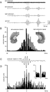
CA1 ripple and associated neuronal events in the ipsilateral and contralateral subiculum. A, Single 400 msec sweep with ripple doublets recorded at three sites and subicular unit.B, Cross-correlograms of ipsilateral (black) and contralateral (gray) subicular ripples to the peak of the CA1 reference ripple (n = 224 CA1 ripples). Note the prominent wave-by-wave coherence on the ipsilateral side and its absence in the contralateral subiculum. Insets inB illustrate position of electrodes in the dorsal subiculum (figures as from Swanson, 1992). C, Averaged subicular ripple (n = 193) and its relation to single (black) and multiunit (gray) activity recorded at the same site. D, Zero reference was negative peak of the local subicular ripple. Inset in D,autocorrelogram of single unit.
Fig. 4.
Fig. 4.
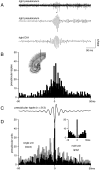
CA1 ripple and associated neuronal events in the deep layers of the ipsilateral presubiculum. A, Single 400 msec sweeps recorded from CA1 and the ipsilateral presubiculum.Top trace illustrates the discharge of a single (*) and multiunit presubicular neurons. B, Cross-correlogram of ipsilateral presubicular ripple to the peak of the CA1 reference ripple (n = 257 CA1 ripples). Inset in Billustrates position of electrode in the dorsal presubiculum.C, Averaged presubicular ripple (n = 213).D, Cross-correlograms of single (black) and multiunit (gray) activity and presubicular ripples; zero reference was negative peak of local presubicular ripple.Inset in D, Autocorrelogram of single unit.
Fig. 5.
Fig. 5.
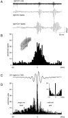
CA1 ripple and associated neuronal events in the deep layers of the ipsilateral entorhinal cortex. A, Single 400 msec sweeps with concurrent ripples in CA1 and the ipsilateral EC.Top trace, discharge of entorhinal neurons at the site where the entorhinal ripple was recorded. B, Cross-correlograms of the ipsilateral entorhinal ripple with the peak of CA1 ripple as zero reference (n = 211). Note the absence of ripple-related modulation in the cross-correlogram. Inset in Billustrates position of electrodes in the rostral EC. C, Averaged entorhinal ripple (n = 213). D, Cross-correlograms of single (black) and multiunit(gray) activity and entorhinal ripples; zero reference was negative peak of entorhinal ripple. Inset in D, Autocorrelogram of single unit.
Fig. 6.
Fig. 6.
Depth profile of an entorhinal sharp wave and its relation to a CA1 ripple. A 16-channel silicon probe (arrow) was used to record concurrently at multiple laminar sites within the entorhinal cortex. The figure illustrates the relation of this large depolarizing input to the dendritic fields of layers V–VI and III neurons throughout the broad expanse of layers IV and III of the entorhinal cortex. Image (right) illustrates the position of the silicon probe with its tip near the superficial border of theEC.ab, Angular bundle.
Fig. 7.
Fig. 7.
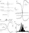
CA1 ripple and associated high-frequency oscillations in the EC. A, Single 400 msec sweeps with concurrent ripples in the CA1 region and both hemispheres of the EC. Note the time lag and lower frequency of the entorhinal oscillations.B, Averaged extracellular fields (wide band) in the hippocampus and ipsilateral EC triggered by the negative peak of CA1 ripples (n = 243). Top trace is average extracellular fields in stratum oriens. Bottom traces are averaged entorhinal sharp waves illustrating reversal near the border of layers II–III. C, Average extracellular fields in the same EC locations triggered by the negative peak of the entorhinal high-frequency oscillation. Top and bottom traces(1–5 kHz) are layers III and II sites as shown in B,respectively. Middle traces are the same sites filtered (100–400 Hz) for ripples. Note reversal of high-frequency oscillation.D, Relation of contralateral entorhinal ripples (black) and ipsilateral CA1 ripples (gray) to the negative peak of the ipsilateral entorhinal ripple (same zero reference as C).

References

    1. Amaral DG, Witter MP. Hippocampal formation. In: Paxinos G, editor. The rat nervous system, 2nd Ed. Academic; Los Angeles: 1995. pp. 443–493.
    1. Bartesaghi R, Gessi T, Sperti L. Electrophysiological analysis of the hippocampal projections to the entorhinal area. Neuroscience. 1989;30:51–62. - PubMed
    1. Bliss TVP, Lømo T. Long-lasting potentiation of synaptic transmission in the dentate area of the anaesthetized rabbit following stimulation of the perforant path. J Physiol (Lond) 1973;232:331–356. - PMC - PubMed
    1. Bliss TVP, Collinridge GL. A synaptic model of memory: long-term potentiation in the hippocampus. Nature. 1993;361:31–39. - PubMed
    1. Boeijinga PH, Lopes da Silva FH. Differential distribution of β and ø EEG activity in the entorhinal cortex of the cat. Brain Res. 1988;448:272–286. - PubMed

Publication types

LinkOut - more resources