. 1996 May 15;16(10):3130-8.
doi: 10.1523/JNEUROSCI.16-10-03130.1996.

cGMP-dependent protein kinase in dorsal root ganglion: relationship with nitric oxide synthase and nociceptive neurons

Affiliations

cGMP-dependent protein kinase in dorsal root ganglion: relationship with nitric oxide synthase and nociceptive neurons

Y Qian et al. J Neurosci. .

Abstract

Nitric oxide and cGMP influence plasticity of nociceptive processing in spinal cord. However, effectors for cGMP have not been identified in sensory pathways. We now demonstrate that cGMP-dependent protein kinase I (cGKl) occurs in the DRGs at levels comparable to that in cerebellum, the richest source of cGKl in the body. Immunohistochemical studies reveal that cGKl is concentrated in a subpopulation of small- and medium-diameter DRG neurons that partially overlap with substance P and calcitonin gene-related polypeptide containing cells. During development, cGKl expression throughout the embryo is essentially restricted to sensory neurons and to the spinal floor and roof plates. Neuronal nitric oxide synthase (nNOS) is coexpressed with cGKl in sensory neurons during embryonic development and after peripheral nerve axotomy. The primary target for cGKl in cerebellum, G-substrate, is not present in developing, mature, or regenerating sensory neurons, indicating that other proteins serve as effectors for cGKl in sensory processing. These data establish sensory neurons as a primary locus for cGMP actions during development and suggest a role for cGKl in plasticity of nociception.


Figures

Fig. 1.
Fig. 1.
cGMP-dependent protein kinase but not nNOS or G-substrate is enriched in dorsal root ganglion extracts. A, Western blotting shows that cGKI occurs at similar levels in crude extracts of DRGs as in cerebellum (Cb) but is only weakly detectable in cerebrocortical (Cx) tissue. B, nNOS is most enriched in cerebellum, is moderately expressed in cerebral cortex, and is only faintly detectable in DRG. C, G-substrate is only detectable in cerebellar extracts and does not occur in cortex or DRG. A, B, Crude tissues were homogenized in 25 mm Tris-HCl buffer, pH 7.4, containing 1 mm EDTA, 100 μm PMSF, and 0.5% Triton X-100. Soluble proteins were resolved by 10% SDS-PAGE (80 μg/lane). ForC, acid-soluble extracts were separated on a 15% SDS-PAGE gel (50 μg/lane). Immunoreactive bands were detected by ECL. Positions of molecular weight standards (in kDa) are indicated on theleft.
Fig. 2.
Fig. 2.
cGKI is enriched in small- and medium-diameter DRG neurons, and expression does not change after axotomy. A, cGKI is heterogeneously expressed in specific neurons of the rat L5 DRG. Examples of densely stained small- and medium-diameter neurons are indicated with a solid arrow. Unstained large cells are identified by an open arrow (magnification 200×).B, C, Expression of cGKI does not dramatically change 18 d after unilateral sciatic nerve axotomy as a similar pattern and density of staining of DRG cells is noted contralateral (CN) and ipsilateral (IP) to the lesion (100×).D, E, nNOS expression is dramatically induced in ipsilateral L5 DRG neurons 18 d after sciatic nerve axotomy (100×). Ten micrometer cryostat sections of DRG were processed for cGKI or nNOS immunohistochemistry and photographed with light-field microscopy.
Fig. 3.
Fig. 3.
cGKI partially overlaps with substance P and CGRP in DRG neurons. Staining of adjacent 6 μm sections shows that cGKI (A) occurs in a greater fraction of DRG cells than substance P (A’). In certain cells, cGKI coexists with substance P (solid arrow), whereas others contain cGKI but not substance P (open arrow) (200×). A cell labeled for cGKI and CGRP (solid arrow) is shown in the middle panels(B, B’). These panels also show a medium-diameter neuron stained for cGKI but not CGRP (open arrow) (400×). Double-immunofluorescent labeling shows colocalization of cGKI (C) and substance P (C’) in a single neuron.
Fig. 4.
Fig. 4.
cGKI is colocalized with substance P in dorsal spinal cord. A, cGKI is restricted to neuronal fibers in laminae I and II of the spinal cord. cGKI is not present in white matter tracks of the dorsal columns (DC) or in spinal neuron cell bodies (50×). B, cGKI also stains the vascular smooth muscle of blood vessels (BV, 100×). C, nNOS occurs in neuronal processes and throughout the dorsal horn (DH) and is present in occasional spinal interneurons (100×). D, Substance P is colocalized with cGKI in laminae I and II of the dorsal horn (100×).
Fig. 5.
Fig. 5.
cGKI is uniquely enriched in DRG tissue extracts during embryonic development. A, Western blotting shows that cGKI is present in DRG, but not brain tissue, in E15 rat extracts. cGKI from E15 DRG comigrates with a similar 74 kDa band from adult cerebellum (Cb). B, Probing a duplicate blot shows that nNOS is expressed at comparable levels in E15 brain and DRG extracts. Solubilized tissue extracts were separated by 10% SDS-PAGE. Five times more protein was loaded from embryonic tissues (100 μg/lane) than from cerebellum (20 μg/lane). Note that E15 DRG extracts were unavoidably contaminated with adjacent spinal tissues and may therefore underestimate true enrichment of cGKI.
Fig. 6.
Fig. 6.
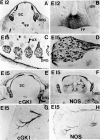
cGKI and nNOS are colocalized in embryonic sensory neurons. At E15, cGKI is apparent only in cells and axonal processes of sensory neurons. Intense staining of the DRG, dorsal cord (DC), and peripheral cutaneous axons (Ax) is noted. nNOS is present in DRG but also occurs in olfactory epithelium (O), cerebrocortical plate (CP), kidney (K), adrenal gland (A), and intestine (In), all of which lack cGKI. Twenty micrometer cryostat sections of E15 rat embryos were stained with antisera to cGKI or nNOS. In these dark-field images, positive staining appears white. Note that fixation conditions for cGKI and nNOS immunohistochemistry are somewhat different and do not allow staining of adjacent sections from the same embryo.
Fig. 7.
Fig. 7.
cGKI expression in embryonic spinal ganglia and floor plate. A, At E12, cGKI is specifically enriched in DRG neurons (100×) and cells of the floor plate (FP) and roof plate (B; 400×). Spinal neurons themselves are unstained, and weak labeling of blood vessels (BV) is detected at this stage. C, D, Parasagittal sections of E15 embryos show that cGKI is enriched in a subset of DRG neurons and is present in both peripheral axons (AX) and central projections to the dorsal spinal cord (DSC). E, F, At E15, nNOS is colocalized with cGKI in DRG and in axonal projections to the dorsal spinal cord (50×). Note that nNOS occurs in certain spinal neurons such as those of the intermediolateral column (IL) that lack cGKI. G, H, Both nNOS cGKI also colocalize in cutaneous branches of sensory axons such as those depicted here near the snout that derive from the trigeminal ganglia.
Fig. 8.
Fig. 8.
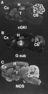
G-substrate and cGKI are colocalized in cerebellum and hippocampus. Immunohistochemistry demonstrates that (A) cGKI is expressed at highest levels in Purkinje cell processes of the molecular layer of the cerebellum (Cb). Lower levels of cGKI are apparent in dentate neurons and pyramidal cells of the hippocampus.B, G-substrate is also enriched in the cerebellum but is restricted to a linear layer of cells that correspond to Purkinje cell soma. The thalamus (T) contains G-substrate immunoreactivity but lacks cGKI. The cerebral cortex and the rest of the forebrain are essentially devoid of cGKI or G-substrate. C, nNOS is also most enriched in cerebellum but displays a broader forebrain distribution than cGKI or G-substrate. Forty micrometer free-floating rat brain sections were stained as described above. In these dark-field images, positive labeling appears white.
Fig. 9.
Fig. 9.
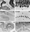
G-substrate and cGKI are coexpressed in Purkinje cell soma. C, D, Immunoperoxidase staining of sagittal sections of rat cerebellum shows that G-substrate is specifically restricted to Purkinje (P) cell bodies and does not stain dendritic processes in the molecular layer (M) or axons in the granule cell layer (G). A,B, cGKI is also uniquely expressed in Purkinje cells but stains dendrites and axons as well as the cell body. E,F, nNOS shows a complimentary distribution to that of G-substrate and cGKI and is uniquely absent from Purkinje cells but, instead, stains granule cells in the granule cell layer and basket cells in the molecular layer.

References

    1. Aanonsen LM, Wilcox GL. Nociceptive action of excitatory amino acids in the mouse: effects of spinally administered opioids, phencyclidine and sigma agonists. J Pharmacol Exp Ther. 1987;243:9–19. - PubMed
    1. Aimi Y, Fujimura M, Vincent SR, Kimura H. Localization of NADPH-diaphorase-containing neurons in sensory ganglia of the rat. J Comp Neurol. 1991;306:382–392. - PubMed
    1. Arancio O, Kandel ER, Hawkins RD. Activity-dependent long-term enhancement of transmitter release by presynaptic 3′,5′-cyclic GMP in cultured hippocampal neurons. Nature. 1995;376:74–80. - PubMed
    1. Aswad DW, Greengard P. A specific substrate from rabbit cerebellum for guanosine 3′:5′-monophosphate-dependent protein kinase. I. Purification and characterization. J Biol Chem. 1981;256:3487–3493. - PubMed
    1. Bredt DS, Snyder SH. Isolation of nitric oxide synthetase, a calmodulin-requiring enzyme. Proc Natl Acad Sci USA. 1990;87:682–685. - PMC - PubMed

Publication types

LinkOut - more resources