. 1996;33(2):106-14.
doi: 10.1002/cm.970330202.

Actin-based motility of isolated axoplasmic organelles

Affiliations

Actin-based motility of isolated axoplasmic organelles

E L Bearer et al. Cell Motil Cytoskeleton. 1996.

Abstract

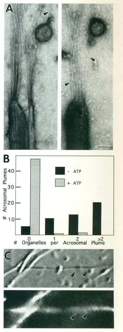
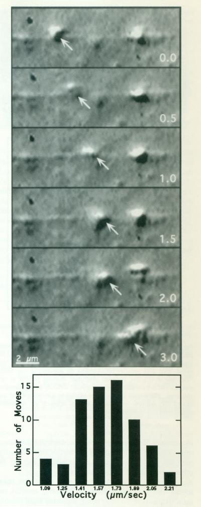
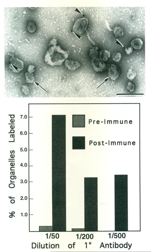
We previously showed that axoplasmic organelles from the squid giant axon move toward the barbed ends of actin filaments and that KI-washed organelles separated from soluble proteins by sucrose density fractionation retain a 235-kDa putative myosin. Here, we examine the myosin-like activities of KI-washed organelles after sucrose density fractionation to address the question whether the myosin on these organelles is functional. By electron microscopy KI-washed organelles bound to actin filaments in the absence of ATP but not in its presence. Analysis of organelle-dependent ATPase activity over time and with varying amounts of organelles revealed a basal activity of 350 (range: 315-384) nmoles Pi/mg/min and an actin-activated activity of 774 (range: 560-988) nmoles/mg/min, a higher specific activity than for the other fractions. By video microscopy washed organelles moved in only one direction on actin filaments with a net velocity of 1.11 +/- .03 microns/s and an instantaneous velocity of 1.63 +/- 0.29 microns/s. By immunogold electronmicroscopy, 7% of KI-washed organelles were decorated with an anti-myosin antibody as compared to 0.5% with non-immune serum. Thus, some axoplasmic organelles have a tightly associated myosin-like activity.


Figures

Fig. 1
Fig. 1
ATP-sensitive binding of KI-washed organelles to acrosomal plumes. A: Actin filaments nucleated off the actin bundle of acrosomal processes of Limulus sperm on EM grids were incubated with mM Mg-ATP. In the absence of ATP, membrane-bound vesicles were frequently associated with the newly polymerized actin filaments (arrowheads) extending in a plume from the end of individual acrosomal processes. Scale bar = 50 nm. B: The histogram compares the number of organelles attached to plumes selected at random from preparations with and without ATP. Assays were performed in 1/2× motility buffer [Vale et al., 1985a]. C: DIC (top) and corresponding fluorescence (bottom) image of a field of rhodamine-phalloidin–stained acrosomal processes with actin filaments polymerized off their ends. Example shows a typical preparation containing a type of long actin-containing plume (arrowheads) developed for the motility assay (Fig. 2).
Fig. 2
Fig. 2
KI-washed organelles move on actin filaments. Upper: High-resolution DIC video sequence of a KI-washed organelle moving along an actin plume. This organelle moves, slows down at 1.5 s, and then speeds up again. Overall, it moves approximately 3 μm in 3 s. The actin plume was nucleated off the end of a KCl-washed acrosomal process, KI-washed, isolated organelles in 1/2× buffer were combined with pre-formed actin filaments in polymerization buffer containing ATP. The numbers in the right lower corner of each image represent the time in seconds. Lower: Movements of seven individual organelles moving on two different extended actin plumes were analyzed by measuring the distanced traveled during a ten-frame sequence in order to determine an instantaneous as opposed to an overall velocity. The ten frames were selected for inclusion if the organelle moved continuously from the first through the tenth frame; 69 such sequences were measured. Velocities were divided into categories according to the smallest incremental distance that could be measured on the video screen.
Fig. 3
Fig. 3
Colloidal gold labeling of KI-washed isolated organelles with anti-myosin antibody. Top: Micrograph of a representative field of negatively stained organelles defined by their limiting membrane (arrows). Four gold particles (arrowheads) are apparent in this field. Scale bar = 0.5 μm. Bottom: Histogram showing percent of organelles which were labeled by anti-myosin antibody or non-immune antiserum. The percent of organelles labeled by anti-myosin did not exceed 7%. Whereas very little label was apparent on organelles treated with non-immune antiserum.

References

    1. Allen RD, Metuzals J, Tasaki I, Brady ST, Gilbert SP. Fast axonal transport in squid giant axon. Science. 1982;218:1127–1128. - PubMed
    1. Bearer EL. Direct observation of actin filament severing by gelsolin and binding by gCap39 and CapZ. J. Cell Biol. 1991;115:1629–1638. - PMC - PubMed
    1. Bearer EL. Cytoskeletal domains in the activated platelet. Cell Motil. Cytoskeleton. 1995;30:50–66. - PMC - PubMed
    1. Bearer EL, DeGiorgis JA, Bodner RA, Kao AW, Reese TS. Evidence for myosin motors on organelles in squid axoplasm. Proc. Natl. Acad. Sci. U.S.A. 1993;90:11252–11256. - PMC - PubMed
    1. Bement WM, Hasson T, Wirth JA, Cheney RE, Mooseker MS. Identification and overlapping expression of multiple unconventional myosin genes in vertebrate cell types. Proc. Natl. Acad. Sci. U.S.A. 1994;91:6549–6553. - PMC - PubMed

Publication types