. 1996 May 17;272(5264):1026-9.
doi: 10.1126/science.272.5264.1026.

Retinal degeneration in mice lacking the gamma subunit of the rod cGMP phosphodiesterase

Affiliations

Retinal degeneration in mice lacking the gamma subunit of the rod cGMP phosphodiesterase

S H Tsang et al. Science. .

Abstract

The retinal cyclic guanosine 3',5'-monophosphate (cGMP) phosphodiesterase (PDE) is a key regulator of phototransduction in the vertebrate visual system. PDE consists of a catalytic core of alpha and beta subunits associated with two inhibitory gamma subunits. A gene-targeting approach was used to disrupt the mouse PDEgamma gene. This mutation resulted in a rapid retinal degeneration resembling human retinitis pigmentosa. In homozygous mutant mice, reduced rather than increased PDE activity was apparent; the PDEalphabeta dimer was formed but lacked hydrolytic activity. Thus, the inhibitory gamma subunit appears to be necessary for integrity of the photoreceptors and expression of PDE activity in vivo.


Figures

Fig. 1
Fig. 1
Targeted disruption of the Pdeg gene in ES cells, and germline transmission of the mutant allele. (A) Physical map of the targeting vector; the wild-type Pdeg locus and the mutant allele are illustrated. The coding exons are indicated by black boxes. Restriction enzyme sites: S, Sal I; Sm, Sma I; B, Bgl II; X, Xba I; Sc, Sac I; A, Acc I; N, Not I; E, Eco RI. N3 and N4 indicate the position of PCR primers. The hatched box shows the position of probe A used to screen for homologous recombination events. (B) Southern blot of ES cell DNAs. After Sac I digestion, hybridization with a full-length Pdeg cDNA probe (18) detects fragments diagnostic of the wild-type (w) allele (4.5 kb) and the targeted (m) allele (6.0 kb). After Xba I digestion, hybridization with probe A detects a wild-type (w) DNA fragment (12.5 kb) and a targeted (m) fragment (14 kb). (C) Southern blot with Sac I–digested tail DNA from offspring of a germline chimera, hybridized with Pdeg cDNA probe (18). (D) Protein immunoblot analysis of retinal homogenate (80 μg) from postnatal day 12 Pdegtml/Pdegtml mutant (−/−) and normal (+/+) offspring obtained from a heterozygote intercross. PDEγ-specific antiserum (HUEY, 1:500 dilution) was used for immunodetection; proteins as small as 3 kD were retained on the blots. Bovine rod outer segment (20 μg) is also shown as a control (ROS).
Fig. 2
Fig. 2
Physiological and morphological characterization of Pdegtml/Pdegtml homozygous mutant mice. (A) Corneal ERGs of a normal mouse (left) compared to those of a mutant mouse (right) at 2 and 8 weeks after birth. Each trace is 0.7 in duration and represents the average of 100 responses of a 1-Hz square-wave flickering stimulus of equal duty cycle. The numbers on the left indicate the log relative energy of the light stimulus. Corneal positivity is shown as an upward deflection. Vertical calibration: 25 μV for the upper trace, 12 μV for the second and third traces, 8 μV for the fourth trace, and 6 μV for the lower two traces the left and all the traces on the right; horizontal calibration: 75 ms for all traces. (B) The relation between the amplitude of the b-wave of the ERG (ordinate) in microvolts and the light intensity of the stimulus (abscissa) in relative logarithmic units for normal (○) and Pdegtml/Pdegtml mutants at 2 (△) and 8 (▲) weeks of age. Each point represents the mean (±SE) of 27 mice for the normal function, 39 mice for the 8 week function, and 6 mice for the 2 week function. (C and D) Transmission electron micrographs of the photoreceptor layer (bar, 2 μm) of 13-day-old control mouse (C) and Pdegtml/Pdegtml mutant mouse (D). The outer segments (indicated by an arrow) of Pdegtml/Pdegtml mutants became disorganized and shortened at this age compared with wild-type mice. (E and F) Light micrograph of the retina from 8-week-old control mouse (E) and Pdegtml/Pdegtml mutant mouse (F). OS, outer segments; IS, inner segments; ONL, outer nuclear layer; INL, inner nuclear layer; GC, ganglion cell layer. The retina of the Pdegtml/Pdegtml mutant mouse (F) has lost the photoreceptor layer (OS, IS, ONL) completely by 8 weeks of age. The inner nuclear and ganglion cell layers (F) appear to be unaffected in the Pdegtml/Pdegtml mouse.
Fig. 3
Fig. 3
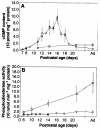
Developmental cGMP content and PDE activity in the retinas of control and mutant mice. (A) cGMP content in the freshly dissected posterior poles of Pdegtml/Pdegtml and wild-type animals during postnatal development (22). The results indicated are means ± SEM of three to nine samples. (B) PDE activity in freshly dissected retinas of Pdegtml/Pdegtml mutant and wild-type mice (23). The results indicated are means ± SEM of three to nine samples. Light- (○) or dark-adapted (●) Pdegtml/Pdegtml mutant retinas and light-adapted control retinas (□) are indicated.
Fig. 4
Fig. 4
Analysis of PDEα and PDEβ subunits in mutant retinal homogenates. (A) Protein immunoblot of PDEα and PDEβ subunits. Proteins from retinal homogenate (80 μg) were separated by SDS-PAGE, transferred to nitrocellulose, and immunoblotted with MOE polyclonal antibody to PDE (1:2000 dilution). (+/+) Wild type; (−/−) Pdegtml/Pdegtml mice; rd/rd, rdl/rdl mutants; Dbl, double homozygous Pdegtml/Pdegtmlrdl/rdl mice. The upper band of the doublet is PDEα and the lower band is PDEβ. (B) Elution profile of PDE activity from an extract of 20 retinas of Pdegtml/Pdegtml mutants at postnatal day 12. The sample was loaded on Sephacryl S-200 HR column (1.5 by 100 cm) and eluted at 6 ml/hour with 1.0-ml fractions collected. Fractions were assayed for PDE activity (23) in the presence of 50 μM cGMP, and total cpm minus background was plotted versus fraction number. The column was calibrated with dextran blue 2000 (V0), sweet potato β-amylase (200 kD), and yeast alcohol dehydrogenase (150 kD). (C) Protein immunoblot of PDEα and PDEβ subunits in fractions pooled from the indicated regions of the column profile shown in (B) and concentrated on a Centricon-1 00 microconcentrator (Amicon). (+/+) Wild-type retinal homogenate (30 μg).

References

    1. Humphries P, Kenna P, Farrar GJ. Science. 1992;256:804. - PubMed
    1. Dryja TP, et al. Nature. 1990;343:364. - PubMed
    2. Sung CH, et al. Proc. Natl. Acad. Sci. U.S.A. 1991;88:6481. - PubMed
    3. Sheffield VC, Fishman GA, Beck JS, Kimura AE, Stone EM. Am. J. Hum. Genet. 1991;49:699. - PMC - PubMed
    1. Connell G, et al. Proc. Natl. Acad. Sci. U.S.A. 1991;88:723. - PubMed
    2. Farrar GJ, et al. Nature. 1991;354:478. - PubMed
    3. Kajiwara K, et al. ibid. :480.
    4. Travis GH, Sutcliffe JG, Bok D. Neuron. 1991;6:61. - PubMed
    1. Dryja TP, et al. Proc. Natl. Acad. Sci. U.S.A. 1995;92:10177. - PMC - PubMed
    2. McInnes RR, Bascom RA. Nature Genet. 1992;1:155. - PubMed
    1. Kuhn H, Hargrave PA. Biochemistry. 1981;20:2410. - PubMed

Publication types

MeSH terms

Substances