

IRK(1-3) and GIRK(1-4) inwardly rectifying K+ channel mRNAs are differentially expressed in the adult rat brain
- PMID: 8642402
- PMCID: PMC6578832
- DOI: 10.1523/JNEUROSCI.16-11-03559.1996
IRK(1-3) and GIRK(1-4) inwardly rectifying K+ channel mRNAs are differentially expressed in the adult rat brain
Abstract
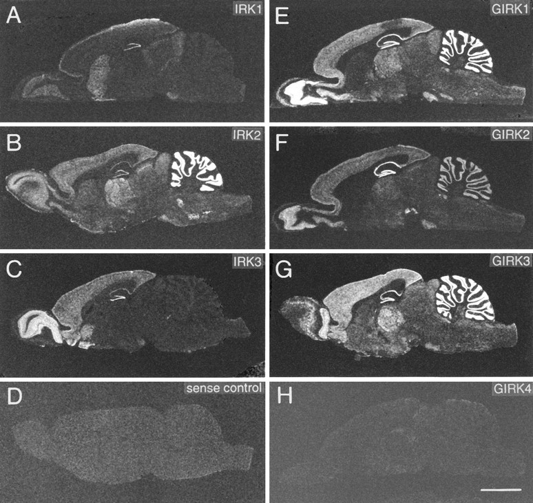
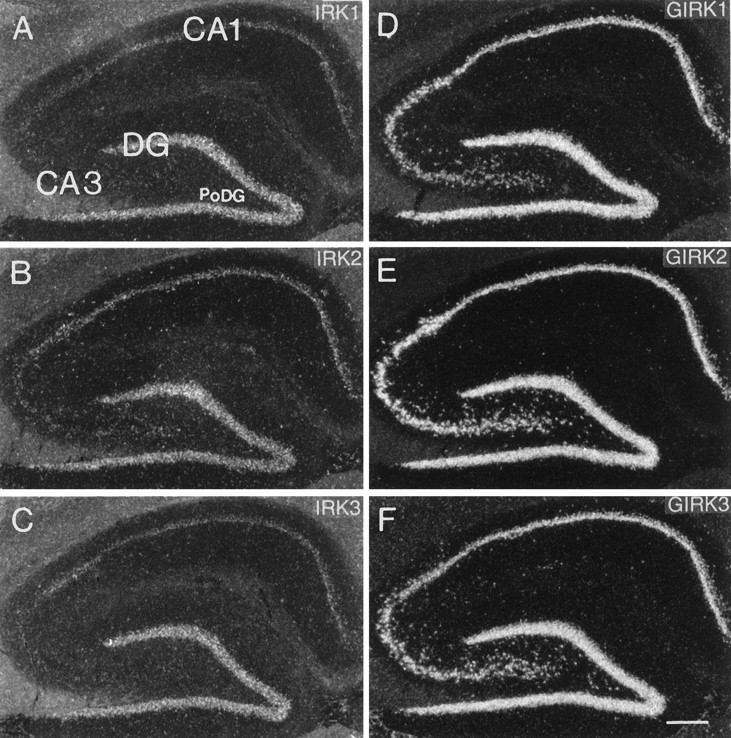
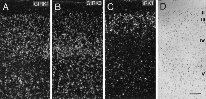
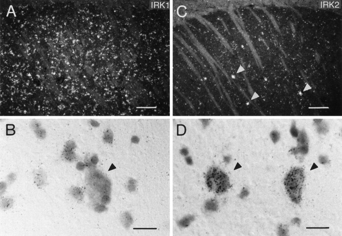
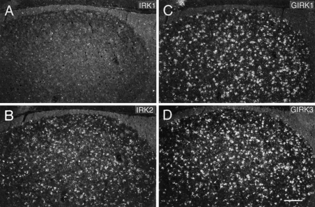
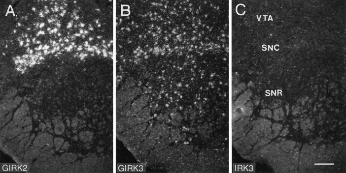
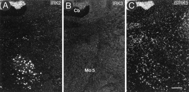
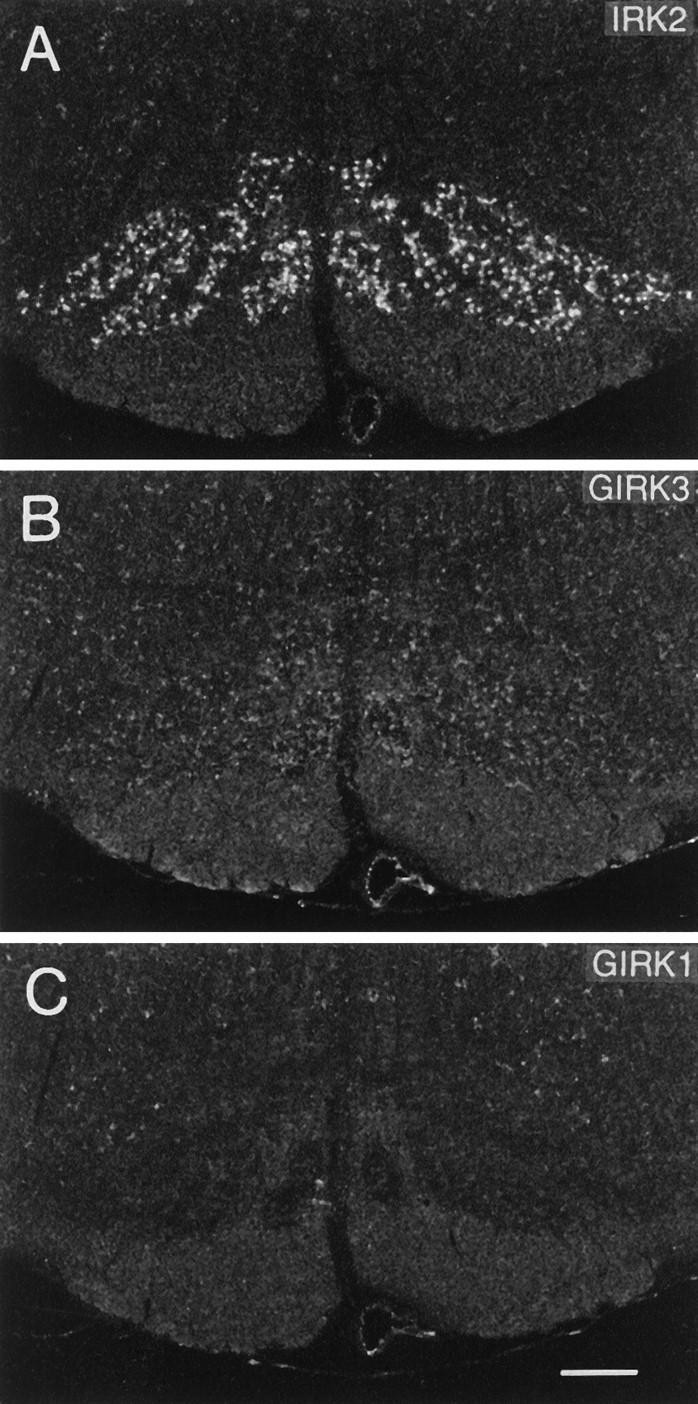
Molecular cloning together with functional characterization has shown that the newly identified family of inwardly rectifying K+ channels consists of several closely related members encoded by separate genes. In this report we demonstrate the differential mRNA expression and detailed cellular localization in the adult rat brain of seven members of the IRK and GIRK subfamilies. Using both radiolabeled cRNA riboprobes and specific oligonucleotide probes directed to nonconserved regions of both known and newly isolated rat brain cDNAs, in situ hybridization revealed wide distribution with partly overlapping expression of the mRNAs of IRK1-3 and GIRK1-4. Except for the low levels of GIRK4 transcripts observed, the overall distribution patterns of the other GIRK subunits were rather similar, with high levels of expression in the olfactory bulb, hippocampus, cortex, thalamus, and cerebellum. Marked differences in expression levels existed only in some thalamic, brainstem, and midbrain nuclei, e.g., the substantial nigra, superior colliculus, or inferior olive. In contrast, IRK subunits were expressed more differentially: all mRNAs were abundant in dentate gyrus, olfactory bulb, caudate putamen, and piriform cortex. IRK1 and IRK3 were restricted to these regions, but they were absent from most parts of the thalamus, cerebellum, and brainstem, where IRK2 was expressed predominantly. Because channel subunits may assemble as heteromultimers, additional functional characterization based on overlapping expression patterns may help to decipher the native K+ channels in neurons and glial cells.
Figures
References
-
- Aguilar-Bryan L, Nichols CG, Wechsler SW, Clement JP, IV, Boyd AE, III, González G, Herrera-Sosa H, Nguy K, Bryan J, Nelson DA. Cloning of the β cell high-affinity sulfonylurea receptor: a regulator of insulin secretion. Science. 1995;268:423–426. - PubMed
-
- Barres BA, Chun LL, Corey DP. Ion channel expression by white matter glia: I. Type II astrocytes and oligodendrocytes. Glia. 1988;1:10–30. - PubMed
-
- Boim MA, Ho K, Shuck ME, Bienkowski MJ, Block JH, Slightom JL, Yang Y, Brenner BM, Hebert SC. ROMK inwardly rectifying ATP-sensitive K+ channel. II. Cloning and distribution of alternative forms. Am J Physiol. 1995;268:F1132–F1140. - PubMed
-
- Bond CT, Ämmälä C, Ashfield R, Blair TA, Gribble F, Khan RN, Lee K, Proks P, Rowe ICM, Sakura H, Ashford MJ, Adelman JP, Ashcroft FM. Cloning and functional expression of the cDNA encoding an inwardly rectifying potassium channel expressed in pancreatic β-cells and the brain. FEBS Lett. 1995;367:61–66. - PubMed
Publication types
MeSH terms
Substances
Associated data
- Actions
LinkOut - more resources
Full Text Sources
Other Literature Sources
Molecular Biology Databases