. 1996 Jun 1;16(11):3652-60.
doi: 10.1523/JNEUROSCI.16-11-03652.1996.

Actions of endogenous opioids on NMDA receptor-independent long-term potentiation in area CA3 of the hippocampus

Affiliations

Actions of endogenous opioids on NMDA receptor-independent long-term potentiation in area CA3 of the hippocampus

S H Williams et al. J Neurosci. .

Abstract

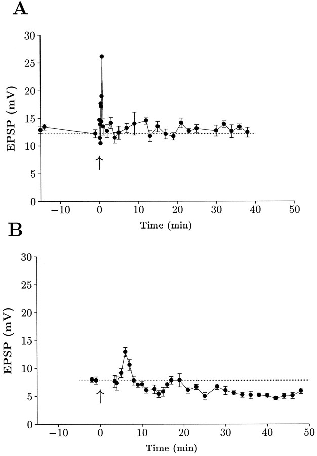
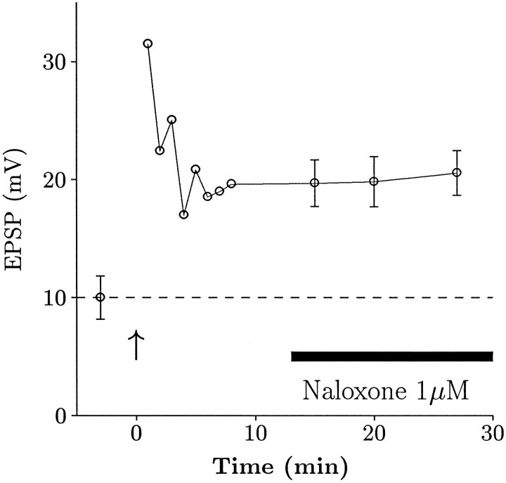
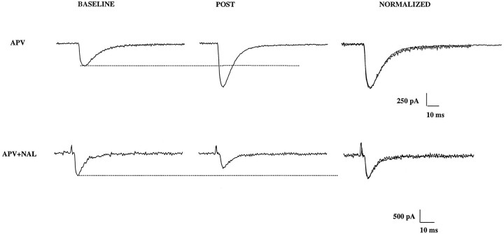
The opioid peptides represent a major class of neurotransmitter in the vertebrate nervous system and are prevalent in the hippocampus. There is considerable interest in the physiological function of the opioids contained in the mossy fiber pathway. The release of opioids from mossy fibers shows a strong frequency dependence. Long-term potentiation (LTP) at this synapse, an NMDA receptor-independent form of LTP, also depends on high-frequency synaptic activity, and this has led to speculation that endogenous opioids may be a critical factor in LTP induction. Previous reports using extracellular recordings have provided evidence for and against a role for opioids in mossy fiber LTP. Using single-cell recording techniques, we have tested the hypothesis that endogenous opioids are required for mossy fiber LTP induction. We recorded from a defined population of synapses that had EPSCs with fast rise times, short latencies, and monophasic decays, consistent with a proximally terminating synapse. The opioid antagonist naloxone prevented mossy fiber LTP in the rat, but had no effect on the commissural/associational system, a nonopioid-containing pathway. The action of naloxone was not mediated through disinhibition because GABAA receptors were pharmacologically blocked in these experiments. We also tested the hypothesis that variations in postsynaptic receptor subtype distribution between species might explain previous controversies regarding the role of endogenous opioids. In contrast to the rat, LTP of the mossy fiber field potential in guinea pig was not blocked by naloxone. Our data suggest that opioids may be the presynaptically released, frequency-dependent, associative factor for mossy fiber LTP induction.


Figures

Fig. 1.
Fig. 1.
Naloxone blocks mossy fiber LTP. Example recordings from a control cell and a neuron bathed in 1 μm naloxone using voltage recording (traces labeled PSP) or voltage-clamp (traces labeled PSC). After a period of recording EPSPs and EPSCs at 0.2 Hz (Baseline), three 1 sec, 100 Hz stimulus trains were delivered. After 15 min, the control EPSP and ESPC clearly exhibited LTP (Post), whereas the naloxone-treated neuron did not. Twenty micromolar d-APV was present in both recordings. All measurements were made at approximately −80 mV, and traces represent averages of 4–10 consecutive traces.
Fig. 2.
Fig. 2.
Average time course of mossy fiber LTP. Data have been grouped together and normalized to illustrate the time course of potentiation in the control group (n = 17) and naloxone group (n = 13). Data are from EPSP measurements that were normalized to the time point just before tetanic stimulation (1.0), illustrated by the dotted horizontal line. Time was normalized such that for each experiment, 0 represents the time at which high-frequency stimulation was delivered (denoted by anarrow). Each point is a mean value and bars are SEs. Data were collected continuously during the first 15 min after tetanus and subsequently were sampled every 5–10 min. Periodic determinations of EPSCs and passive membrane properties were made throughout the experiment. Both groups were clearly potentiated for a brief period after high-frequency stimulation, but only the control group exhibited a sustained potentiation.
Fig. 3.
Fig. 3.
Naloxone can prevent LTP or cause a depression of response. A, Data from a single representative experiment in which there was no change in EPSP after tetanic stimulation. Points represent mean ± SEM recorded over 30–60 sec periods, except during the PTP phase in which points represent single measures (no error bars). B, Data from a cell that exhibited a persistent depression of the EPSP after tetanic stimulation. Note that the EPSP remained depressed for the duration of the recording. (EPSP points were not measured during the PTP phase in this cell). The input resistance of this cell was 100 MΩ before tetanus and 109 MΩ after tetanus.
Fig. 4.
Fig. 4.
Naloxone does not affect established LTP. These data are from an experiment in which naloxone was added after LTP was established. Points with error bars represent a mean value from data collected over a 1–2 min period. Naloxone clearly did not occlude LTP that was already established.
Fig. 5.
Fig. 5.
Naloxone does not affect NMDA-dependent LTP of the C/A input to CA3. LTP of both the EPSP and EPSC were obtained in the presence of 1 μm naloxone. Note that in this experiment no APV was present.
Fig. 6.
Fig. 6.
The kinetics of mossy fiber responses are unaltered after LTP. Top traces(d-APV) show the EPSC from a control cell before LTP induction (BASELINE) and after tetanic stimulation (POST). Clearly there was no change in the 10–90% rise-time or decay-time constant. This is illustrated in the far right panel in which the two responses are normalized to the same amplitude to allow a closer comparison. Similar data are shown for a naloxone-treated cell (d-APV+NAL) shown at thebottom. In this case, the EPSC was depressed after tetanus, but again the kinetics were unchanged.
Fig. 7.
Fig. 7.
Dynorphin does not affect the rat mossy fiber EPSP. Intracellularly recorded mossy fiber LTP time course. LTP was elicited in a cell that had been bathed in 10 μm picrotoxin, 10 μmbicuculline, and 20 μmd-APV. Subsequent perfusion of dynorphin A (1–17) clearly did not affect the EPSP. This particular cell had an input resistance of 77 ± 1 MΩ before drug application and 78 ± 3 MΩ during drug application.
Fig. 8.
Fig. 8.
κ opioids depress synaptic transmission in the guinea pig, but not in the rat. Mossy fiber field potentials were recorded in the stratum lucidum area of CA3 using paired-pulse stimulation protocol, and a baseline level of synaptic activity was measured (Base). To determine whether these field potentials were truly mossy fiber in origin, we elicited LTP in the presence of 20 μmd-APV (LTP), demonstrating that they were capable of showing NMDA-independent LTP. Application of U-69593 (U69593) at 2 μm clearly depressed the guinea pig (B) response but had no effect in the rat (A). The depression was reversed by the κ-selective antagonist nor-BNI (nBNI) at 1 μm. C, Time course of U-69593 (U69593) action in the guinea pig. Plot of pEPSP (normalized) over time. LTP was induced in the presence of 20 μmd-APV, and, after a new baseline was established, 2 μm U-69593 was applied by bath perfusion. A dramatic reduction was observed in the response. The depression was reversed by nor-BNI (nBNI) at 1 μm. (In the absence of antagonist, responses were poorly reversible at these doses.)
Fig. 9.
Fig. 9.
Naloxone has no effect on LTP in the guinea pig. Summary time course data from slices that exhibited LTP in control (open circles) and in the presence of 1 μm naloxone (filled circles). Data are pEPSP slope measurements that were normalized to baseline values before tetanic stimulation (1.0), illustrated by the dotted horizontal line. Time was normalized such that for each experiment, 0 represents the time at which high-frequency stimulation was delivered, denoted by an arrow. Each point is a mean value, and bars are SEs. Both groups were clearly potentiated to a very similar degree.

References

    1. Abraham WC, Goddard GV. Asymmetric relationship between homosynaptic long-term potentiation and heterosynaptic long-term depression. Nature. 1983;305:717–719. - PubMed
    1. Amaral DG, Dent JA. Development of the mossy fibers of the dentate gyrus. I. A light and electron microscopic study of the mossy fibers and their expansions. J Comp Neurol. 1981;195:51–86. - PubMed
    1. Amaral DG, Ishizuka N, Claiborne B. Neurons, numbers and the hippocampal network. In: Storm-Mathisen J, Zimmer J, Ottersen OP, editors. Understanding the brain through the hippocampus: the hippocampal region as a model for studying structure and function, Vol. 83. Elsevier; Amsterdam: 1990. pp. 1–11. - PubMed
    1. Bashir ZI, Bortolotto ZA, Davies CH, Berretta N, Irving AJ, Seal AJ, Henley JM, Jane DE, Watkins JC, Collingridge GL. Induction of LTP in the hippocampus needs synaptic activation of glutamate metabotropic receptors. Nature. 1993;363:347–350. - PubMed
    1. Bear MF, Malenka RC. Synaptic plasticity: LTP and LTD. Curr Opin Neurobiol. 1994;4:389–399. - PubMed

Publication types

LinkOut - more resources