

Functional anatomy of a prelearned sequence of horizontal saccades in humans
- PMID: 8642414
- PMCID: PMC6578842
- DOI: 10.1523/JNEUROSCI.16-11-03714.1996
Functional anatomy of a prelearned sequence of horizontal saccades in humans
Abstract
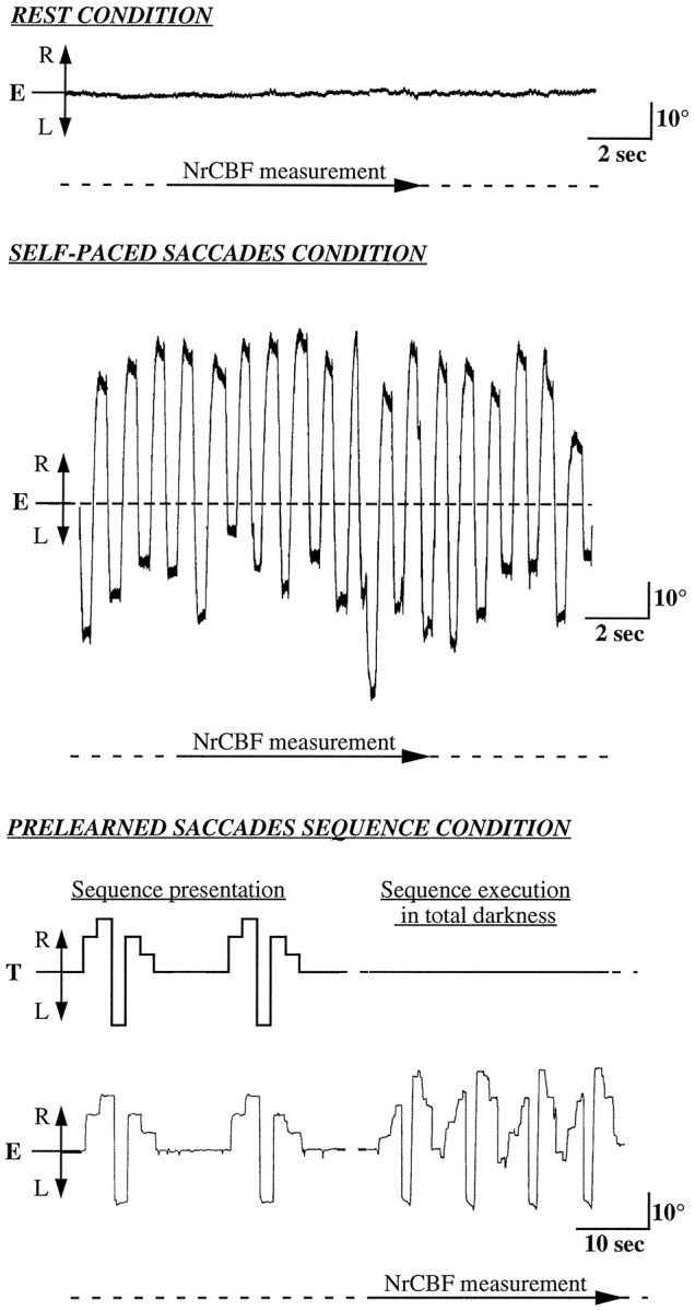
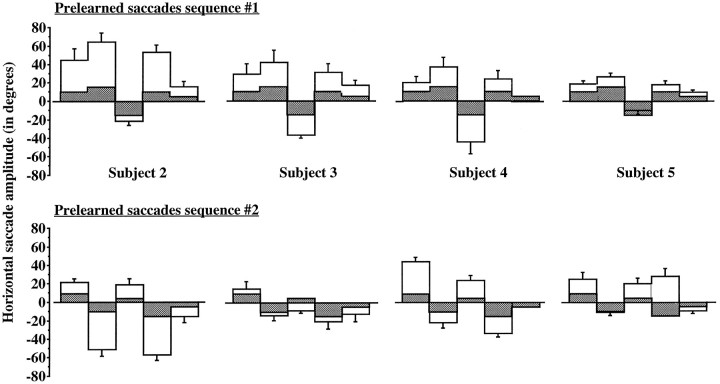
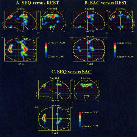
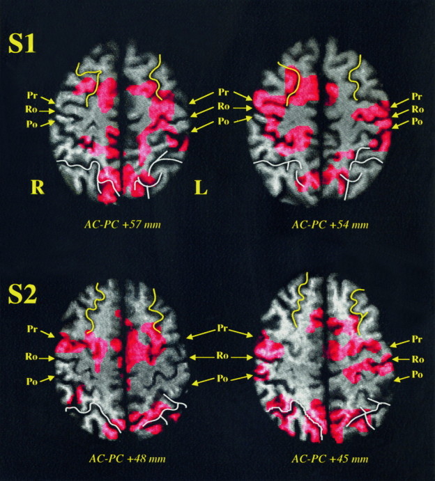
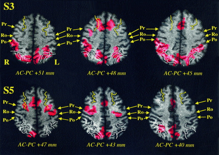
We have used positron emission tomography (PET) to study the functional anatomy of the repetition of a prelearned sequence of horizontal saccadic eye movements. Five subjects had to memorize a sequence of six successive horizontal saccades. The subjects were scanned in total darkness under three different conditions: at rest, during the execution of self-paced horizontal saccades, and while repeating a prelearned saccades sequence. The repetition of the prelearned saccades sequence led to specific normalized regional cerebral blood flow (NrCBF) increases at the depth of the superior frontal sulcus as well as at the rostral part of the supplementary motor area, whereas at the parietal level an important activation was observed in the intraparietal sulcus extending up to the precuneus. In addition, it was noticed that compared with the resting control condition, both oculomotor tasks activated a common set of cortical and subcortical areas. At the cortical level, this network was composed of the frontal eye fields, the supplementary eye fields, the median part of the cingulate gyrus, and the insula. At the subcortical level, the lenticular nucleus and the thalamus as well as the cerebellar vermis were activated consistently. A direct comparison of our results with those of other PET studies on spatial vision suggest that the dorsal visuospatial pathway could be extended toward the frontal premotor region. In such a scheme, visuospatial information computed in the intraparietal sulcus would be transmitted to the frontal premotor cortex to optimize a spatial-oriented behavior. This is consistent with the early proposal that perceptual and intentional components of spatial information are mediated through superior parietal and frontal areas, respectively.
Figures
Similar articles
-
Activation of frontoparietal cortices during memorized triple-step sequences of saccadic eye movements: an fMRI study.Eur J Neurosci. 2001 Mar;13(6):1177-89. doi: 10.1046/j.0953-816x.2001.01472.x. Eur J Neurosci. 2001. PMID: 11285015
-
Positron emission tomography study of voluntary saccadic eye movements and spatial working memory.J Neurophysiol. 1996 Jan;75(1):454-68. doi: 10.1152/jn.1996.75.1.454. J Neurophysiol. 1996. PMID: 8822570
-
PET study of voluntary saccadic eye movements in humans: basal ganglia-thalamocortical system and cingulate cortex involvement.J Neurophysiol. 1993 Apr;69(4):1009-17. doi: 10.1152/jn.1993.69.4.1009. J Neurophysiol. 1993. PMID: 8492144
-
Cortical control of saccades.Exp Brain Res. 1998 Nov;123(1-2):159-63. doi: 10.1007/s002210050557. Exp Brain Res. 1998. PMID: 9835405 Review.
-
Parietal and hippocampal contribution to topokinetic and topographic memory.Philos Trans R Soc Lond B Biol Sci. 1997 Oct 29;352(1360):1437-48. doi: 10.1098/rstb.1997.0130. Philos Trans R Soc Lond B Biol Sci. 1997. PMID: 9368932 Free PMC article. Review.
Cited by
-
Visual avoidance in phobia: particularities in neural activity, autonomic responding, and cognitive risk evaluations.Front Hum Neurosci. 2013 May 31;7:194. doi: 10.3389/fnhum.2013.00194. eCollection 2013. Front Hum Neurosci. 2013. PMID: 23754994 Free PMC article.
-
Attention and cognitive control as emergent properties of information representation in working memory.Cogn Affect Behav Neurosci. 2004 Dec;4(4):501-16. doi: 10.3758/cabn.4.4.501. Cogn Affect Behav Neurosci. 2004. PMID: 15849893 Review.
-
Functional anatomy of spatial mental imagery generated from verbal instructions.J Neurosci. 1996 Oct 15;16(20):6504-12. doi: 10.1523/JNEUROSCI.16-20-06504.1996. J Neurosci. 1996. PMID: 8815928 Free PMC article.
-
Dissociation and convergence of the dorsal and ventral visual working memory streams in the human prefrontal cortex.Neuroimage. 2013 Jan 15;65:488-98. doi: 10.1016/j.neuroimage.2012.10.002. Epub 2012 Oct 12. Neuroimage. 2013. PMID: 23063444 Free PMC article.
-
An fMRI study of optokinetic nystagmus and smooth-pursuit eye movements in humans.Exp Brain Res. 2005 Aug;165(2):203-16. doi: 10.1007/s00221-005-2289-7. Epub 2005 Apr 29. Exp Brain Res. 2005. PMID: 15864563
References
-
- Alexander GE, Delong MR, Strick PL. Parallel organization of functionally segregated circuits linking basal ganglia and cortex. Annu Rev Neurosci. 1986;9:357–381. - PubMed
-
- Andersen RA, Gnadt JW. Posterior parietal cortex. In: Wurtz RH, Goldberg ME, editors. The neurobiology of saccadic eye movements. Elsevier; Amsterdam: 1989. pp. 315–335. - PubMed
-
- Anderson TJ, Jenkins IH, Brooks DJ, Hawken MB, Frackowiak RSJ, Kennard C. Cortical control of saccades and fixation in man: a PET study. Brain. 1994;117:1073–1084. - PubMed
-
- Balint R. Seelenlähmung des “Schauens,” optische Ataxie, räumliche Störung der Aufmerksamkeit. Monatsschr Psychiatr Neurol. 1909;25:51–81.
-
- Barash S, Bracewell RM, Fogassi L, Gnadt JW, Andersen RA. Saccade-related activity in the lateral intraparietal area. 1. Temporal properties: comparison with area 7a. J Neurophysiol. 1991;66:1095–1108. - PubMed
Publication types
MeSH terms
LinkOut - more resources
Full Text Sources