

Specific involvement of human parietal systems and the amygdala in the perception of biological motion
- PMID: 8642416
- PMCID: PMC6578830
- DOI: 10.1523/JNEUROSCI.16-11-03737.1996
Specific involvement of human parietal systems and the amygdala in the perception of biological motion
Abstract
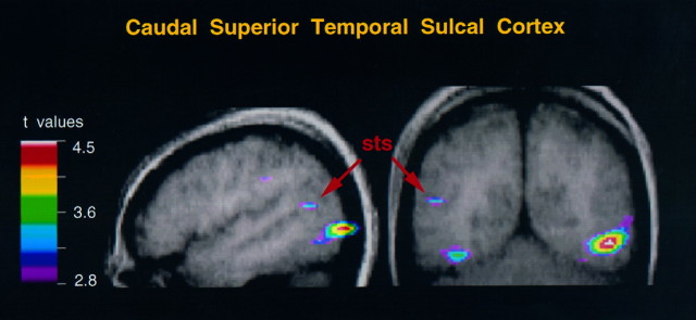
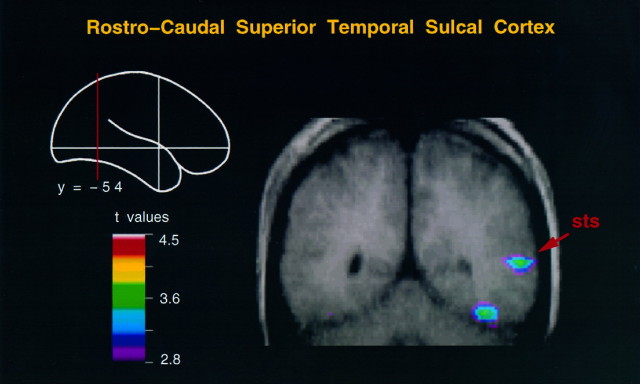
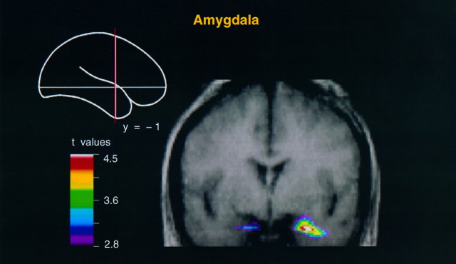
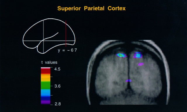
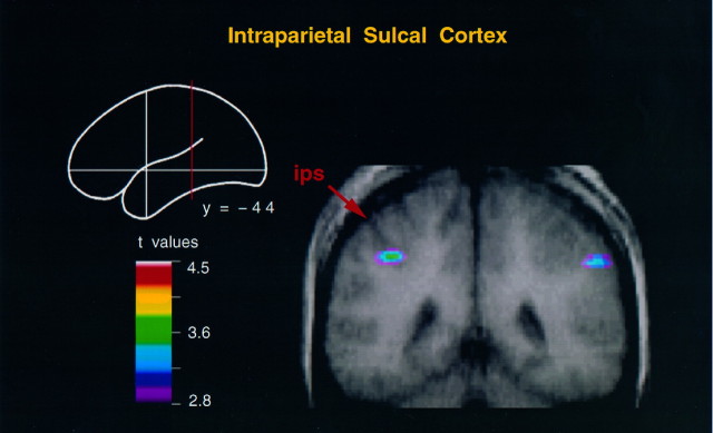
To explore the extent to which functional systems within the human posterior parietal cortex and the superior temporal sulcus are involved in the perception of action, we measured cerebral metabolic activity in human subjects by positron emission tomography during the perception of simulations of biological motion with point-light displays. The experimental design involved comparisons of activity during the perception of goal-directed hand action, whole body motion, object motion, and random motion. The results demonstrated that the perception of scripts of goal-directed hand action implicates the cortex in the intraparietal sulcus and the caudal part of the superior temporal sulcus, both in the left hemisphere. By contrast, the rostrocaudal part of the right superior temporal sulcus and adjacent temporal cortex, and limbic structures such as the amygdala, are involved in the perception of signs conveyed by expressive body movements.
Figures
References
-
- Adolphs R, Tranel D, Damasio H, Damasio A. Impaired recognition of emotion in facial expressions following bilateral damage to the human amygdala. Nature. 1994;372:669–672. - PubMed
-
- Aggleton JP, Burton MJ, Passingham RE. Cortical and subcortical afferents to the amygdala of the rhesus monkey (Macaca mulatta ). Brain Res. 1980;190:347–368. - PubMed
-
- Amaral DG, Price JL. Amygdalo-cortical projections in the monkey (Macaca fascicularis ). J Comp Neurol. 1984;230:465–496. - PubMed
-
- Andersen RA. Inferior parietal lobule function in spatial perception and visuomotor integration. In: Plum F, Mountcastle VB, Geiger SR, editors. Handbook of physiology, The nervous system, Vol. 5. American Physiological Society; Bethesda, MD: 1987. pp. 483–518.
-
- Andersen RA, Asanuma C, Essik G, Siegel RM. Corticocortical connections of anatomically and physiologically defined subdivisions within the inferior parietal lobule. J Comp Neurol. 1990;269:65–113. - PubMed
Publication types
MeSH terms
LinkOut - more resources
Full Text Sources
Other Literature Sources