. 1996 Jun 15;16(12):3798-806.
doi: 10.1523/JNEUROSCI.16-12-03798.1996.

Determinants of competitive antagonist sensitivity on neuronal nicotinic receptor beta subunits

Affiliations

Determinants of competitive antagonist sensitivity on neuronal nicotinic receptor beta subunits

S C Harvey et al. J Neurosci. .

Abstract

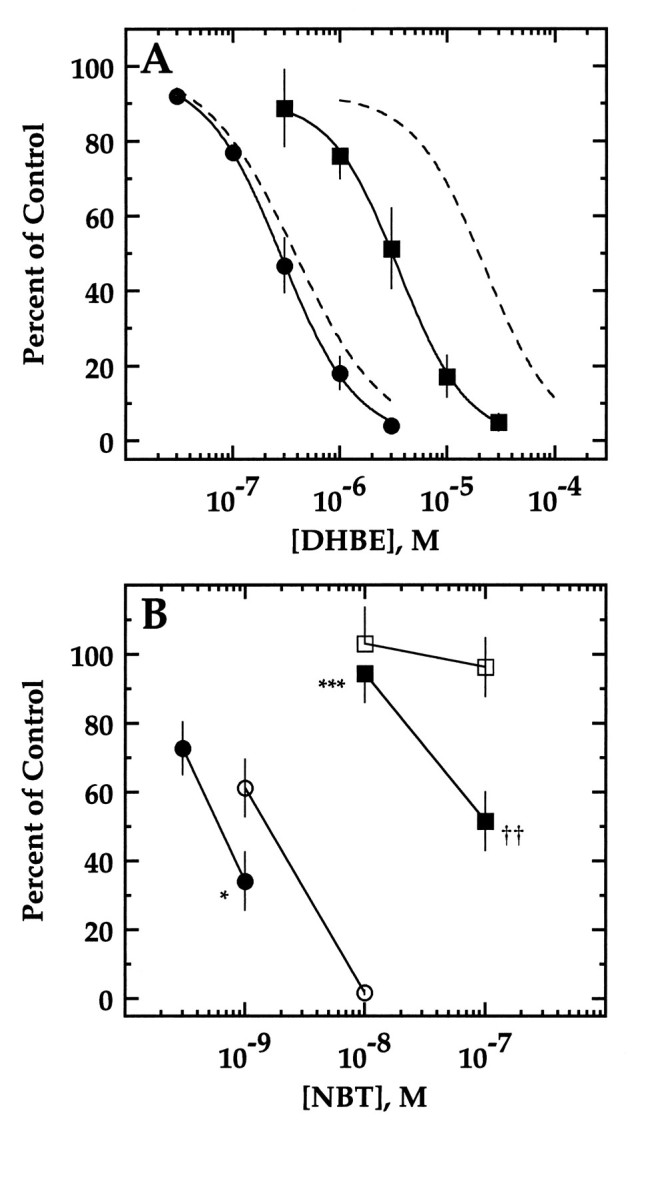
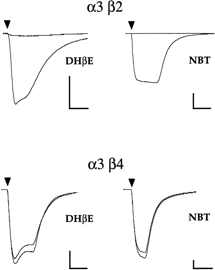
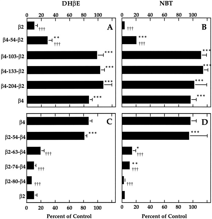
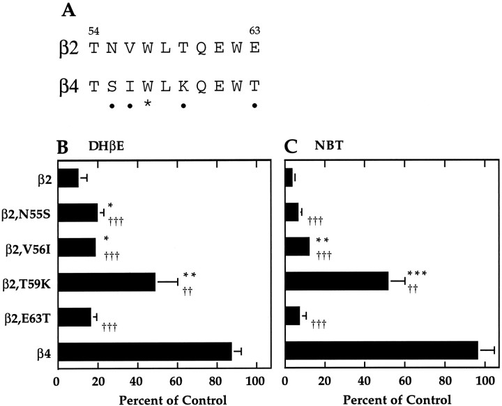
We constructed a series of chimeric and mutant neuronal nicotinic acetylcholine receptor beta subunits to map amino acid residues that determine sensitivity to competitive antagonists. The beta 2 and beta 4 subunits form pharmacologically distinct receptors when expressed in combination with the alpha 3 subunit in Xenopus oocytes. At equipotent acetylcholine concentrations, alpha 3 beta 2 is 56-fold more sensitive to blockage by dihydro-beta-erythroidine than is alpha 3 beta 4. The alpha 3 beta 2 combination is also sensitive to long-term blockade by neuronal bungarotoxin, whereas alpha 3 beta 4 is not. Pharmacological analysis of receptors formed by chimeric beta subunits reveals that amino acid residues that determine both dihydro-beta-erythroidine and neuronal bungarotoxin sensitivity are located within several sequence segments. The major determinant of sensitivity to both competitive antagonists is located between residues 54 and 63. A minor determinant of sensitivity to both antagonists lies between residues 1 and 54, whereas a minor determinant of NBT sensitivity lies between residues 74 and 80. Within region 54-63 of beta 2, mutant beta 2 subunits were used to identify threonine 59 as a residue critical in determining competitive antagonist sensitivity. Changing threonine 59 to lysine, as occurs in beta 4, causes a 9-fold decrease in dihydro-beta-erythroidine sensitivity and a 71-fold decrease in neuronal bungarotoxin sensitivity. Changing polar threonine 59 to negatively charged aspartate causes a 2.5-fold increase in neuronal bungarotoxin sensitivity and has no effect on dihydro-beta-erythroidine sensitivity.


Figures

Fig. 5.
Fig. 5.
Effect of mutations of threonine 59 on DHβE and NBT sensitivity. A, DHβE sensitivity of α3β2,T59K (filled squares) and α3β2,T59D (filled circles). Current in response to an EC20 concentration of ACh in the presence of various concentrations of DHβE is presented as a percent of the response to ACh alone (mean ± SD of 3–6 oocytes). The lines are fits to a Hill equation (see Materials and Methods). IC50 values are 3.8 ± 0.9 μm for α3β2,T59K and 0.30 ± 0.07 μm for α3β2,T59D. Inhibition curves for α3β2 and α3β4 from Figure 2B are shown as dashed lines for reference. B, NBT sensitivity of α3β2,T59K (filled squares), α3β2,T59D (filled circles), α3β2 (open circles), and α3β4 (opensquares). Current in response to an ACh concentration at or below the EC50 after 30 min incubation with various concentrations of NBT is presented as a percentage of the response to ACh alone (mean ± SD of 3 separate oocytes). Significant differences from β2 are denoted by asterisks(*p < 0.02; ***p < 0.001). Significant differences from β4 are denoted by daggers(††p < 0.01). Some error bars are obscured by symbols.
Fig. 1.
Fig. 1.
α3β2 and α3β4 are pharmacologically distinct. Top traces, Current responses of an α3β2-expressing oocyte to 10 μm ACh alone and in combination with 3 μmDHβE (left), and current responses of a different α3β2-expressing oocyte to 1 μm ACh before and after 30 min incubation with 100 nmNBT (right).Bottom traces, Current responses of an α3β4-expressing oocyte to 100 μm ACh alone or in combination with 3 μmDHβE (left), and current responses of a different α3β4-expressing oocyte to 10 μm ACh before and after 30 min incubation with 100 nmNBT (right). ACh application of ∼10 sec is indicated by arrowheads. Scale bars: 150 nA, 10 sec.
Fig. 2.
Fig. 2.
A, Acetylcholine dose–response curves for α3β2 (circles) and α3β4 (squares). Symbols are the mean normalized responses ± SEM of three separate sets of oocytes, each set consisting of three to four separate oocytes. The lines are fits to a Hill equation (see Materials and Methods). EC50 and n values are 70.8 ± 19.6 μm and 0.74 ± 0.11 for α3β2, respectively, and 209.7 ± 40.7 μm and 1.56 ± 0.02 for α3β4, respectively. B, DHβE inhibition curves for α3β2 (circles) and α3β4 (squares). Increasing DHβE concentrations were coapplied with an EC20 ACh concentration of 100 μm for α3β4 and 10 μm for α3β2. The response in the presence of DHβE is reported as a percent of the response to ACh alone (mean ± SD of 3 oocytes). The lines are fits to a Hill equation (see Materials and Methods). IC50 values are 0.41 ± 0.17 μm for α3β2 and 23.1 ± 10.2 μm for α3β4. Some error bars are obscured by symbols.
Fig. 3.
Fig. 3.
DHβE and NBT sensitivity of receptors formed by chimeric β subunits. A, DHβE sensitivity of receptors formed by each of a series of chimeric subunits in which increasingly larger portions of the N-terminal end of β2 were replaced by the corresponding portion of β4. Current in response to an EC20 concentration of ACh in the presence of 3 μm DHβE is presented as a percent of the response to ACh alone (mean ± SD of 3–4 separate oocytes).B, NBT sensitivity of receptors formed by the chimeras inA. Current in response to an ACh concentration at or below the EC50 after 30 min incubation with 100 nm NBT is presented as a percentage of the response to ACh alone (mean ± SD of 3–4 separate oocytes, except for β4, which is mean ± SEM of 3 separate sets of oocytes, each set consisting of 3–4 separate oocytes). C, DHβE sensitivity of receptors formed by each of a series of chimeric subunits in which increasingly larger portions of β4 were replaced by the corresponding portion of β2. Current in response to an EC20concentration of ACh in the presence of 3 μmDHβE is presented as a percent of the response to ACh alone (mean ± SD of 3–4 separate oocytes). D, NBT sensitivity of receptors formed by the chimeras in C. Current in response to an ACh concentration at or below the EC50after 30 min incubation with 100 nm NBT is presented as a percentage of the response to ACh alone (mean ± SD of 3–4 separate oocytes, except for β2-54-β4, which is mean ± SEM of 3 separate sets of oocytes, each set consisting of 3–4 separate oocytes). Significant differences from β2 are denoted byasterisks (*p < 0.05; **p < 0.01; ***p < 0.001). Significant differences from β4 are denoted by daggers (p < 0.05;†††p < 0.001). Some error bars are too small to appear.
Fig. 4.
Fig. 4.
Threonine 59 of β2 is critical to both DHβE and NBT sensitivity. A, Alignment of β2 and β4 sequences within segment 54–63. Residues that differ are denoted by solid circles. Tryptophan 57 is starred. B, DHβE sensitivity of receptors formed by each of a series of mutant β2 subunits. Current in response to an EC20 concentration of ACh in the presence of 3 μm DHβE is presented as a percent of the response to ACh alone (mean ± SD of 3 separate oocytes).C, NBT sensitivity of receptors formed by the β2 mutants in B. Current in response to an ACh concentration at or below the EC50 after 30 min incubation with 100 nm NBT is presented as a percentage of the response to ACh alone (mean ± SD of 3 separate oocytes, except for β4, which is mean ± SEM of three separate sets of oocytes, each set consisting of 3–4 separate oocytes). Significant differences from β2 are denoted by asterisks (*p < 0.05; **p < 0.01; ***p < 0.001). Significant differences from β4 are denoted by daggers(††p < 0.01;†††p < 0.001). Some error bars are too small to appear.

Similar articles

Cited by

References

    1. Abramson SN, Li Y, Culver P, Taylor P. An analog of lophotoxin reacts covalently with Tyr190 in the α-subunit of the nicotinic acetylcholine receptor. J Biol Chem. 1989;264:12666–12672. - PubMed
    1. Bertrand D, Bertrand S, Ballivet M. Pharmacological properties of the homomeric α7 receptor. Neurosci Lett. 1992;146:87–90. - PubMed
    1. Blount P, Merlie J. Molecular basis of the two nonequivalent ligand binding sites of the muscle nicotinic acetylcholine receptor. Neuron. 1989;3:349–357. - PubMed
    1. Boulter J, Connolly J, Deneris E, Goldman D, Heinemann S, Patrick J. Functional expression of two neuronal nicotinic acetylcholine receptors from cDNA clones identifies a gene family. Proc Natl Acad Sci USA. 1987;84:7763–7767. - PMC - PubMed
    1. Cohen JB, Blanton MP, Chiara DC, Sharp SD, White BH. Structural organization of functional domains of the nicotinic acetylcholine receptor. J Cell Biochem [Suppl] 1992;16E:217.

Publication types

MeSH terms

LinkOut - more resources