. 1996 Jun 15;16(12):3817-26.
doi: 10.1523/JNEUROSCI.16-12-03817.1996.

The taste of monosodium glutamate: membrane receptors in taste buds

Affiliations

The taste of monosodium glutamate: membrane receptors in taste buds

N Chaudhari et al. J Neurosci. .

Abstract

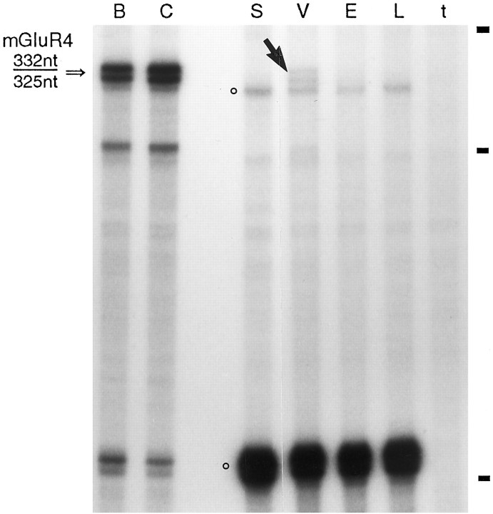
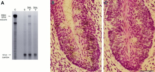
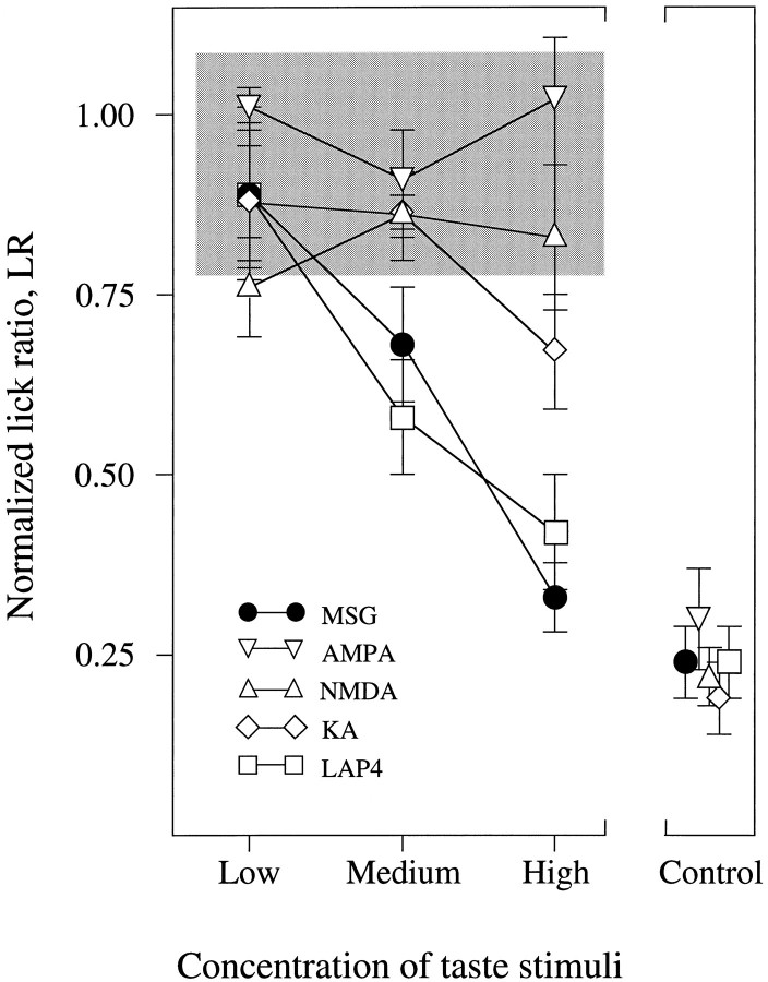
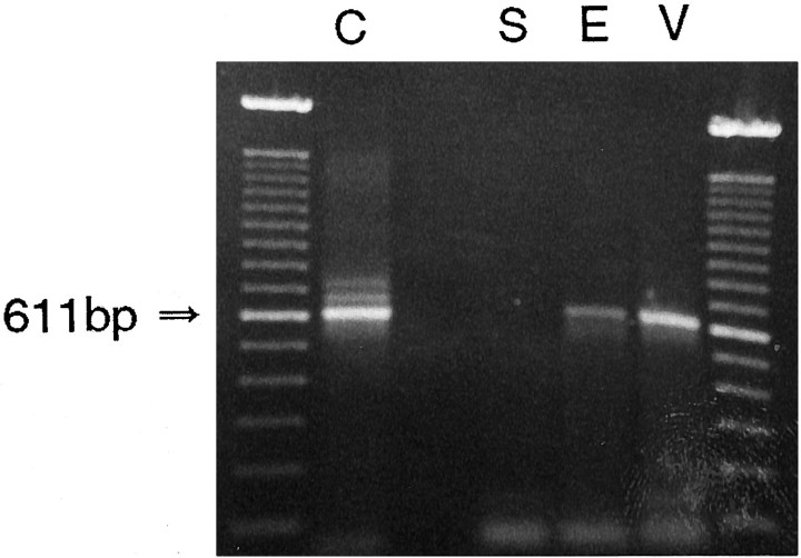
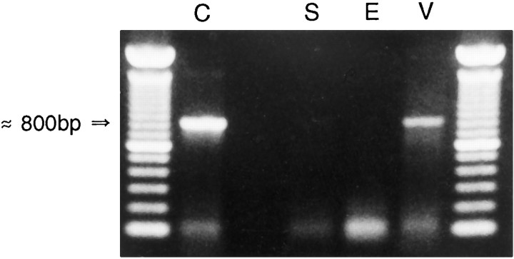
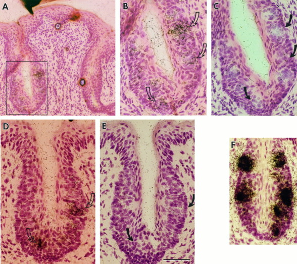
Receptor proteins for photoreception have been studied for several decades. More recently, putative receptors for olfaction have been isolated and characterized. In contrast, no receptors for taste have been identified yet by molecular cloning. This report describes experiments aimed at identifying a receptor responsible for the taste of monosodium glutamate (MSG). Using reverse transcriptase (RT)-PCR, we found that several ionotropic glutamate receptors are present in rat lingual tissues. However, these receptors also could be detected in lingual tissue devoid of taste buds. On the other hand, RT-PCR and RNase protection assays indicated that a G-protein-coupled metabotropic glutamate receptor, mGluR4, also is expressed in lingual tissues and is limited only to taste buds. In situ hybridization demonstrated that mGluR4 is detectable in 40-70% of vallate and foliate taste buds but not in surrounding nonsensory epithelium, confirming the localization of this metabotropic receptor to gustatory cells. Expression of mGluR4 in taste buds is higher in preweaning rats compared with adult rats. This may correspond to the known higher sensitivity to the taste of MSG in juvenile rodents. Finally, behavioral studies have indicated that MSG and L-2-amino-4-phosphonobutyrate (L-AP4), a ligand for mGluR4, elicit similar tastes in rats. We conclude that mGluR4 may be a chemosensory receptor responsible, in part, for the taste of MSG.


Figures

Fig. 3.
Fig. 3.
RNase protection assay to assess the relative concentration of mGluR4 mRNA in taste buds and in surrounding epithelium. Single-strand [32P]RNA antisense probe was synthesized for mGluR4, hybridized in solution, and protected from RNase digestion by 0.2 μg poly(A)RNA from positive control tissues, brain (B) and cerebellum (C). The expected band of a protected probe (a doublet at 332/325 nt) is indicated by an arrow. Hybridization with 9 μg of poly(A)RNA from vallate plus foliate papillae (V) also shows the same doublet band (➘). Hybridization with 9 μg of poly(A)RNA from lingual epithelium that lacked taste buds (E) showed no mGluR4 bands, nor were protected fragments visible in parallel reactions containing 9 μg each of tRNA (t) or poly(A)RNA from liver (L) or skeletal muscle (S). The hybridization reactions also contained a [32P]RNA probe for GAPDH, a ubiquitous glycolytic enzyme, which gives rise to two protected bands, the principal one at 151 nt and a secondary band at 315 nt (○). GAPDH band intensities indicate approximately equal loading in the negative control and test RNAs. The positive controls were underloaded to avoid overexposing the film. Autoradiographic exposure, 15 d. Hatchmarks on the right indicate bands from a single-strand RNA marker (415, 276, and 145 nt).
Fig. 5.
Fig. 5.
Expression of mGluR4 in taste buds during postnatal development. A, RNase protection assays were performed with 9 μg poly(A)RNA from vallate plus foliate papillae (V) from either 19- to 26-d-old preweaning rats (wk.) or 3-month-old adult rats (mo.). Thex-ray film was exposed for 17 d. The radioactivity in the mGluR4 doublet bands was densitometrically quantified and was normalized to the control GAPDH signal in the same lane. In three experiments using separate batches of mRNA, the protected doublet, 332/325 nt, indicated mGluR4 mRNA was found at an average of two- to threefold higher concentration in the younger animals. B,C, In situ hybridizations were run identically in parallel on vallate papillae from a 27-d-old juvenile rat (B) and a 120-d-old adult rat (C). Antisense probes for mGluR4, labeled with 33P, demonstrate the higher grain density (indicating higher mRNA levels) obtained in taste buds from juvenile rats. Autoradiographic exposure, 8 weeks. Scale bar, 50 μm.
Fig. 6.
Fig. 6.
Conditioned taste aversion indicates thatl-AP4, an agonist for mGluR4, mimics the taste of MSG in rats. Rats were conditioned to avoid solutions containing MSG and then were presented with three concentrations of MSG, AMPA, KA, NMDA, or l-AP4, as described in Materials and Methods. Ordinate, Lick rate in experimental rats normalized to rate in control rats. Abscissa, Concentration of test substances, as specified in Materials and Methods. Each point is the mean ± SEM (n = 6–8 conditioned and 6–8 control rats per data point). The shaded area represents the range of responses to 25 mm KCl, a neutral distractor, to indicate nonspecific aversive behavior (see Materials and Methods).Control, For each animal a single, maximal concentration of MSG (150 mm) was tested also to verify that complete aversion to MSG had been established. The mean ± SEM of normalized LR for each group is shown.
Fig. 1.
Fig. 1.
RT-PCR to detect NMDA-glutamate receptor expression in lingual epithelium. Poly(A)RNAs from several tissues were reverse-transcribed and subjected to PCR using primers specific for NMDAR1. Tissues analyzed included cerebellum (C), which expresses NMDARs; skeletal muscle (S), which is not known to express any GluRs; lingual epithelium (E), which lacks taste buds; and vallate plus foliate papillae (V), which contain abundant taste buds. The gel was loaded with 5 μl of each reaction. With cerebellar RNA, the expected 611 bp PCR product (arrow) was consistently obtained, as well as faint higher bands that correspond to alternative splicing variants within the amplified region (Durand et al., 1992; Nakanishi et al., 1992). The far leftand right lanes contain a 100 bp ladder.
Fig. 2.
Fig. 2.
RT-PCR to identify mGluR expression in lingual epithelium. Poly(A)RNAs from several tissues were reverse-transcribed and subjected to PCR using degenerate primers for the family of mGluRs. Tissues analyzed included cerebellum (C), which expresses mGluRs; skeletal muscle (S), which is not known to express mGluRs; lingual epithelium (E), which lacks taste buds; and vallate plus foliate papillae (V), which contain abundant taste buds. The gel was loaded with 5 μl from the cerebellum reaction and 50 μl (concentrated by ethanol precipitation) of the other reactions. The arrow indicates the expected amplification product at ∼800 bp. The far left and rightlanes contain a 100 bp ladder.
Fig. 4.
Fig. 4.
In situ hybridization for mGluR4 in rat vallate and foliate papillae. [33P]RNA probes for mGluR4 were synthesized using T7 RNA polymerase. Adjacent sections of vallate (AC) and foliate (DF) papillae were hybridized in parallel with probes in antisense (A, B, D) or sense (C, E) orientation. The boxedarea in A is shown at higher magnification inB. AC, Papillae were from 19-d-old rats. D, E, Papillae were from 27-d-old rats. Autoradiographic exposure on Kodak NTB3 emulsion was for 6 1/2 weeks (AC) or 8 weeks (D, E).B, D, Open arrows indicate taste buds with concentrations of silver grains, indicating mGluR4 mRNA. C,E, Filled arrows indicate the same taste buds as in B and D, respectively, but which remain unlabeled after hybridization with the sense probe. Control hybridizations with a gustducin antisense (F) probe in rat foliate papillae demonstrate that grain densities are substantially higher than those for mGluR4. The hybridization was performed in parallel on adjacent sections to those shown in D andE. Autoradiographic exposure, 8 weeks. Scale bar, 50 μm.

Similar articles

Cited by

References

    1. Abe T, Sugihara H, Nawa H, Shigemoto R, Mizuno N, Nakanishi S. Molecular characterization of a novel metabotropic glutamate receptor mGluR5 coupled to inositol phosphate/Ca2+ signal transduction. J Biol Chem. 1992;267:13361–13368. - PubMed
    1. Abe K, Kusakabe Y, Tanemura K, Emori Y, Arai S. Primary structure and cell-type specific expression of a gustatory G protein-coupled receptor related to olfactory receptors. J Biol Chem. 1993;268:12033–12039. - PubMed
    1. Akabas MH. The molecular biology of chemotransduction. In: Simon SA, Roper SD, editors. Mechanisms of taste transduction. CRC; Boca Raton, FL: 1993. pp. 175–200.
    1. Akabas MH, Dodd J, Al-Awqati Q. A bitter substance induces a rise in intracellular calcium in a sub-population of rat taste cells. Science. 1988;242:1047–1050. - PubMed
    1. Avenet P, Hofmann F, Lindemann B. Transduction in taste receptor cells requires cAMP-dependent protein kinase. Nature. 1988;331:51–54. - PubMed

Publication types

MeSH terms

LinkOut - more resources