. 1996 Jun 15;16(12):3848-61.
doi: 10.1523/JNEUROSCI.16-12-03848.1996.

Muscarinic activation of a voltage-dependent cation nonselective current in rat association cortex

Affiliations

Muscarinic activation of a voltage-dependent cation nonselective current in rat association cortex

S Haj-Dahmane et al. J Neurosci. .

Abstract

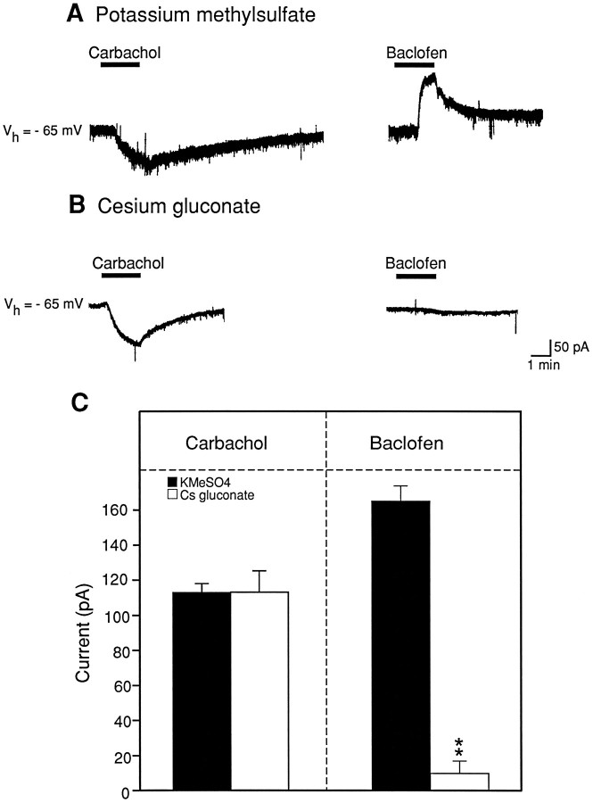
The ionic mechanism underlying the acetylcholine-induced depolarization of layer V pyramidal neurons of rat prefrontal cortex was examined using whole-cell recording in in vitro rat brain slices. Consistent with previous results, pressure application of acetylcholine to layer V pyramidal neurons elicited a strong depolarization. Pharmacological analysis of this response indicated that it was mediated by the stimulation of muscarinic receptors as it was mimicked by muscarinic agonists, but not by nicotine, and was blocked by atropine. The inward current responsible for the depolarization resulted from the activation of a voltage-dependent, cation nonselective current. Thus, the amplitude of the current was critically dependent on extracellular sodium concentration but not on extracellular potassium or chloride concentration. Examination of the I-V relationship for the muscarinic current using voltage clamp revealed that the current reversed near -15 mV and exhibited a strong voltage dependence, turning off rapidly in the subthreshold range. The voltage dependence of the current led to the appearance of a current associated with a conductance decrease when examined using steady-state voltage- or current-clamp measurements. This might have led to earlier misidentification of this response as mediated by a decrease in potassium conductance. These results question the traditional interpretation that muscarinic depolarization in cortex is mediated by a decrease in potassium conductance. They indicate that the fundamental mechanism responsible for muscarinic depolarization in prefrontal cortex involves the activation of a voltage-dependent, cation nonselective current. This current might represent a previously unsuspected mechanism capable of mediating slow depolarization in the central nervous system.


Figures

Fig. 1.
Fig. 1.
Acetylcholine depolarizes layer V pyramidal neurons of rat medial prefrontal cortex. A, Toptrace, Pressure application of acetylcholine depolarized this neuron and triggered a period of sustained spiking activity.Bottom trace, Two minutes after recovery, a second application of acetylcholine induced a comparable effect. B, After bath administration of 100 nm atropine, the acetylcholine response was almost completely inhibited. These recordings were obtained using potassium methylsulfate-based intracellular solution containing 20 μmEGTA.
Fig. 2.
Fig. 2.
Carbachol elicits a concentration-dependent depolarization of resting membrane potential of the cell. Current-clamp recording of the pyramidal neuron was obtained using the nystatin perforated-path recording technique. Bath administration of carbachol resulted in a concentration-dependent depolarization. In this and the subsequent figures, the bar above the traceindicates the period of the carbachol application.
Fig. 3.
Fig. 3.
The carbachol-induced depolarization behaves as if mediated by a decrease in potassium conductance when it is examined using a variety of traditional electrophysiological tests.A, Recording from a pyramidal neuron in current-clamp mode. Hyperpolarizing current pulses (140 ms, 0.2 nA) were applied periodically to monitor the input resistance. Administration of carbachol (30 μm) in the presence of TTX (1 μm) to this cell elicited a membrane depolarization associated with an apparent increase in membrane resistance. B1, Carbachol administration (30 μm) induced an inward current in a different pyramidal neuron voltage-clamped at −65 mV in the presence of TTX (1 μm). B2, Current–voltage (IV) relationship obtained using a voltage ramp from −120 to −30 mV taken before (○) and during the superfusion with carbachol (•) from the cell illustrated in B1(holding current = 0.16 nA). C, Carbachol-induced current in a different neuron voltage-clamped at −65 mV (holding current = 0.11 nA). Superfusion with barium (2 mm) induced an inward shift of the holding current and a reduction of the amplitude of the carbachol-induced current.
Fig. 4.
Fig. 4.
The inward current caused by carbachol fails to reverse polarity even at potentials more negative thanEK in the majority of cells studied.A, In most cells studied, the net carbachol current failed to reverse at EK. B, In a small proportion of cells, the net carbachol current reversed nearEK. The holding current for the cell illustrated in A was 0.15 nA and in B was 0.12 nA.
Fig. 5.
Fig. 5.
Carbachol elicits an inward current atEK as well as belowEK. A, The lefttrace illustrates the inward current induced by carbachol (30 μm), and the right traceshows the outward current elicited by baclofen (30 μm) in a cell held at −65 mV in the control condition (2.5 mm potassium) and in the presence of TTX (1 μm). B, Carbachol-induced currents (left traces) and baclofen-induced currents (right traces) recorded at different holding potentials (−65, −75, and −85 mV) in 6 mmpotassium and 1 μm TTX. Note that carbachol still induced an inward current even at potentials more negative thanEK. C, Recovery of carbachol and baclofen currents in 2.5 mm potassium. All of the traces represented in A, B, andC are from the same cell. The holding current at −65 mV in 2.5 mm potassium was 0.07 nA at the beginning of the experiment and 0.06 nA after recovery from 6 mm potassium. In 6 mmextracellular potassium, the holding current at −65 mV was 0.04 nA, at −75 mV was −0.17 nA, and at −85 mV was −0.27 nA.
Fig. 6.
Fig. 6.
The carbachol current is dependent on the extracellular concentration of sodium. A, Effect of reducing extracellular sodium on the amplitude of the carbachol-induced current on a cell clamped at −65 mV (holding current = 0.17 nA) in the presence of TTX (1 μm). The lefttrace illustrates the carbachol response in control sodium (146 mm), the middle traceillustrates the response to carbachol observed in low sodium (26 mm), and the right traceillustrates the recovery. Dashed lines indicate the baseline current. B, IV curves of the carbachol current recorded under control conditions (a), in 26 mm sodium (b), and recovery (c) from the same neuron illustrated in A.C, Summary plot of the amplitude of the carbachol-induced current recorded under control conditions (solid bar) and in 26 mm sodium (open bar). Error bars illustrate the SEM from five different determinations. **p < 0.01 versus control.
Fig. 7.
Fig. 7.
Voltage dependence of muscarinic-activated inward current. A, Carbachol current recorded with cesium gluconate-based intracellular solution in the presence of cesium (2 mm), barium (100 μm), cadmium (100 μm), and TTX (1 μm) at −40 mV. B, Hyperpolarizing voltage step commands from −40 to −120 mV (protocol represented inupper right traces) were applied before and during carbachol application. B, Net carbachol current tracings were obtained by subtracting the currents obtained in control conditions from those obtained during carbachol application. The instantaneous muscarinic current increases in amplitude with hyperpolarization. However, the hyperpolarizing steps also resulted in a fast outward relaxation, reflecting the voltage-dependent turning-off of the muscarinic current. C, Comparison of the instantaneous and steady-state IV curves for the carbachol-induced current obtained from the cell represented inA. The holding current at −40 mV was 0.05 nA.
Fig. 8.
Fig. 8.
The carbachol-induced inward current reverses polarity near −15 mV. A, Current–voltage (IV) plots were obtained using a linear voltage ramp from −80 to +10 mV before and during carbachol application in a neuron recorded with a cesium gluconate-based intracellular solution in the presence of cadmium (100 μm) and TTX (1 μm). B, Net carbachol current obtained by subtracting the IV plots represented in A. The carbachol-induced current reversed polarity at a mean potential of −16 mV ± 2 mV (n = 4).
Fig. 9.
Fig. 9.
The carbachol-induced current is not reduced by intracellular injection of cesium. A, Carbachol- and baclofen-induced currents were recorded using a potassium methylsulfate-based intracellular solution in the presence of TTX (1 μm). The left traceillustrates the inward current caused by carbachol (30 μm) at a holding potential of −65 mV. Theright trace illustrates the outward current elicited by baclofen (30 μm) in the same cell (holding current = 0.11 nA). B, Carbachol-induced (lefttrace) and baclofen-induced (right trace) currents were recorded using a cesium gluconate-based intracellular solution. Intracellular injection of cesium failed to reduce the carbachol current but suppressed the outward current elicited by baclofen. The holding current for this cell was −0.08 nA.C, Summary plot comparing the absolute amplitude of the carbachol- and baclofen-induced currents recorded using potassium methylsulfate-based (solid bar; n = 6) or cesium gluconate-based (open bar; n = 6) intracellular solutions. Error bars illustrate the SEM determined for each group. **p < 0.01 versus control.
Fig. 10.
Fig. 10.
Effect of elevated extracellular potassium on theIV curves for the carbachol-induced inward current. A, Control and carbachol IVrelationships obtained in the presence of 2.5 mmextracellular potassium. B, Control and carbacholIV relationships obtained in the presence of 5 mm extracellular potassium. C,IV relationship for the net carbachol current observed in the presence of 2.5 and 5 mmextracellular potassium (holding current = 0.06 nA).
Fig. 11.
Fig. 11.
The carbachol current is insensitive to changes in the chloride gradient. A, Carbachol-induced current recorded at a holding potential of −65 mV in the presence of TTX (1 μm), under control conditions (126.5 mm extracellular chloride, lefttrace), in low extracellular chloride (7.5 mm, middle trace), and during recovery (right trace). Lowering the extracellular concentration of chloride failed to induce changes in either the polarity or the amplitude of the carbachol current. B, Inhibitory postsynaptic potential evoked by stimulation of layer III (25–30 V for 500 μsec) in the presence of DNQX (10 μm) and APV (10 μm) in control conditions and in low chloride. Chloride substitution induced a change in the polarity of GABAA synaptic potential, indicating that this manipulation was effective in altering the chloride gradient across the cell membrane (holding current = 0.11 nA).
Fig. 12.
Fig. 12.
TEA inhibits the carbachol current without by itself eliciting an inward current. Administration of carbachol (30 μm) in the presence of TTX (1 μm) elicited an inward current in a cell clamped at −65 mV. Bath administration of TEA (2 mm) after recovery from the carbachol failed to induce any inward current. However, a second application of carbachol in the presence of TEA elicited a greatly reduced inward current. Partial recovery of the carbachol current was observed after removal of the TEA from the bath (holding current = 0.17 nA).

Similar articles

Cited by

References

    1. Andrade R. Cell excitation enhances muscarinic cholinergic responses in rat association cortex. Brain Res. 1991;548:81–93. - PubMed
    1. Araneda R, Andrade R. 5-HT2 and 5-HT1A receptors mediate opposing responses on membrane excitability in rat association cortex. Neuroscience. 1991;40:399–412. - PubMed
    1. Benardo LS, Prince DA. Ionic mechanisms of cholinergic excitation in mammalian hippocampal pyramidal cells. Brain Res. 1982;249:333–344. - PubMed
    1. Benham CD. Acetylcholine activates an inward current in single mammalian smooth muscle cells. Nature. 1985;316:345–346. - PubMed
    1. Benson DM, Blitzer RD, Landau EM. An analysis of the depolarization produced in guinea-pig hippocampus by cholinergic receptor stimulation. J Physiol (Lond) 1988;404:479–496. - PMC - PubMed

Publication types

MeSH terms

LinkOut - more resources