. 1996 Jun 15;16(12):3900-11.
doi: 10.1523/JNEUROSCI.16-12-03900.1996.

Characterization of Drosophila tyramine beta-hydroxylase gene and isolation of mutant flies lacking octopamine

Affiliations

Characterization of Drosophila tyramine beta-hydroxylase gene and isolation of mutant flies lacking octopamine

M Monastirioti et al. J Neurosci. .

Abstract

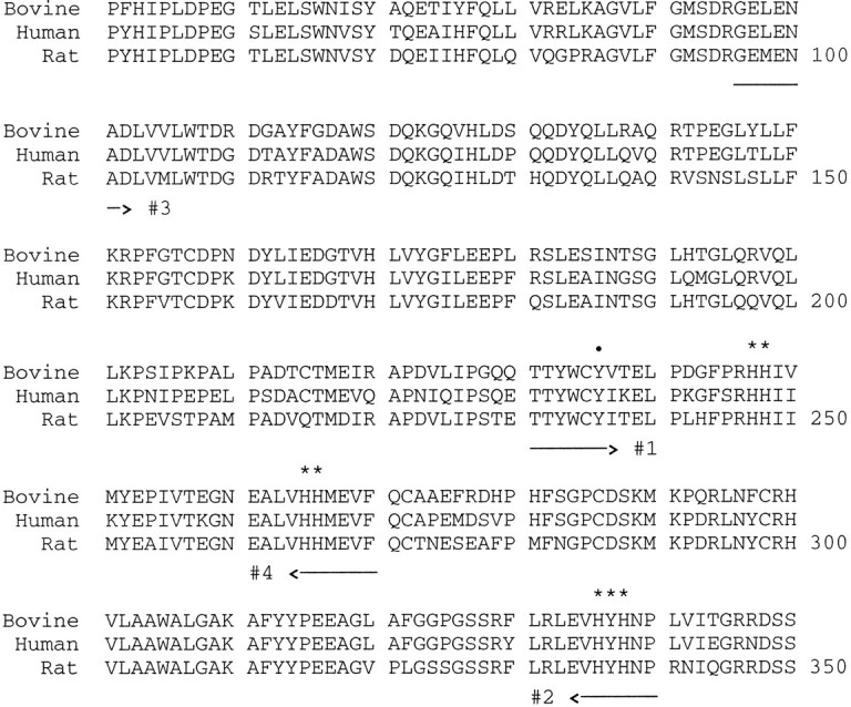
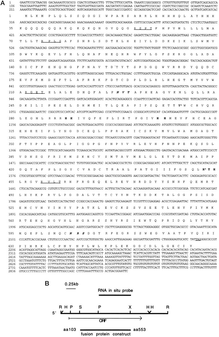
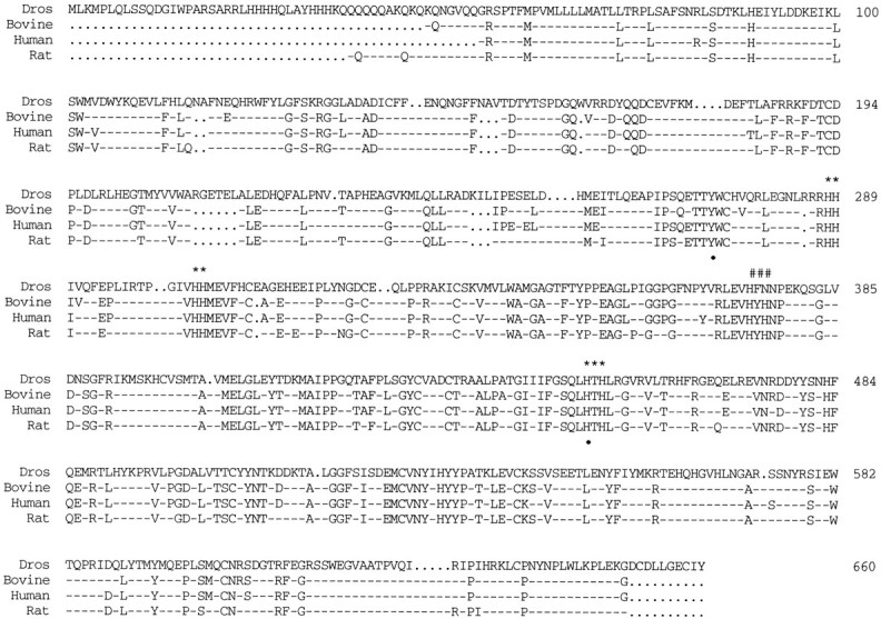
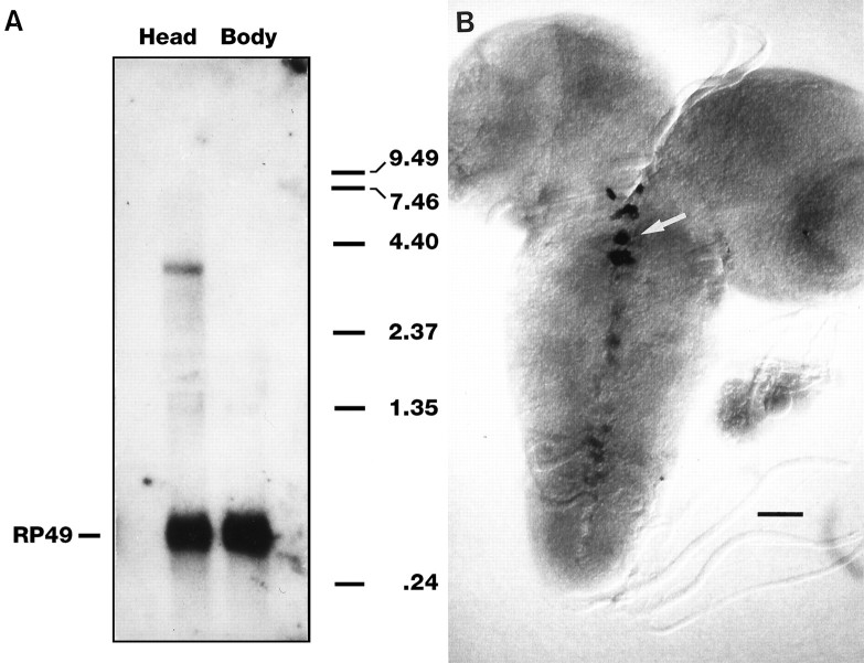
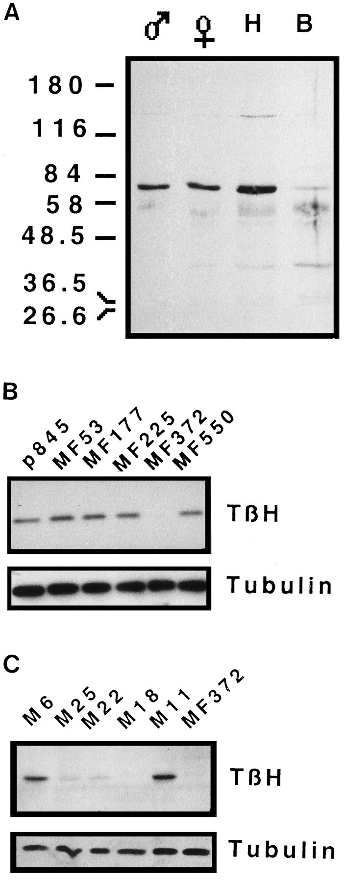
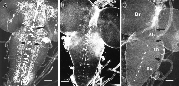
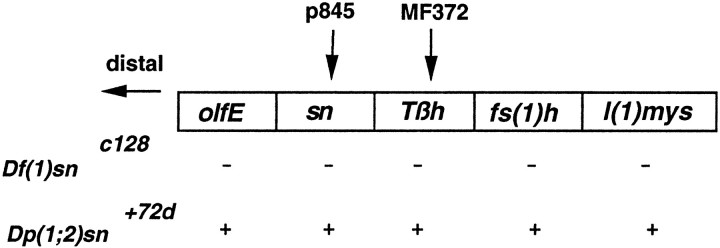
Octopamine is likely to be an important neuroactive molecule in invertebrates. Here we report the molecular cloning of the Drosophila melanogaster gene, which encodes tyramine beta-hydroxylase (TBH), the enzyme that catalyzes the last step in octopamine biosynthesis. The deduced amino acid sequence of the encoded protein exhibits 39% identity to the evolutionarily related mammalian dopamine beta-hydroxylase enzyme. We generated a polyclonal antibody against the protein product of T beta h gene, and we demonstrate that the TBH expression pattern is remarkably similar to the previously described octopamine immunoreactivity in Drosophila. We further report the creation of null mutations at the T beta h locus, which result in complete absence of TBH protein and blockage of the octopamine biosynthesis. T beta h-null flies are octopamine-less but survive to adulthood. They are normal in external morphology, but the females are sterile, because although they mate, they retain fully developed eggs. Finally, we demonstrate that this defect in egg laying is associated with the octopamine deficit, because females that have retained eggs initiate egg laying when transferred onto octopamine-supplemented food.


Figures

Fig. 1.
Fig. 1.
Alignment using LINEUP program of a portion of DBH amino acid sequence from three mammalian species (bovine, human, rat) in the histidine-rich regions. The numbers to the rightcorrespond to amino acid coordinates of the rat DBH.Asterisks indicate the paired histidines or the H-X-H residues; dot indicates Tyr236 of the rat sequence, conserved in all three species. The regions selected for primer design are underlined with arrowsindicating the 5′−3′ direction of the primer.
Fig. 2.
Fig. 2.
A, Nucleotide and deduced amino acid sequences of Drosophila Tβh cDNA (EMBL Data Library numberZ70316). Amino acid residues in bold indicate paired histidines or the H-X-H sites. Residues in bold with anasterisk correspond to those amino acids of the bovine DBH protein modified by mechanism-based inhibitors (see text, Fig. 3). Residues in bold italics correspond to consensus for potential N-glycosylation sites, and underlined tetrads of amino acids correspond to potential phosphorylation sites. The stop codon is underlined. B, Restriction map of theDrosophila Tβh cDNA. Restriction sites: H,HindIII; P, PstI; R,EcoRI; S, SalI; X,XhoI. The open reading frame region of the cDNA is indicated by the arrow below the restriction map. The fragments used for the RNA in situ probe and the fusion protein construct are represented with solid lines above andbelow the restriction map, respectively. The first and last amino acid included in the fusion protein construct are indicated at the end of the corresponding line.
Fig. 3.
Fig. 3.
Alignment using LINEUP program ofDrosophila TBH protein sequence to the DBH protein from three mammalian species. The top line corresponds to theDrosophila TBH amino acids 1–660. Only the identical amino acids are shown, whereas gaps are marked with dots and nonconserved residues are marked with dashed lines.Asterisks indicate conserved His-His or His-X-His residues, and # indicates an His-X-His site of the mammalian DBH not conserved in the Drosophila TBH. The residues Tyr273 and His452 (TBH coordinates), which correspond to those modified by mechanism-based inhibitors (DeWolf et al., 1988, 1989), are indicated with a dotbelow them.
Fig. 4.
Fig. 4.
Tβh transcript analysis. A, Total Canton-S RNA (12 μg/lane) from adult head and body tissue was hybridized with a 32P-labeled RNA probe corresponding to the PstXho fragment ofTβh cDNA and subsequently with a32P-labeled DNA probe of RP49. The size of the transcript was determined relative to RNA markers (RBL) shown on theright. B, Cellular distribution of theTβh transcript. Confocal image of whole-mount third instar larval CNS hybridized with a digoxigenin-labeled DNA probe corresponding to the PstXho fragment of theTβh cDNA. Anterior is to the top. The CNS is composed of the paired brain lobes and a fused ventral ganglion composed of the subesophageal ganglion, thoracic ganglion, and abdominal ganglion (see legend to Fig. 6C). The in situ hybridization signal as detected with an alkaline phosphatase-conjugated antidigoxigenin antibody was present in the ventral ganglion in a cluster of cells in the subesophageal ganglion (white arrow) and in cells along the midline of the thoracic and abdominal ganglia. Scale bar, 50 μm.
Fig. 5.
Fig. 5.
Immunoblot analysis of wild-type and mutant lines. Affinity-purified anti-TBH antiserum was used as the primary antibody; signal was visualized by chemiluminescent detection. A, Immunoblot analysis of head and body homogenates from Canton-S male and female flies. Protein (30 μg) from each sample was analyzed by SDS-PAGE on a 7.5% gel. B, TBH immunodetection in a sample of lines produced by the P element mutagenesis. Protein, equivalent of one head homogenate, from the original p845 line (insert in thesn locus) and five independent new insertion lines was loaded. To control for sample loading, the blot was stripped and reprobed using an anti-Tubulin antibody. A total of 250 lines were screened by this method. Note the lack of signal in MF372 lane.C, TBH immunodetection in a sample of lines in which the transposon in MF372 was excised. The procedure was the same as inB. A total of 67 lines were screened. Note the lack of signal in M18 and MF372 lanes and normal signal in M6 and M11 lanes.
Fig. 6.
Fig. 6.
TBH immunoreactivity in representative samples of larval CNSs from wild-type (Canton-S) and Tβh mutant strains. Third instar larval CNSs were immunoreacted with affinity-purified anti-TBH antiserum primary antibody and an anti-rat FITC-conjugated secondary antibody. These images were collected as Z series at the same laser level and gain settings. Care was taken to photograph and develop the negatives using similar conditions, but the images from the mutant brains were developed longer to show the residual signal. Each image represents a superimposition of 8 to 10 confocal sections taken at a Z step of 2.16 μm. In all images, the anterior is to the top. A, Confocal image of wild-type larval CNS. Note the intense immunoreactivity in the neuropil and the characteristic foci (white arrows), one in each brain lobe. In the ventral ganglion, note the cells in the subesophageal region (black arrow) and along the midline in the thoracic and abdominal ganglia and the paramedial cells (black arrowheads). B, Confocal image of a larval CNS of theTβhMF372mutant. Note the near absence of the neuropil staining and the reduced signal in the TBH-immunoreactive cells compared with the wild type. (Because of the low TBH signal, the background signal is high). C, Confocal image of a larval CNS from the TβhnM18 excision mutant. Note the complete absence of immunoreactivity from both the neuropil and the TBH-expressing cells. Black arrow points to the subesophageal ganglion; black arrowheads delineate thoracic ganglia. (We believe that the dotted pattern along the midline of the ventral ganglion is caused by nonspecific staining, because we have observed such staining in dorsal midline using other antibodies, especially when the antibody concentration is high. This pattern is confined to the dorsal-most sections, which are not included in the wild-type CNS representation in A. Br, Brain lobes; sb, subesophageal ganglion; th: thoracic ganglion; ab: abdominal ganglion. Scale bar, 50 μm inAC.
Fig. 7.
Fig. 7.
Genetic map of the 7D1–2 region of the X chromosome. Four known loci besides Tβh are represented byboxes. The signs below each box indicate the loci uncovered by Df(1)snC128(−) or covered by the Dp(1;2)sn+72d(+), two of the chromosomal rearrangements corresponding to the area. Arrowspoint to the position of p845 and MF372 transposon insertions.olfE, Olfactory E; sn, singed; Tβh,Tyramine β-hydroxylase; fs(1)h, female sterile homeotic; l(1)mys, lethal (1) myospheroid.

References

    1. Altschul SF, Lipman DJ. Protein database searches for multiple alignments. Proc Natl Acad Sci USA. 1990;87:5509–5513. - PMC - PubMed
    1. Arakawa S, Gocoyne JD, McCombie WR, Urquhart DA, Hall LM, Fraser CM, Venter JC. Cloning, localization, and permanent expression of a Drosophila octopamine receptor. Neuron. 1990;2:343–354. - PubMed
    1. Bicker G, Menzel R. Chemical codes for the control of behaviour in arthropods. Nature. 1989;337:33–39. - PubMed
    1. Bier E, Vaessin H, Shepherd S, Lee K, McCall K, Barbel S, Ackerman L, Carretto R, Uemura T, Grell E, Jan LY, Jan YN. Searching for pattern and mutation in the Drosophila genome with a P-lacZ vector. Genes Dev. 1989;3:1273–1287. - PubMed
    1. Braun G, Bicker G. Habituation of an appetitive reflex in the honeybee. J Neurophysiol. 1992;67:588–598. - PubMed

Publication types

MeSH terms

Associated data