. 1996 Jun 15;16(12):3925-33.
doi: 10.1523/JNEUROSCI.16-12-03925.1996.

Cloning and functional characterization of a novel dopamine receptor from Drosophila melanogaster

Affiliations

Cloning and functional characterization of a novel dopamine receptor from Drosophila melanogaster

G Feng et al. J Neurosci. .

Abstract

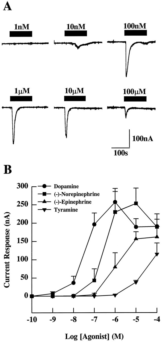
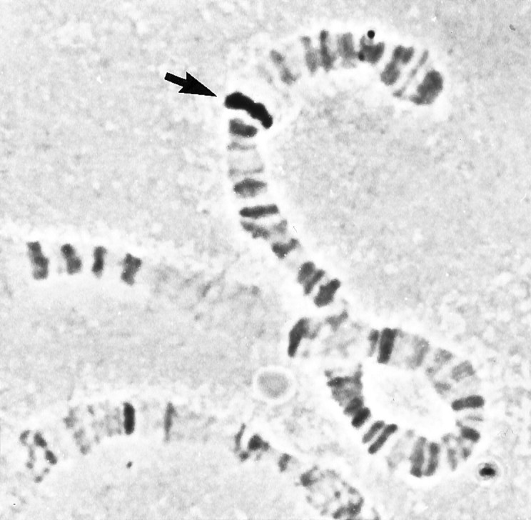
A cDNA clone is described that encodes a novel G-protein-coupled dopamine receptor (DopR99B) expressed in Drosophila heads. The DopR99B receptor maps to 99B3-5, close to the position of the octopamine/tyramine receptor gene at 99A10-B1, suggesting that the two may be related through a gene duplication. Agonist stimulation of DopR99B receptors expressed in Xenopus oocytes increased intracellular Ca2+ levels monitored as changes in an endogenous inward Ca2+-dependent chloride current. In addition to initiating this intracellular Ca2+ signal, stimulation of DopR99B increased cAMP levels. The rank order of potency of agonists in stimulating the chloride current is: dopamine > norepinephrine > epinephrine > tyramine. Octopamine and 5-hydroxytryptamine are not active (< 100 microM). This pharmacological profile plus the second-messenger coupling pattern suggest that the DopR99B receptor is a D1-like dopamine receptor. However, the hydrophobic core region of the DopR99B receptor shows almost equal amino acid sequence identity (40-48%) with vertebrate serotonergic, alpha 1- and beta-adrenergic, and D1-like and D2-like dopaminergic receptors. Thus, this Drosophila receptor defines a novel structural class of dopamine receptors. Because DopR99B is the second dopamine receptor cloned from Drosophila, this work establishes dopamine receptor diversity in a system amenable to genetic dissection.


Figures

Fig. 1.
Fig. 1.
The DopR99B sequence. A, Nucleotide sequence and deduced amino acid sequence of theDrosophila dopamine receptor DopR99B. In-frame stop codons are indicated by asterisks. The sequence of a potential ribosome binding site is boxed. The seven transmembrane domains are underlined and numbered. Potential PKC phosphorylation sites are indicated by Δ. Potential N-glycosylation sites are marked by ♦. B, Hydrophobicity plot of the deduced DopR99B amino acid sequence. Regionsabove the line are hydrophobic. Transmembrane domains arenumbered. The hydropathy plot was done using the method ofKyte and Doolittle (1982) and the Geneworks software (Intelligenetics). The Genbank accession number for DopR99B is U34383.
Fig. 2.
Fig. 2.
Structural relationship of DopR99B with other G-protein-coupled receptors. Sequence alignment was done with the PILEUP program of the Wisconsin Genetic Computer Group (GCG) software (Devereux et al., 1984) using the hydrophobic cores of each receptor listed. The hydrophobic core was defined as the region between and including transmembrane domains I and V plus the region between and including transmembrane domains VI and VII. The lengths of the horizontal lines are inversely proportional to the percentages of sequence similarity between receptors or groups of receptors. In the receptor names listed, the first three to four letters refer to the general receptor type: Adr, adrenergic; Dop, dopamine; 5ht, serotonin; Octy, octopamine/tyramine. The next letters in the name refer to the species:dro, Drosophila; hum, human;rat, rat; xen, Xenopus. The exceptions to this nomenclature are three Drosophilareceptors (DopR35EF, DopR99B,OctyR99AB). For these, the type name is followed byR (receptor) and then the salivary gland chromosome map position. Pathway refers to the second-messenger coupling reported in the literature for each receptor: PLC, phospholipase C;AC, adenylyl cyclase. An upward arrowindicates stimulation of the indicated second-messenger system, and adownward arrow indicates inhibition.Accession # refers to the Genbank database locator number.
Fig. 3.
Fig. 3.
Inward current response after agonist stimulation mediated by the DopR99B receptor expressed in Xenopusoocytes. Two minute pulses of agonists were given to Xenopusoocytes 3 d after the injection of DopR99B cRNA. A, Typical responses of a single oocyte to various concentrations of dopamine.B, Dose–response curves for dopamine, (−)-norepinephrine, (−)-epinephrine, and tyramine. The results (from at least 6 oocytes) are expressed as the mean peak inward current ± SE initiated by each amine.
Fig. 4.
Fig. 4.
Dose–response curves for increase in cAMP levels in oocytes expressing DopR99B. Five days after injection withDrosophila DopR99B receptor cRNA, injected and uninjected (control) oocytes were treated with the indicated dopamine concentrations for 30 min in the presence of 100 μm IBMX after preincubation for 30 min in 100 μm IBMX. The results are expressed as the mean oocyte cAMP level (pmol/oocyte) ± SE (n = 5 oocytes).B, Basal levels.
Fig. 5.
Fig. 5.
Northern blot analysis of DopR99B. A Northern blot of poly(A)+ RNA isolated from heads (H), bodies (B), and legs/antennae (L/A) was probed with the32P-labeled 0.68 kb PCR fragment. To control for mRNA recovery, the blot was reprobed with ribosomal protein cDNA (rp49), which is expressed throughout the organism (O’Connell and Robash, 1984).
Fig. 6.
Fig. 6.
Chromosome localization of DopR99B. In situ hybridization to Drosophila salivary gland polytene chromosomes using the biotinylated 0.68 kb PCR fragment. Thearrow indicates the site of hybridization at 99B3–5 on the right arm of chromosome 3.

Similar articles

Cited by

References

    1. Ali DW, Orchard I. Characterization of dopamine and serotonin receptors on the salivary glands of the locust, Locusta migratoria . Biog Amines. 1994;10:195–212.
    1. Arakawa S, Gocayne JD, McCombie WR, Urquhart DA, Hall LM, Fraser CM, Venter JC. Cloning, localization, and permanent expression of a Drosophila octopamine receptor. Neuron. 1990;4:343–354. - PubMed
    1. Barnard EA, Miledi R, Sumikawa K. Translation of exogenous messenger RNA coding for nicotinic acetylcholine receptors produces functional receptors in Xenopus oocytes. Proc R Soc Lond [Biol] 1982;215:241–246. - PubMed
    1. Bodnaryk RP. Identification of specific dopamine- and octopamine-sensitive adenylate cyclases in the brain of Mamestra configurata Wlk. Insect Biochem. 1979;9:155–162. - PubMed
    1. Brown CS, Nestler C. Catecholamines and indolalkylamines. In: Kerkut GA, Gilbert LI, editors. Comprehensive insect physiology, biochemistry and pharmacology, Vol. 11. Pergamon; Oxford: 1985. pp. 436–497.

Publication types

MeSH terms

Associated data