. 1996 Jun 15;16(12):3934-42.
doi: 10.1523/JNEUROSCI.16-12-03934.1996.

Biosynthesis of an endogenous cannabinoid precursor in neurons and its control by calcium and cAMP

Affiliations

Biosynthesis of an endogenous cannabinoid precursor in neurons and its control by calcium and cAMP

H Cadas et al. J Neurosci. .

Abstract

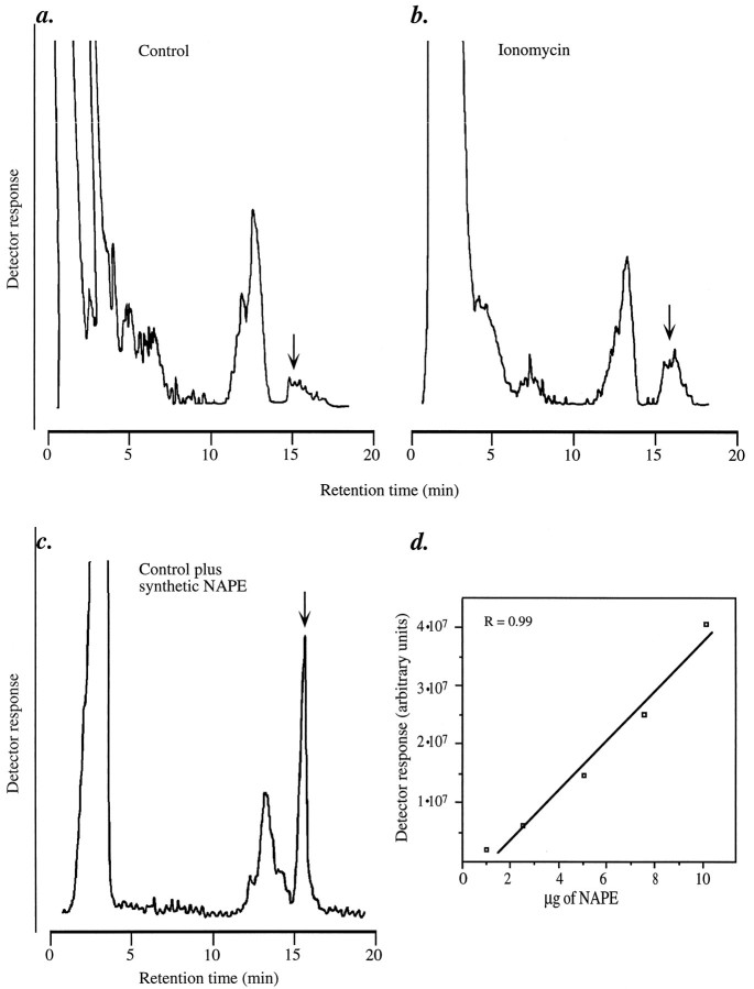
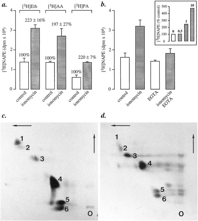
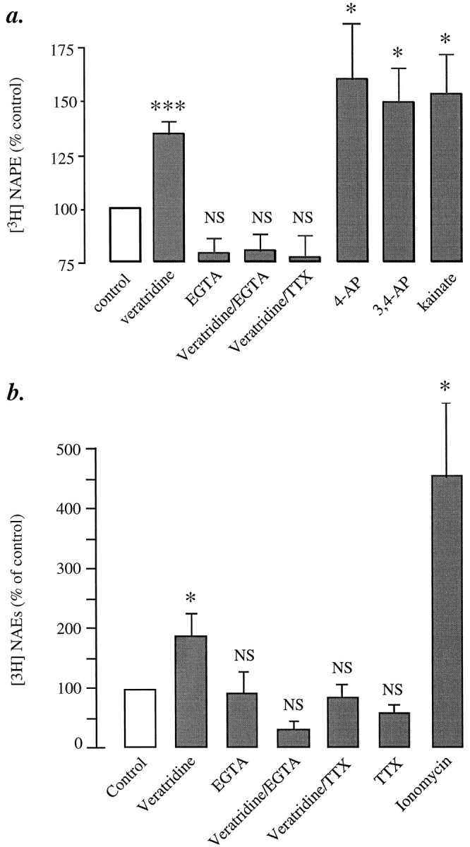
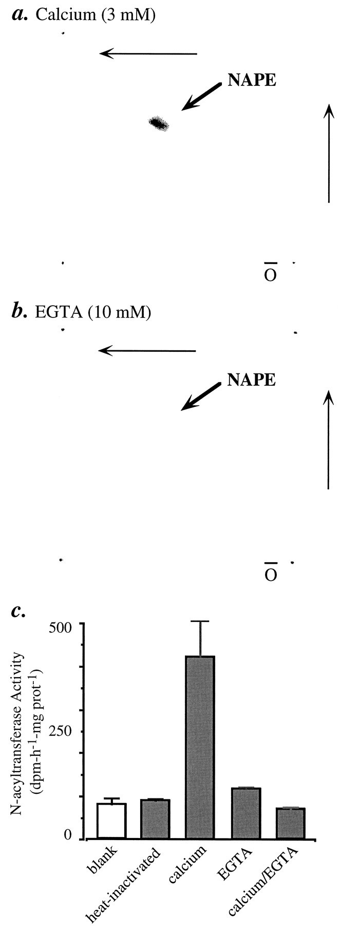
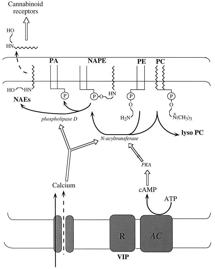
Understanding the mechanisms involved in the biogenesis of N-arachidonoylethanolamine (anandamide) and N-palmitoylethanolamine is important in view of the possible role of these lipids as endogenous cannabinoid substances. Anandamide (which activates cannabinoid CB1 receptors) and N-palmitoylethanolamine (which activates a CB2-like receptor subtype in mast cells) may both derive from cleavage of precursor phospholipid, N-acylphosphatidylethanolamine (NAPE), catalyzed by Ca(2+)-activated D-type phosphodiesterase activity. We report here that the de novo biosynthesis of NAPE is enhanced in a Ca(2+)-dependent manner when rat cortical neurons are stimulated with the Ca(2+)-ionophore ionomycin or with membrane-depolarizing agents such as veratridine and kainate. This reaction is likely to be mediated by a neuronal N-acyltransferase activity, which catalyzes the transfer of an acyl group from phosphatidylcholine to the ethanolamine moiety of phosphatidylethanolamine. In addition, we show that Ca2+-dependent NAPE biosynthesis is potentiated by agents that increase cAMP levels, including forskolin and vasoactive intestinal peptide. Our results thus indicate that NAPE levels in cortical neurons are controlled by Ca2+ ions and cAMP. Such regulatory effect may participate in maintaining a supply of cannabimimetic N-acylethanolamines during synaptic activity, and prime target neurons for release of these bioactive lipids.


Figures

Fig. 2.
Fig. 2.
Quantitative analysis of neuronal NAPE by HPLC coupled to evaporative light-scattering detection. Representative chromatograms from (a) control neurons, (b) 1 μm ionomycin-stimulated neurons, and (c) control neurons to which synthetic NAPE (10 μg) was added before extraction, to verify its HPLC coelution with native NAPE. The arrows indicate the retention time of synthetic NAPE. Lipids were extracted and fractionated by column chromatography. The NAPE-containing fractions from two culture dishes were pooled and subjected to reversed-phase HPLC, as described in Materials and Methods. d, Response of the light-scattering detector as a function of injected synthetic NAPE (in 40 μl of chloroform). Photomultiplier voltage was set at 800 V, and nebulization temperature was set at 90°C.
Fig. 1.
Fig. 1.
Biosynthesis of [3H]NAPE in primary cultures of rat brain cortical neurons. a, Ionomycin (1 μm, 10 min) stimulates [3H]NAPE biosynthesis in neurons labeled by incubation with [3H]ethanolamine (Eth), [3H]arachidonic acid (AA), or [3H]palmitic acid (PA). NAPE was fractionated by column chromatography, and NAPE-containing fractions were analyzed by monodimensional TLC. Results are expressed as mean ± SEM (dpm/dish) of six separate experiments.b, The effect of ionomycin is prevented by chelating extracellular Ca2+ with EGTA (10 mm) and is concentration-dependent (inset, μm concentrations). In these experiments, the neurons were labeled with [3H]ethanolamine. c, d, Ionomycin stimulates NAPE biosynthesis in neurons (c), but not in astrocytes (d). Cultures were labeled by overnight incubation with [14C]arachidonic acid. NAPE was analyzed by bidimensional TLC and autoradiography. On these representative chromatograms, the numbers indicate the positions of the following lipids: (1) NAPE; (2) unknown; (3) cerebrosides; (4) PE; (5) PC; (6) phosphatidylserine plus phosphatidylinositol.O, Origin.
Fig. 3.
Fig. 3.
Effects of various depolarizing agents on [3H]NAPE biosynthesis and [3H]NAE formation in cortical neurons.a, [3H]NAPE biosynthesis: veratridine (20 μm), 4-aminopyridine (4-AP; 3 mm), 3,4-diaminopyridine (3,4-AP; 3 mm), and kainate (0.1 mm). EGTA and tetrodotoxin (TTX) were used at 10 mm and 1 μm, respectively. b, [3H]NAE formation was determined in the same or parallel experiments. The ability of 4-AP, 3,4-AP, and kainate to stimulate [3H]NAE formation was documented previously (Di Marzo et al., 1994). The effect of ionomycin (1 μm, 10 min) is shown for comparison. Results are expressed as percent of control and represent the mean ± SEM of five to eight separate experiments; *p < 0.05, ***p < 0.001 (ANOVA); NS, nonsignificantly different from unstimulated cultures.
Fig. 4.
Fig. 4.
Potentiation of ionomycin (iono)-stimulated [3H]NAPE biosynthesis by forskolin (FSK; 10 μm) and vasoactive intestinal peptide (VIP; 1 μm) in cortical neurons. ***p < 0.001; a, versus control; b, versus ionomycin; NS, nonsignificant.
Fig. 5.
Fig. 5.
Ca2+-dependent [14C]NAPE formation andN-acyltransferase activity in homogenates of cortical neurons. Homogenates were incubated with [14C]dioleoyl-PC or [14C]dipalmitoyl-PC in the presence of either Ca2+ (3 mm) (a) or 10 mm EGTA (b). The incubation mixtures were subjected to lipid extraction and analyzed by bidimensional TLC. Radioactivity on the plates was visualized with a PhosphorImager. c, N-acyltransferase activity in neuron homogenates. Assay conditions and analysis by column chromatography are described in Materials and Methods. Results (mean ± SEM) are from one experiment, performed in triplicate and typical of four experiments.
Fig. 6.
Fig. 6.
Model of biosynthesis and regulation of the endogenous cannabinoid precursor NAPE in rat cortical neurons. [Ca2+]i rises produced by neuronal depolarization may stimulate an N-acyltransferase activity that catalyzes the intermolecular transfer of a fatty acyl group from a glycerophospholipid [e.g., phosphatidylcholine (PC)], to the ethanolamine moiety of phosphatidylethanolamine (PE), forming NAPE and lysophospholipid (e.g., lyso PC). Neuromodulators (e.g., VIP) may enhance Ca2+-dependent NAPE biosynthesis by activating a membrane receptor (R) coupled to the activation of adenylyl cyclase (AC) and to the subsequent stimulation of cAMP-dependent protein kinase (PKA) activity. The broken arrow indicates that the molecular target of PKA leading to enhanced NAPE biosynthesis remains to be determined. NAPE is composed of several molecular species, differing in the fatty acyl group linked to the ethanolamine moiety (Schmid et al., 1990; Cadas et al., 1996). Therefore, cleavage of NAPE by a D-type phosphodiesterase activity [phospholipase D (PLD)] may give rise to multipleN-acylethanolamines (NAEs), including anandamide (which activates cannabinoid CB1-type receptors) andN-palmitoylethanolamine (which activates CB2-type receptors in certain cell types). Although cannabimimetic NAEs may be recovered in the extracellular fluid of stimulated neurons in culture (Di Marzo et al., 1994; Hansen et al., 1995), the mechanism of extrusion of these lipids (indicated schematically as a broken arrow) is still unknown.

Similar articles

Cited by

References

    1. Cadas H, Schinelli S, Piomelli D (1996) Membrane localization of N-acylphosphatidylethanolamine in central neurons: studies with exogenous phospholipases. J Lipid Med, in press. - PubMed
    1. Chapman K, Moore JT. Catalytic properties of a newly discovered acyltransferase that synthesizes N -acylphosphatidylethanolamine in cottonseed ( Gossipuim hirsutum L .) microsomes. Plant Physiol. 1993;102:761–769. - PMC - PubMed
    1. Colodzin M, Bachur N, Weissbach H, Udenfriend S. Enzymatic formation of fatty acid amides of ethanolamine by rat liver microsomes. Biochem Biophys Res Commun. 1963;10:165–170. - PubMed
    1. Christie W. Pergamon; Oxford: 1987. HPLC and lipids: a practical guide, pp 23–25. .
    1. Desarnaud F, Cadas H, Piomelli D. Anandamide amidohydrolase activity in rat brain microsomes: identification and partial characterisation. J Biol Chem. 1995;270:6030–6035. - PubMed

MeSH terms

Substances