. 1996 Jun 15;16(12):3979-90.
doi: 10.1523/JNEUROSCI.16-12-03979.1996.

Schwann cell apoptosis during normal development and after axonal degeneration induced by neurotoxins in the chick embryo

Affiliations

Schwann cell apoptosis during normal development and after axonal degeneration induced by neurotoxins in the chick embryo

D Ciutat et al. J Neurosci. .

Abstract

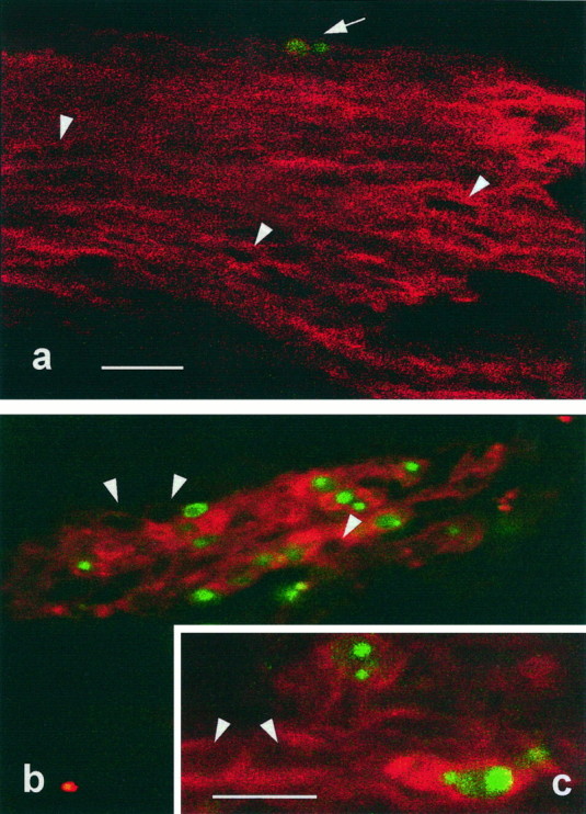
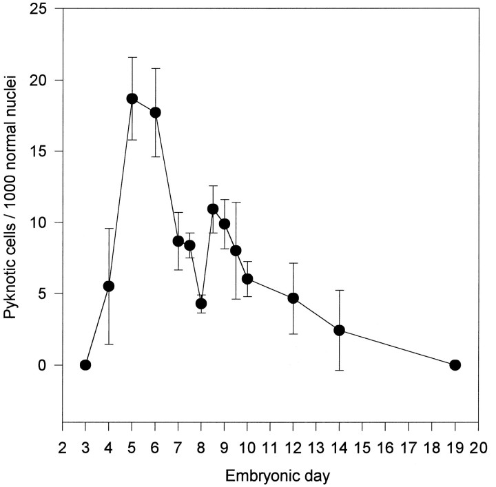
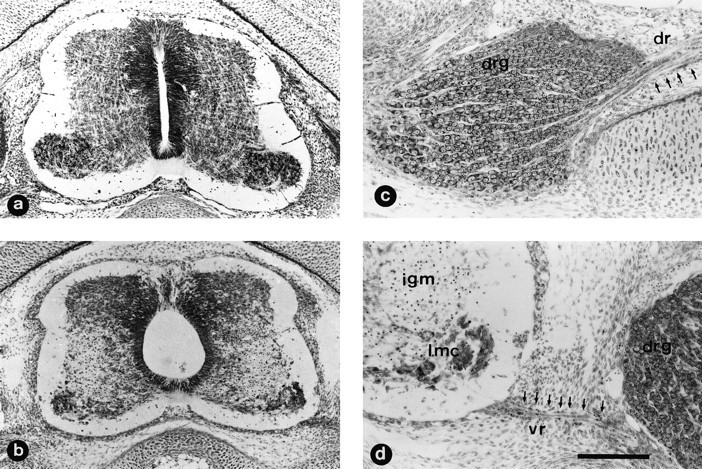
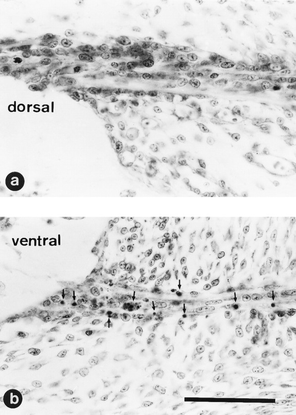
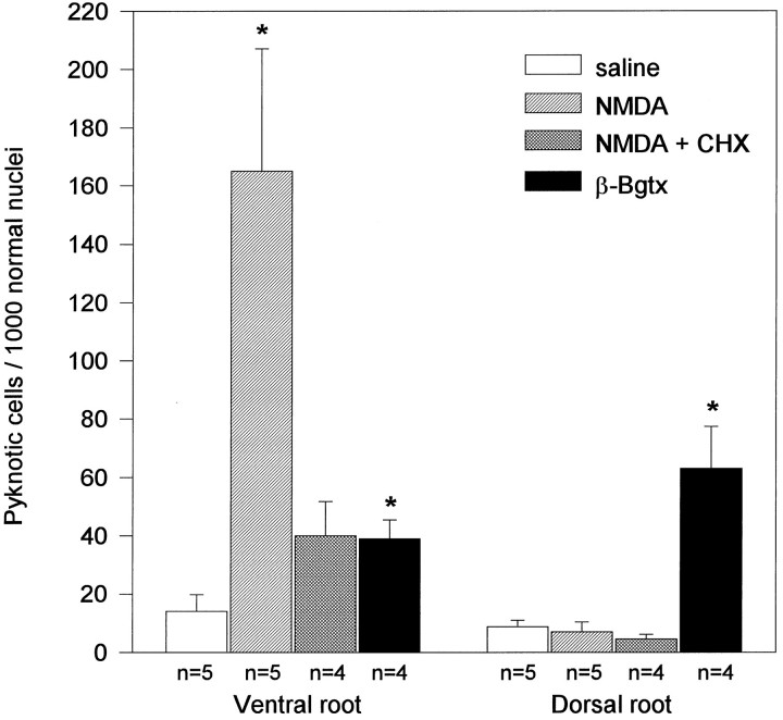
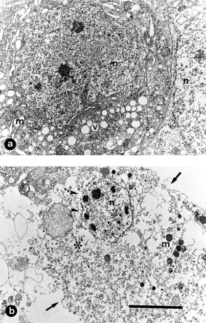
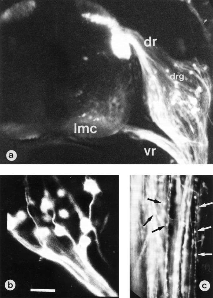
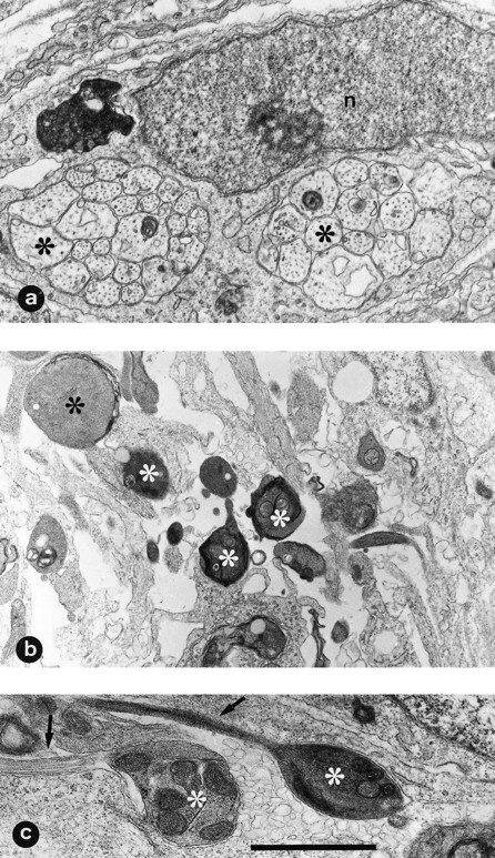
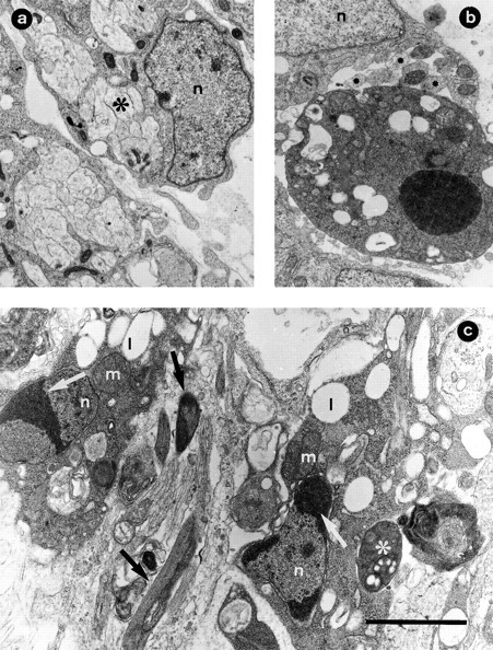
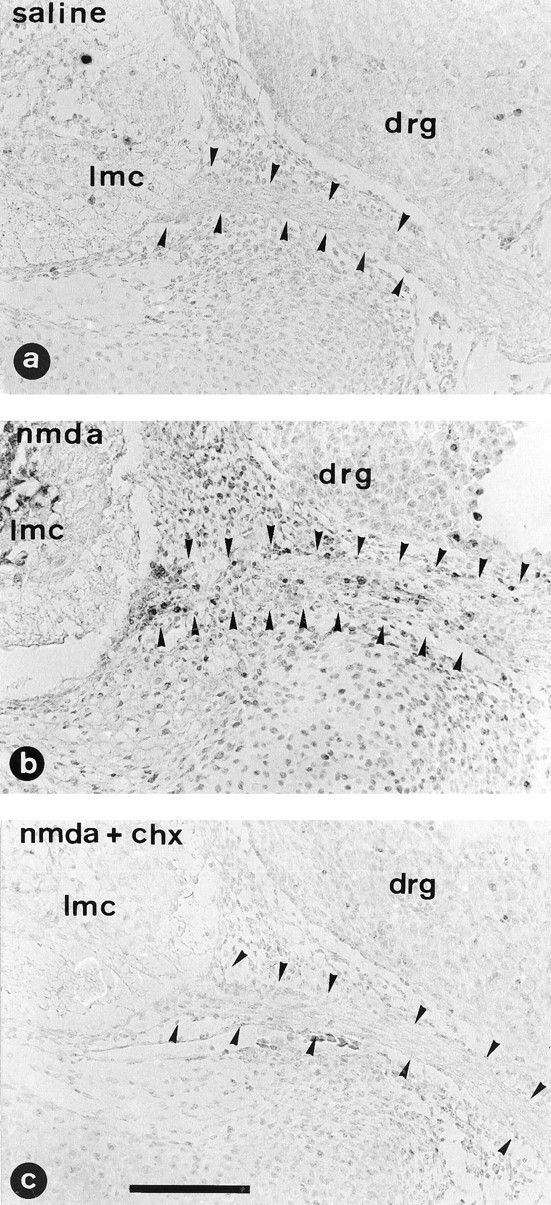
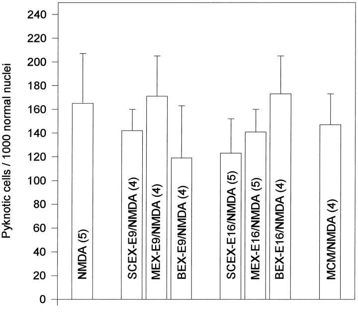
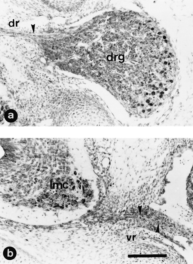
In the present work, we show that chick embryo Schwann cells die by apoptosis both during normal development and after axonal degeneration induced by neurotoxin treatment. Schwann cell apoptosis during development takes place during a period roughly coincidental with normally occurring motoneuron death. Administration of NMDA to chick embryos on embryonic day 7 induces extensive excitotoxic motoneuronal damage in the spinal cord without any apparent effects on neurons in the dorsal root ganglia (DRG). The death of Schwann cells in ventral nerve roots after NMDA treatment causes degenerative changes that display ultrastructural features of apoptosis and exhibit in situ detectable DNA fragmentation. By contrast, NMDA treatment does not increase the death of Schwann cells in dorsal nerve roots. In situ detection of DNA fragmentation in combination with the avian Schwann cell marker 1E8 antibody demonstrates that dying cells in ventral nerve roots are in the Schwann cell lineage. Administration of cycloheximide does not prevent the toxic effects of NMDA on motoneurons, but dramatically reduces the number of pyknotic Schwann cells and DNA fragmentation profiles in the ventral nerve roots. In ovo administration of various tissue extracts (muscle, brain, and spinal cord) from the chick embryo or of the motoneuron conditioned medium fails to prevent Schwann cell apoptosis in NMDA-treated embryos. Intramuscular administration of the snake toxin beta-bungarotoxin produces a massive death of both lateral motor column motoneurons and DRG neurons, resulting in a substantial increase in the number of pyknotic Schwann cells in both ventral and dorsal nerve roots. It is concluded that during development, axonal-derived trophic signals are involved in the regulation of Schwann cell survival in peripheral nerves.


Figures

Fig. 1.
Fig. 1.
Double-fluorescent labeling to demonstrate immunoreactivity to 1E8 antibody and DNA fragmentation in ventral nerve roots from E7.5 chick embryos. Confocal laser-scanning micrographs from cryostat sections in which 1E8-positive labeling (red) shows that some Schwann cells (arrow) contain fragmented DNA (green) in embryos treated with saline (a) and 12 hr after NMDA treatment (b, c). Note that the number of dying Schwann cells is substantially increased after NMDA treatment; arrowheads indicate 1E8-positive cells that contain negative TUNEL nuclear profiles. Scale bars: a, b, 20 μm; c, 10 μm.
Fig. 2.
Fig. 2.
Developmental regulation of normally occurring Schwann cell death in L3 ventral nerve roots from chick embryo. The number of pyknotic Schwann cells was plotted as a function of the developmental age. Each point represents the mean ± SEM of 3–10 different embryos.
Fig. 3.
Fig. 3.
NMDA treatment produces severe damage in the spinal cord but not in DRG. Transverse paraffin sections of lumbar spinal cord from E7.5 chick embryos 12 hr after in ovotreatment with a single injection of either saline solution (a) or 1 mg of NMDA (b–d). Sections were stained with thionin. Note in b and d the severe cellular depletion in the lateral motor column (lmc) that extends through the intermediate gray matter (igm); the central canal appears distended, and many pyknotic cells are present in the ventral nerve root (vr) (identified by arrows ind). No damage is observed in the dorsal region of the gray matter or in dorsal root ganglia (drg) or dorsal nerve root (dr) (identified by arrows in c). Scale bar (shown in d): a, b, 100 μm; c, d, 50 μm.
Fig. 4.
Fig. 4.
NMDA treatment induces Schwann cell death in ventral but not in dorsal nerve roots. Higher magnification view of a section through L3 dorsal (a) or ventral (b) nerve roots from an E7.5 chick embryo 12 hr after treatment with 1 mg of NMDA. Note the large number of pyknotic cells (arrows) exclusively present in the ventral nerve roots; no pyknotic cells are present in the dorsal nerve root. Scale bar (shown in b): 50 μm.
Fig. 10.
Fig. 10.
Increased Schwann cell death after treatment with neurotoxins. Number (mean ± SEM) of pyknotic cells in ventral and dorsal nerve roots from E7.5 chick embryos treated with saline solution, 1 mg of NMDA, 1 mg of NMDA and CHX (2 μg every 4 hr), and 100 ng of β-Bgtx. Counts were made in each condition 12 hr after the single injection of either saline, NMDA, or β-Bgtx. n, Sample size. *p ≤ 0.05 versus saline, Student’st test.
Fig. 5.
Fig. 5.
Ultrastructural demonstration of acute motoneuronal damage induced by NMDA. Samples were taken from E7.5 chick embryo 12 hr after treatment with 1 mg of NMDA. a, Motoneuron cell body showing accumulation of small membrane-bounded vacuoles (v), dilatation of rough endoplasmic reticulum, swollen mitochondria (m), and nucleus (n) with chromatin aggregated in fine granules. In b, a more advanced stage of motoneuronal degeneration showing extreme organelle disruption (∗), condensed mitochondria (m), and disruption of plasma membrane (arrows) and nuclear envelope (arrowheads). Scale bar (shown in b):a, 5 μm; b, 7 μm.
Fig. 6.
Fig. 6.
Neuronal DiI tracing in an NMDA-treated embryo. Confocal laser-scanning micrographs of a transverse section from E7.5 chick embryo spinal cord 150 min after treatment with 1 mg of NMDA.a, Embryo was processed for visualization of ventral (vr) and dorsal (dr) nerve fibers by means of DiI labeling; some ganglionic cells in dorsal root ganglion (drg) and motoneuronal cell bodies in lateral motor column (lmc) are also labeled. b, c, High-magnification micrographs of axonal profiles present in dorsal (b) or ventral (c) nerve roots showing the presence of large numbers of varicosities only in the ventral axons (arrows). Scale bar (shown in b): a, 125 μm; b, 25 μm; c, 50 μm.
Fig. 7.
Fig. 7.
Ultrastructural morphology of ventral nerve root axons from normal and NMDA-treated embryos. Electron micrographs were taken from E7.5 chick embryos 12 hr after treatment with either saline (a) or 1 mg of NMDA (b, c). Compare the normal morphology of axons (∗) in a surrounded by an immature Schwann cell with the dark appearance of degenerated axons inb; some degenerated axons (arrows) showing varicosities with accumulation of degenerated organelles can be seen (c). n, Schwann cell nucleus. Scale bar (shown in c): a, 1.5 μm; b, 2.90 μm; c, 1.45 μm.
Fig. 8.
Fig. 8.
Ultrastructural morphology of ventral nerve root Schwann cells from normal and NMDA-treated embryos. Electron micrographs were taken from E7.5 chick embryos 12 hr after treatment with either saline (a) or 1 mg of NMDA (b,c). In a, an immature Schwann cell (n) is seen surrounding densely packed fascicles of axons (∗). b, One Schwann cell with typical apoptotic morphology is seen near clustered nerve fibers (∗) and adjacent to a normal Schwann cell (n). In c, two Schwann cells with initial apoptotic changes are seen. Note the marginal clustering of condensed chromatin (white arrows) in the Schwann cell nucleus (n) and the dark appearance of the cytoplasm containing lipid droplets (l) and mitochondria (m). Degenerating axons are seen adjacent (black arrows) or in close contact (∗) with apoptotic Schwann cells. Scale bar (shown in c): a, 2 μm; b, 2.6 μm; c, 1.80 μm.
Fig. 9.
Fig. 9.
In situ detection of DNA fragmentation. Transverse paraffin sections of lumbar spinal cord from E7.5 chick embryos treated in ovo with saline solution (saline) (a), 1 mg of NMDA (nmda) (b), or both CHX (2 μg every 4 hr) and NMDA (1 mg) (nmda + chx) (c). Embryos were killed 12 hr after the single dose of either saline or NMDA. Arrowheadsdelimitate the ventral nerve roots. Note the absence of labeled cells in the embryos treated with saline and both NMDA and CHX. Normal nuclei are lightly labeled because of the methyl green counterstain.lmc, Lateral motor column; drg, dorsal root ganglion. Scale bar, 100 μm.
Fig. 11.
Fig. 11.
Treatment with embryonic extracts or motoneuron conditioned media does not rescue Schwann cell death after NMDA treatment. Number (mean ± SEM) of pyknotic Schwann cells in ventral nerve roots on E7.5 NMDA-injected embryos after treatment with saline, spinal cord extract (SCEX, 150 μg), muscle extract (MEX, 150 μg), brain extract (BEX, 150 μg), and motoneuron conditioned medium (MCM, 250 μg). Tissue extracts were obtained from either E9 or E16 chick embryos; MCM was obtained from 6 d primary cultures of motoneurons purified from E5.5 chick embryos. For details, see Materials and Methods. Numbersin parentheses indicate sample size.
Fig. 12.
Fig. 12.
Injection of β-Bgtx induces cell death in both dorsal root ganglion and lateral motor column neurons. a, Transverse paraffin section stained with thionin showing the massive death of dorsal root ganglion (drg) ventrolateral neurons 12 hr after a single intramuscular injection of β-Bgtx (100 ng) on E7 chick embryos. In b, a section of spinal cord from the same embryo in which the extensive spaces observed within the lateral motor column (lmc) were induced by the acute motoneuron loss, whereas some degenerating motoneurons can be observed as dark-stained profiles. Although the photos were taken at low magnification, some pyknotic Schwann cells can be identified in both dorsal (a, dr) and ventral (b, vr) nerve roots (arrowheads). Scale bar, 100 μm.

References

    1. Aguayo AJ, Terry LC, Bray GM. Spontaneous loss of axons in sympathetic unmyelinated nerve fibres of the rat during development. Brain Res. 1973;54:360–364. - PubMed
    1. Bhattacharyya A, Frank E, Ratner N, Brackenbury R. P0 is an early marker of the Schwann cell lineage in chickens. Neuron. 1991;7:831–844. - PubMed
    1. Barde Y-A. Trophic factors and neuronal survival. Neuron. 1984;2:1525–1534. - PubMed
    1. Barde Y-A. Neurotrophic factors: an evolutionary perspective. J Neurobiol. 1994;25:1329–1333. - PubMed
    1. Barres BA, Raff MC. Control of oligodendrocyte number in the developing rat optic nerve. Neuron. 1994;12:935–942. - PubMed

Publication types

MeSH terms

LinkOut - more resources