. 1996 Jun 15;16(12):4005-16.
doi: 10.1523/JNEUROSCI.16-12-04005.1996.

Analysis of the globose basal cell compartment in rat olfactory epithelium using GBC-1, a new monoclonal antibody against globose basal cells

Affiliations

Analysis of the globose basal cell compartment in rat olfactory epithelium using GBC-1, a new monoclonal antibody against globose basal cells

B J Goldstein et al. J Neurosci. .

Abstract

The olfactory epithelium (OE) supports ongoing neurogenesis throughout life and regenerates after experimental injury. Although evidence indicates that proliferative cells within the population of globose (light) basal cells (GBCs) give rise to new neurons, little is known about the biology of GBCs. Because GBCs have been identifiable only by an absence of staining with reagents that mark other cell types in the epithelium, we undertook to isolate antibodies that specifically react against GBCs and to characterize the GBC compartment in normal and regenerating OE. Monoclonal antibodies were produced using mice immunized with regenerating rat OE, and a monoclonal antibody designated GBC-1, which reacts against GBCs of the rat OE, was isolated. In immunohistochemical analyses, antibody GBC-1 was found to label GBCs in both normal and regenerating OE as we are currently able to define them: basal cells that incorporate the mitotic tracer bromodeoxyuridine and fail to express cytokeratins or neural cell adhesion molecule. During epithelial reconstitution after direct experimental injury with methyl bromide, expression of the GBC-1 antigen overlaps to a limited extent with expression of cell-specific markers for horizontal basal cells, Bowman's gland and sustentacular cells, and neurons. These data suggest that GBC-1 may mark multipotent cells residing in the GBC compartment, which are prominent during regeneration. However, a limited number of cells in the regenerating OE with other phenotypic characteristics of GBCs lack expression of the GBC-1 antigen. GBC-1 has revealed novel aspects of GBC biology and will be useful for studying the process of olfactory neurogenesis.


Figures

Fig. 1.
Fig. 1.
Monoclonal antibody GBC-1 labels GBCs. GBCs are defined as mitotically active (BrdU-incorporating) basal cells that lack expression of NCAM. A, B, The same section of normal OE stained with anti-BrdU and anti-NCAM, respectively; arrowsindicate BrdU (+)/NCAM (−) GBCs. C, The staining with GBC-1 in normal OE is confined primarily to the zone occupied by GBCs.D, Monoclonal antibody SUS-4 labels sustentacular cells and cells of Bowman’s glands. Single arrowheads mark the basal lamina. Magnification: 380× for A and B; 160× for C; 320× for D.
Fig. 2.
Fig. 2.
In normal OE, GBC-1 does not label HBCs, and many GBC-1 (+) cells are not neurons. A, B, A single section was stained with GBC-1 (immunoperoxidase label inB), BS-I (red fluorescence in A) to label HBCs, and anti-NCAM (green fluorescence inA) to label neurons. Examples of GBC-1-labeled basal cells are indicated by the arrows in A andB. Comparison of A and B indicates some overlap between anti-NCAM and GBC-1; for example, the more superficial of the two GBC-1 (+) cells above the right arrow is NCAM (+). Arrowheads mark the basal lamina. Magnification, 320×.
Fig. 4.
Fig. 4.
GBC-1 stains many cells in the OE during reconstitution after experimental injury. A, At 2 d after direct injury with MeBr gas. The regenerating epithelium contains a thin layer of cells, many of which are GBC-1 (+). B, At 7 d after MeBr exposure. GBC-1 staining extends through much of the height of the epithelium. C, At 4 d after olfactory bulb ablation. GBC-1 (+) cells are numerous and located deep in the epithelium. The more superficially placed GBC-1 (+) cells in B andC are neurons (compare with Fig. 5). Arrowheadsindicate the basal lamina. Magnification, 320×.
Fig. 5.
Fig. 5.
In addition to GBCs, GBC-1 labels some neurons. Tissue taken 4 d after olfactory bulb ablation, when neurogenesis is upregulated, and stained with GBC-1 (brown) and the neuronal marker anti-NCAM (blue-gray). GBC-1 (+)/NCAM (+) double-labeled cells are indicated by thin arrows.Short open arrows designate examples of cells labeled only by GBC-1, whereas the curved arrow indicates a cell labeled only by anti-NCAM. Arrowhead indicates the basal lamina. Magnification, 565×.
Fig. 8.
Fig. 8.
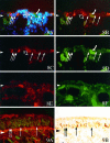
GBC-1 (−) globose cells are found in MeBr-lesioned and regenerating OE. A–D, A 3-μm-thick section harvested from ventral OE 2 d after MeBr-induced lesion. Stained with GBC-1 (red fluorescence inA–C), SUS-4 (green fluorescence inA, B, D), and the general nuclear stain bis-benzimide (blue fluorescence inA). Photomicrograph using dual FITC/Texas Red epifluorescent filter set is shown in B. Double exposure with the dual cube and a UV cube is shown in A. The open arrowdesignates an example of a GBC-1 (+) cell. The curved arrowmarks a SUS-4 (+) cell. Thin arrows indicate nuclei of cells that are not labeled by either marker. E, F, Adjacent section stained with BS-I and anti-NCAM, respectively. No cells in this area of the epithelium are labeled with either the HBC (E) or neuronal marker (F). The SUS-4 (−)/NCAM (−)/BS-I (−) population is classified as GBCs and is heterogeneous with respect to expression of the GBC-1 antigen. Arrowheadsindicate the basal lamina. Magnification, 340×.
Fig. 10.
Fig. 10.
A working model of lineage relationships among cell types in the OE and their definition by the expression of biochemical markers based on the data presented here and the published literature (Graziadei and Monti Graziadei, 1979; Calof and Chikaraishi, 1989; Verhaagen et al., 1990; Schwartz Levey et al., 1991, 1992;Schwob, 1992; Schwob et al., 1992, 1994a, 1995; Caggiano et al., 1994;DeHammer et al., 1994; Gordon et al., 1995; Holbrook et al., 1995).

Similar articles

Cited by

References

    1. Akeson RA, Haines SL. Rat olfactory cells and a central nervous system neuronal subpopulation share a cell surface antigen. Brain Res. 1989;488:202–212. - PubMed
    1. Anderson DJ, Carnahan JF, Michelsohn A, Patterson PH. Antibody markers identify a common progenitor to sympathetic neurons and chromaffin cells in vivo and reveal the timing of commitment to neuronal differentiation in the sympathoadrenal lineage. J Neurosci. 1991;11:3507–3519. - PMC - PubMed
    1. Blaugrund E, Pham TD, Tennyson VM, Lo L, Sommer L, Anderson DJ, Gershon MD. Distinct subpopulations of enteric neuronal progenitors defined by time of development, sympathoadrenal lineage markers and Mash-1 dependence. Development. 1996;122:309–320. - PubMed
    1. Caggiano M, Kauer JS, Hunter DD. Globose basal cells are neuronal progenitors in the olfactory epithelium: a lineage analysis using a replication-incompetent retrovirus. Neuron. 1994;13:339–352. - PubMed
    1. Calof AL, Chikaraishi DM. Analysis of neurogenesis in a mammalian neuroepithelium: proliferation and differentiation of an olfactory neuron precursor in vitro . Neuron. 1989;3:115–127. - PubMed

Publication types

MeSH terms

LinkOut - more resources