

Functional mapping of human learning: a positron emission tomography activation study of eyeblink conditioning
- PMID: 8656296
- PMCID: PMC6578600
- DOI: 10.1523/JNEUROSCI.16-12-04032.1996
Functional mapping of human learning: a positron emission tomography activation study of eyeblink conditioning
Abstract
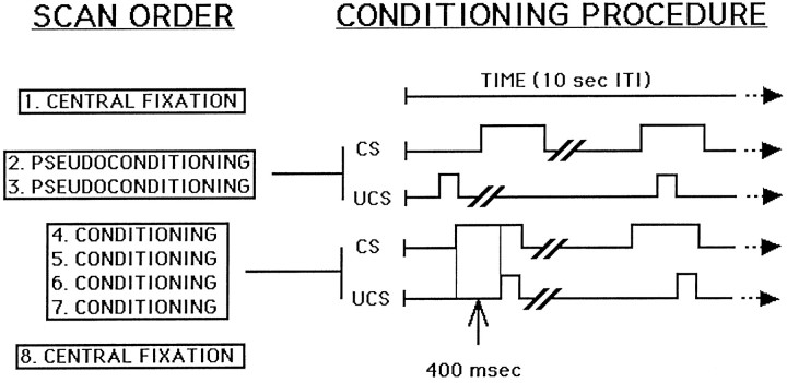
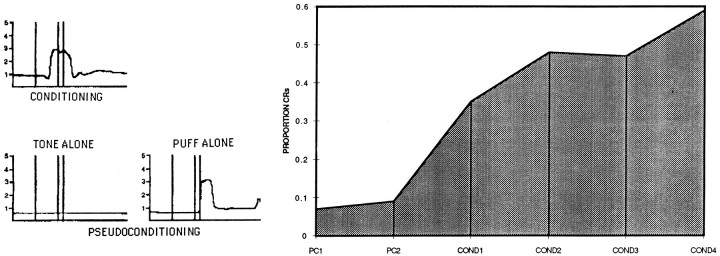
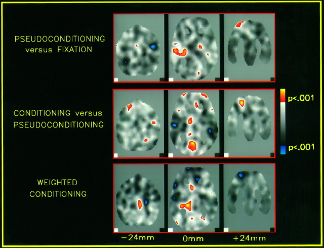
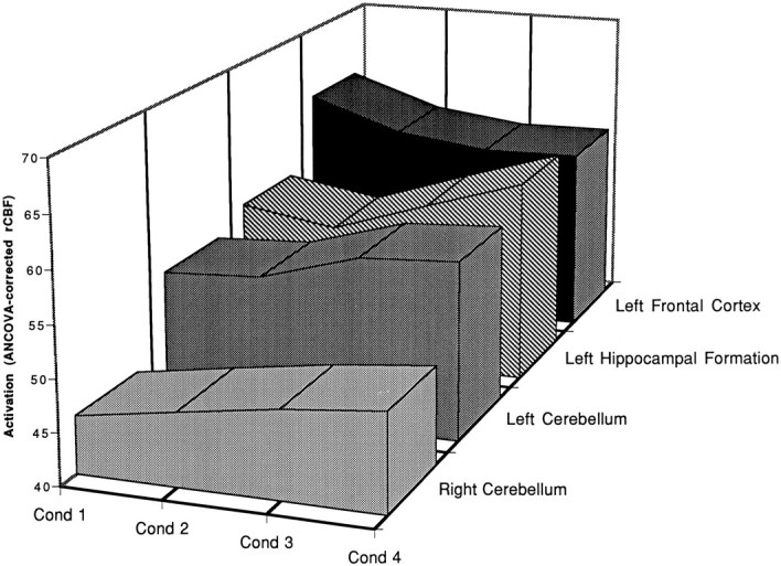
Regional cerebral blood flow (rCBF) was measured using positron emission tomography during eyeblink conditioning in young adults. Subjects were scanned in three experimental conditions: delay conditioning, in which binaural tones preceded air puffs to the right eye by 400 msec; pseudoconditioning, in which presentations of tone and air puff stimuli were not correlated in time; and fixation rest, which served as a baseline control. Compared with fixation, pseudoconditioning produced rCBF increases in frontal and temporal cortex, basal ganglia, left hippocampal formation, and pons. Learning-specific activations were observed in conditioning as compared with pseudoconditioning in bilateral frontal cortex, left thalamus, right medial hippocampal formation, left lingual gyrus, pons, and bilateral cerebellum; decreases in rCBF were observed for bilateral temporal cortex, and in the right hemisphere in putamen, cerebellum, and the lateral aspect of hippocampal formation. Blood flow increased as the level of learning increased in the left hemisphere in caudate, hippocampal formation, fusiform gyrus, and cerebellum, and in right temporal cortex and pons. In contrast, activation in left frontal cortex decreased as learning increased. These functional imaging results implicate many of the same structures identified by previous lesion and recording studies of eyeblink conditioning in animals and humans and suggest that the same brain regions in animals and humans mediate multiple forms of associative learning that give meaning to a previously neutral stimulus.
Figures
References
-
- Akase E, Thompson LT, Disterhoft JF. A system for quantitative analysis of associative learning. 2. Real-time software for MS-DOS microcomputers. J Neurosci Methods. 1994;54:119–130. - PubMed
-
- Anderson BJ (1993) The effects of paired and unpaired eyeblink conditioning on Purkinje cell morphology. PhD thesis, University of Illinois at Urbana Champaign.
-
- Berger TW, Orr WB. Hippocampectomy selectively disrupts discrimination reversal conditioning of the rabbit nictitating membrane response. Behav Brain Res. 1983;8:49–68. - PubMed
-
- Berger TW, Rinaldi PC, Weisz DJ, Thompson RF. Single-unit analysis of different hippocampal cell types during classical conditioning of rabbit nictitating membrane response. J Neurophysiol. 1983;50:1197–1219. - PubMed
-
- Blaxton TA, Bookheimer SY, Zeffiro TA, Figlozzi CM, Gaillard WD, Theodore WH (1996) Functional mapping of human memory using PET: comparisons of conceptual and perceptual tasks. Can J Exp Psychol, in press. - PubMed
Publication types
MeSH terms
Grants and funding
LinkOut - more resources
Full Text Sources