. 1996 Jul 15;16(14):4457-67.
doi: 10.1523/JNEUROSCI.16-14-04457.1996.

Organization of AMPA receptor subunits at a glutamate synapse: a quantitative immunogold analysis of hair cell synapses in the rat organ of Corti

Affiliations

Organization of AMPA receptor subunits at a glutamate synapse: a quantitative immunogold analysis of hair cell synapses in the rat organ of Corti

A Matsubara et al. J Neurosci. .

Abstract

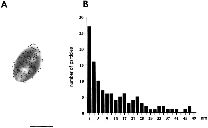
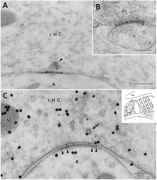
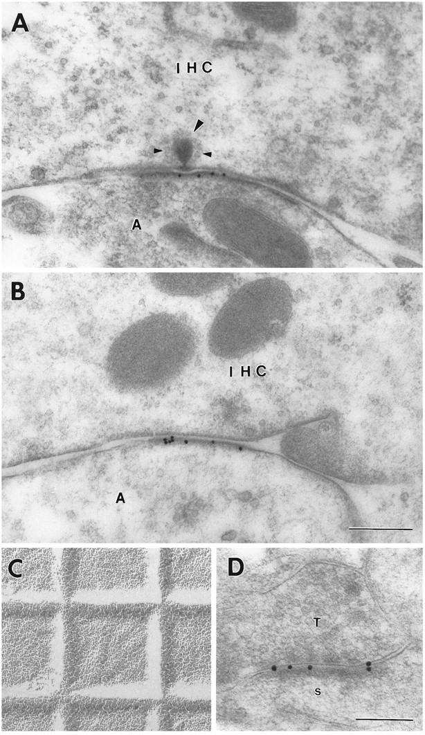
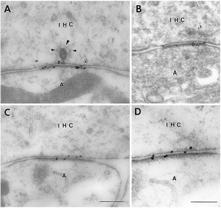
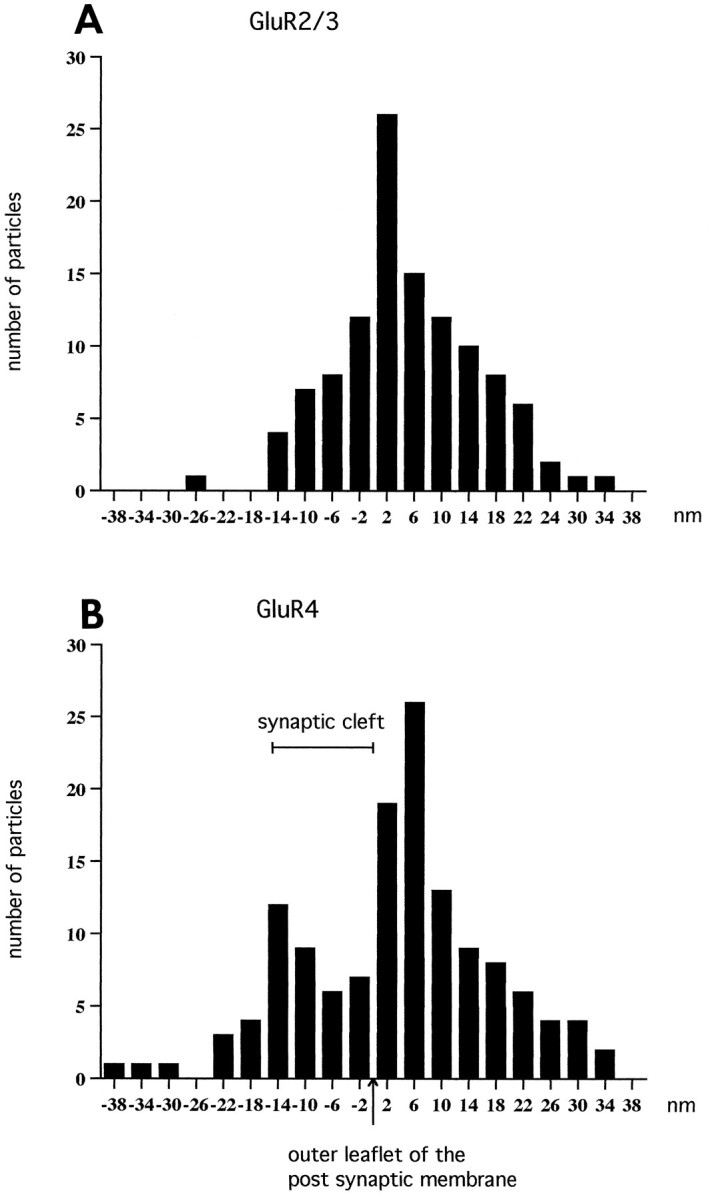
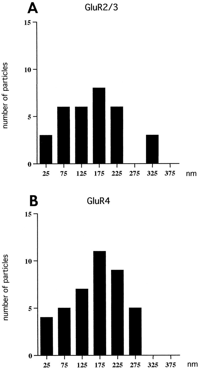
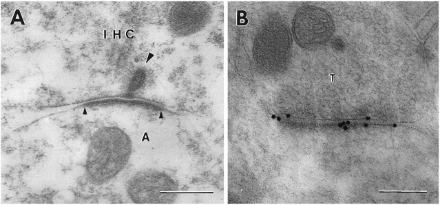
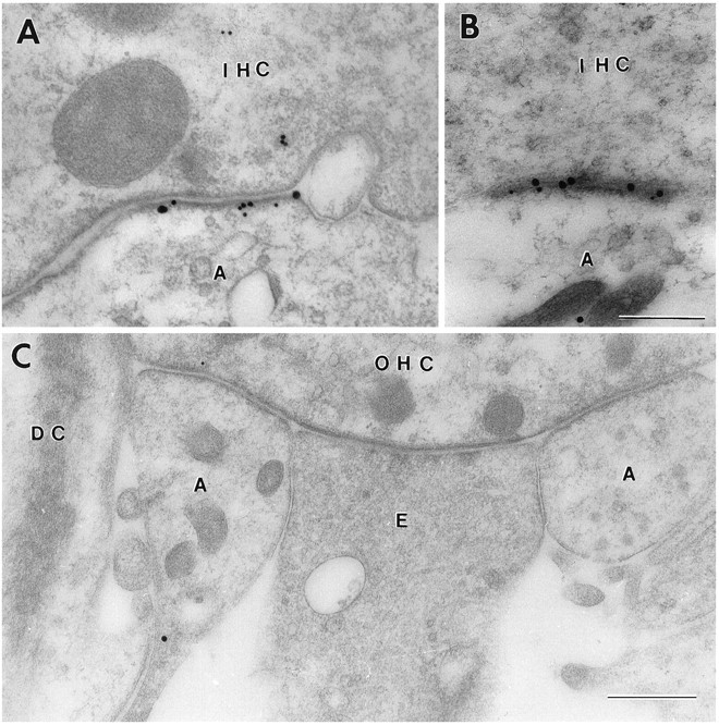
Sensitive and high-resolution immunocytochemical procedures were used to investigate the spatial organization of AMPA receptor subunits (GluR1-4) at the synapse between the inner hair cells and the afferent dendrites in the rat organ of Corti. This is a synapse with special functional properties and with a presynaptic dense body that defines the center of the synapse and facilitates its morphometric analysis. A quantitative postembedding immunocytochemical analysis was performed on specimens that had been embedded in a metachrylate resin at low temperature after freeze substitution. Single- and double-labeling procedures indicated that GluR2/3 and GluR4 subunits were colocalized throughout the postsynaptic density, with a maximum distance of 300 nm from the presynaptic body and with higher concentrations peripherally than centrally. No receptor immunolabeling was found at extrasynaptic membranes, but some GluR4 subunits appeared to be expressed presynaptically. The synapses between outer hair cells and afferent dendrites were devoid of labeling. The present data indicate that AMPA receptor subunits are inserted into the postsynaptic membrane in a very precise manner and that their density increases on moving away from the center of the synapse.


Figures

Fig. 7.
Fig. 7.
Lateral resolution of current immunogold procedure (15 nm gold particles). Values along the x-axis inB denote distance from centers of gold particles to the margin of antigen-containing bodies (A). Particles were included for analysis only where the adjacent margin was sharply defined. Background level of labeling is reached ∼28 nm off the bodies. Bin width, 2 nm (midpoint indicated). The data were based on the analysis of 30 bodies containing glutaraldehyde-fixedl-aspartate as a model antigen. The aspartate antibody (No. 18) has been characterized (Zhang et al., 1990). Scale bar, 0.3 μm.
Fig. 1.
Fig. 1.
Electron micrograph of freeze-substituted specimen of the organ of Corti showing the synaptic region of the inner hair cell (IHC). The section was counterstained with uranyl acetate and lead citrate. The ultrastructure of the IHC and afferent nerve endings (A) in contact with the cell body were well preserved. IPC, Inner pillar cell; IPH, inner phalangeal cell; TC, tunnel of Corti. Fixative No. 2. Scale bar, 3.0 μm.
Fig. 2.
Fig. 2.
Immunoreactivity for GluR2/3 at an inner hair cell (IHC) synapse in the organ of Corti (A, C) and a parallel fiber (Pf) to Purkinje cell synapse in the cerebellum (B). The 1.4 nm gold particles were made visible by silver enhancement. The section in A is not at the center of the synapse because the synaptic body (arrowhead) is cut near its periphery. C, Double immunolabeling. After demonstration of GluR2/3 by silver intensification (small particles,arrowheads), the sections were immunolabeled for glutamate (30 nm gold particles). Some of the large particles appear to be associated with vesicles (arrow) and with mitochondria (M). Inset shows a diagram of the organ of Corti.Frame indicates area represented in this and subsequent illustrations. Asterisk, Inner hair cell contacted by afferent dendrites; 1–3, the three rows of outer hair cells. s, Purkinje cell spine; A, afferent dendrite; TM, tectorial membrane. A, B, Fixative No. 1 (see Materials and Methods). C, Fixative No. 2. Freeze substitution. Scale bars: 0.5 μm in A, 0.2 μm in B and C.
Fig. 3.
Fig. 3.
Immunoreactivity for GluR2/3 at an inner hair cell synapse (A, B) and at a hippocampal synapse (D) as demonstrated by 15 nm gold particles. Large arrowhead, Synaptic body surrounded by synaptic vesicles (small arrowheads). C, Electron micrograph of grid (square width 0.4629 mm) for accurate calibration (same magnification asA and B). T, Presynaptic terminal in stratum oriens of CA1; s, postsynaptic spine. Other abbreviations as in Figure 1. Fixative No. 2; freeze substitution. Scale bars: 0.3 μm in A–C, 0.2 μm inD.
Fig. 4.
Fig. 4.
Immunoreactivity for GluR4 at inner hair cell synapses as demonstrated by 15 nm immunogold particles using different fixatives and tissue preparation methods. A, C, D, Fixative No. 2, freeze substitution. B, Fixative No. 4, method ofPhend et al. (1995). Note that some gold particles are associated with the presynaptic membrane. Large arrowhead, Synaptic body surrounded by synaptic vesicles (small arrowheads).D, Enlargement of C. Abbreviations as in Figure1. Scale bars: 0.3 μm in A–C, 0.2 μm inD.
Fig. 5.
Fig. 5.
Histograms showing the radial distribution of gold particles representing GluR2/3 (A) and GluR4 (B) at inner hair cell synapses. The distances between the centers of the 15 nm gold particles and the outer leaflet of the postsynaptic membrane were grouped into bins 4 nm wide. The values along the abscissa indicate bin centers. Minus signs indicate direction of the presynaptic element. The data were pooled from 25 synapses (A) and 23 synapses (B). The particles signaling GluR2/3 (A) showed essentially a normal distribution with an average of 4.9 nm (SEM 1.0). The histogram of GluR4 distribution was broader than that of GluR2/3 and displayed two peaks. The extent of the synaptic cleft is indicated.
Fig. 6.
Fig. 6.
Histogram showing the tangential distribution of gold particles (15 nm) representing GluR2/3 (A) and GluR4 (B) in the postsynaptic membrane of inner hair cell synapses. Zero is defined as the point opposite the center of the synaptic body. The tangential extent of the synapse was set at 100%, and the synaptic body was localized at 50.9 ± 6.3% (mean ± SD,n = 25), i.e., near the middle of the synapse. Only synapses with distinct synaptic bodies cut at their approximate centers were included in the analysis (13 synapses for GluR2/3 and 12 for GluR4). Bin width, 50 nm. Values along the x-axis indicate centers of bins. Gold particles were omitted if situated >28 nm away from the cytoplasmic aspect of the postsynaptic membrane, i.e., particles, which according to Figure 7, could not represent postsynaptic receptors with a localization corresponding to the position of the peak in Figure 5A. The distance (mean ± SD, n = 25) between the synaptic body and the lateralmost gold particle was 196 ± 64 nm (maximum 301 nm) compared with a radius of the postsynaptic density of 260 ± 44 nm (range 157–382).
Fig. 8.
Fig. 8.
Section of an inner hair cell synapse (A) and of a hippocampal synapse (B) incubated in the same drop of GluR1 antiserum. Only the latter synapse is labeled.Large arrowhead, Synaptic body. Small arrowheadsindicate extent of postsynaptic specialization. T, Presynaptic terminal in stratum oriens of CA1. Other abbreviations as in Figure 1. Fixative No. 1; freeze substitution. Scale bars: 0.3 μm in A, 0.2 μm in B.
Fig. 9.
Fig. 9.
Double-labeled ultrathin sections from the rat organ of Corti. A, B, Inner hair cells (IHC).A, GluR2/3, 30 nm gold particles; GluR4, 15 nm gold particles. B, GluR2/3, 15 nm gold particles; GluR4, 30 nm gold particles. Fixative No. 2. C, No immunoreactivity was detected in the synaptic region of the outer hair cells (OHC). Same combination of antibodies as in B.DC, Deiters cell; E, efferent nerve terminal. Other abbreviations as in Figure 1. Fixative No. 2. Freeze substitution. Scale bars: 0.3 μm in A and B, 0.5 μm in C.

References

    1. Altschuler RA, Sheridan CE, Horn JW, Wenthold RJ. Immunocytochemical localization of glutamate immunoreactivity in the guinea pig cochlea. Hear Res. 1989;42:167–174. - PubMed
    1. Anniko M, Lundquist P-G. Temporal bone morphology after systemic arterial perfusion or intralabyrinthine in situ immersion. 1. Hair cells of the vestibular organs and the cochlea. Micron. 1980;11:73–83.
    1. Baude A, Nusser Z, Molnár E, McIlhinney RAJ, Somogyi P. High-resolution immunogold localization of AMPA type glutamate receptor subunits at synaptic and non-synaptic sites in rat hippocampus. Neuroscience. 1995;69:1031–1055. - PubMed
    1. Bennett JA, Dingledine R. Topology profile for a glutamate receptor: three transmembrane domains and a channel-lining reentrant membrane loop. Neuron. 1995;14:373–384. - PubMed
    1. Boulter J, Hollmann M, O’Shea-Greenfield A, Hartley M, Deneris E, Maron C, Heinemann S. Molecular cloning and functional expression of glutamate receptor subunit genes. Science. 1990;249:1033–1037. - PubMed

Publication types

LinkOut - more resources