. 1996 Aug 2;271(31):18859-68.
doi: 10.1074/jbc.271.31.18859.

Identification and cloning of centaurin-alpha. A novel phosphatidylinositol 3,4,5-trisphosphate-binding protein from rat brain

Affiliations

Identification and cloning of centaurin-alpha. A novel phosphatidylinositol 3,4,5-trisphosphate-binding protein from rat brain

L P Hammonds-Odie et al. J Biol Chem. .

Abstract

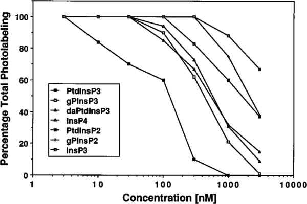
Using an affinity resin and photoaffinity label based on phospholipid analogs of inositol 1,3,4,5-tetrakisphosphate (InsP4), we have isolated, characterized, and cloned a 46-kDa protein from rat brain, which we have named centaurin-alpha. Binding specificity was determined using displacement of 1-O-[3H](3-[4-benzoyldihydrocinnamidyl]propyl)-InsP4 photoaffinity labeling. Centaurin-alpha displayed highest affinity for phosphatidylinositol 3,4,5-trisphosphate (PtdInsP3) (IC50 = 120 nM), whereas InsP4, PtdInsP2, and InsP3 bound with 5-, 12-, and >50-fold lower affinity, respectively. Screening a rat brain cDNA library with a polymerase chain reaction product, generated using partial amino acid sequence from tryptic peptides, yielded a full-length clone. The 2,450-base pair cDNA contained an open reading frame (ORF) encoding a novel protein of 419 amino acids. Northern analysis revealed a 2.5-kilobase transcript that is highly expressed in brain. The deduced sequence contains a novel putative zinc finger motif, 10 ankyrin-like repeats, and shows homology to recently identified yeast and mammalian Arf GTPase-activating proteins. Given the specificity of binding and enrichment in brain, centaurin-alpha is a candidate PtdInsP3 receptor that may link the activation of phosphoinositide 3-kinase to downstream responses in the brain.


Figures

Fig. 1
Fig. 1. Purification and photoaffinity labeling of centaurin-α
Panel A, samples of the crude soluble fraction (S), column flow-through (F), and column eluate (E) obtained during heparin-agarose column chromatography of rat brain as described under “Experimental Procedures” were separated by SDS-PAGE and the gel stained with Coomassie Blue. Panel B, early (E) and late (L) eluting fractions from the aminopropyl-InsP4 affinity column purification. Proteins were separated by SDS-PAGE and stained with Coomassie Blue. The 46-kDa protein is marked with an arrowhead. For panels A and B, the molecular mass standards, 205, 116, 97.4, 66, and 45 kDa, are indicated with dashes. Panel C, photoaffinity labeling of the partially purified (heparin-agarose column eluate) fraction from rat brain supernatant. Molecular mass markers, 112, 84, and 52.5 kDa, are indicated with dashes. Panel D, heparin-agarose purified fractions of soluble and detergent-extracted membranes from rat brain and heart, with the 42–48-kDa region of the gel shown. For panels C and D, total photolabeling (−) with 70 nm [3H]BZDC-InsP4 and nonspecific photolabeling (+) were determined with 70 nm [3H]BZDC-InsP4 plus 10 μm unlabeled InsP4. Typical purification and photolabeling profiles are shown and were repeated five times with similar results.
Fig. 2
Fig. 2. Specificity of photoaffinity labeling of centaurin-α
Competition of photolabeling was done by including increasing concentrations of unlabeled ligand (indicated). The heparin eluate fraction from the supernatant of rat whole brain was used for photolabeling with 20–50 nm [3H]BZDC-InsP4, and the 46-kDa region of the fluorograph was scanned using a densitometer. Results are presented as a percentage of total photolabel (in the absence of unlabeled competitor), and each value represents the average of at least four independent determinations. The abbreviations are defined in Footnote 1.
Fig. 3
Fig. 3. Nucleotide and deduced amino acid sequence of cloned cDNA of centaurin-α
The predicted amino acid sequence is expressed in the single-letter system. The nucleotide residue number is indicated along the left, and the amino acid residue is along the right. Amino acid sequences identical to microsequenced tryptic peptides are underlined.
Fig. 4
Fig. 4
Panel A, schematic presentation of conserved domains in centaurin-α and related proteins. Black bars indicate the positions of the potential Zn2+ coordinating cysteine residues; shaded boxes represent ankyrin repeat domains. In the S. cerevisiae GTS1/LSR1 protein, the region of homology with the Drosophila melanogaster Per proteins (61) is marked, as are the conserved nucleoporin-like FP repeats in the human RIP1/RabCCF protein (68, 69). The minimal domain having full Arf-GAP activity (50) is marked by a broken line under RnArf-GAP. A line beneath centaurin-α indicates a region of homology with TERM proteins (see Fig. 5C). The percentage identity between each protein and centaurin-α (produced by pairwise alignment using the GAP program of GCG8) is presented on the right side of the figure. Panel B, comparison of sequences in putative Zn2+ coordination region of centaurin-α and related proteins. The amino acid residue number marking the beginning of the sequence in each protein is given on the left side of the alignment. The percentage identity between each protein and centaurin-α in this region is presented on the right side of the alignment. Residues conforming to a consensus for the members of this family are marked above the sequence and within the alignment are indicated by bold type. In addition to rat centaurin-α, the sequences represented are: SpAC26a3 10, a putative S. pombe ORF (GenBank accession number Z69240); ScYIE4, the putative product of the S. cerevisiae YIE4 gene (accession number P40529); HsKIAA0041 and HsKIAA0050, two human putative ORFs (accession numbers D26069 and D30758); ScGCS1, the product of the S. cerevisiae GCS1 gene (59); ScGTS1/LSR1, the predicted product of the S. cerevisiae gene cloned both as GTS1 (61) and LSR1 (accession number X91489); ScSPS18, the putative product of the S. cerevisiae SPS18 sporulation-specific gene (60); HsRIP1/RabCCF, the cellular cofactor of retroviral Rev and Rex RNA regulatory proteins (68, 69); RnArf-GAP, the rat liver Arf-GAP (50); ScD971727, the predicted product of the S. cerevisiae putative ORF D71727 (accession number U33057); ScGLO3, the product of the S. cerevisiae GLO3 gene (59).
Fig. 5
Fig. 5
Panel A, alignment of repeat sequences in centaurin-α with repeats 15–24 of human erythrocyte ankyrin. Residues in upper case show conservation of identity between the two proteins. Boxes indicate residues belonging to the same Dayhoff groups (GPAST, KRH, NQED, MILV, FWY, C) conserved between the two proteins. Consensus sequences for centaurin repeats and ankyrin repeats (54) are presented above and below the sequences, respectively. Asterisks indicate the positions of residues that are conserved in both consensus sequences. Panel B, comparison of sequences in second ankyrin repeat of centaurin-α with the inositol polyphosphate binding regions. The second ankyrin repeat of centaurin-α was compared with the C2B domains of mouse synaptotagmin II (MmSynaptotagminII; 11) and human InsP4-regulated ras-GAP (HsInsP4-BP; 12). The suggested consensus sequence for inositol polyphosphate binding is marked; + indicates a conserved positively charged residue, and residues conforming to this sequence are printed in bold in the alignment. The lysine residue demonstrated to be essential for InsP4 binding to synaptotagmin is marked with an * above the sequence. The sequence of repeat 16 of human ankyrin (54) and the consensus sequence for centaurin repeats derived in panel A are also presented. Panel C, alignment of a region including centaurin repeat 6 with homologous sequences in members of the TERM protein family. The repeat 6 region of centaurin-α was compared with some of the TERM family proteins (53): MmMerlin, L28176; HsEzrin, P15311; and HsMoesin, P26038. The consensus sequence for the centaurin-α repeats is shown above the alignment. The residues showing conservation within Dayhoff groups in all proteins are in bold, and a consensus sequence for this homology is presented below the alignment.
Fig. 6
Fig. 6. Northern analysis of centaurin-α mRNA in rat tissues
Each lane of the multiple tissue Northern contained 2 μg of poly(A)+ RNA. H, heart; B, brain; S, spleen; G, lung; V, liver; M, skeletal muscle; K, kidney; and T, testis. Gel locations of size markers of 9.2, 7.5, 4.4, 2.4, and 1.35 kb are indicated with dashes.
Fig. 7
Fig. 7. Characterization of native and recombinant centaurin-α using InsP4 affinity resin and anti-centaurin-α antibodies
Panel A, lysates of COS-7 cells, mock-transfected or centaurin-α-transfected, were purified with InsP4 affinity resin. T, whole cell lysates; F, InsP4 affinity column flow-through; W, InsP4 affinity column wash; E, InsP4 affinity column eluate. A typical purification scheme is shown. The purification was repeated three times with similar results. The arrowhead highlights the purification of recombinant centaurin-α in InsP4 column eluate. Gel locations of molecular mass standards, 205, 116, 97.4, 66, and 45 kDa, are indicated with dashes. Panel B, fractions from rat brain and COS-7 cell lysates were separated by SDS-PAGE, transferred to nitrocellulose, and immunoblotted with anti-centaurin-α COOH-terminal fusion protein antibodies (J4) and anti-peptide NH2-terminal antibodies (J49). Lane 1, postnatal day 14 rat brain homogenate; lane 2, heparin-agarose column eluate from rat brain; lane 3, InsP4 affinity column eluate from rat brain; lane 4, mock-transfected COS-7 cell lysate; lane 5, p63d.2.13 (centaurin-α)-transfected COS-7 cell lysate. Molecular mass standards of 108, 84, 67, 55, and 39.5 kDa are indicated with dashes.
Fig. 8
Fig. 8. Comparison of [3H]BZDC-InsP4 and PtdInsP3 structures
Both molecules have the 3,4,5-trisphosphate recognition element of d-myo-Ins(1,3,4,5)-P4, as well as a P-1 phosphodiester linked to a 3-carbon chain with a hydrophobic appendage.

References

    1. Michell RH. Trends Biochem. Sci. 1992;17:274–276. - PubMed
    1. Berridge MJ. Nature. 1993;361:315–325. - PubMed
    1. Ferris CD, Snyder SH. Annu. Rev. Physiol. 1992;54:469–488. - PubMed
    1. Nakamura S, Nishizuka Y. J. Biochem. 1994;115:1029–1034. - PubMed
    1. Liscovitch M, Cantley LC. Cell. 1994;77:329–334. - PubMed

Publication types

MeSH terms

Associated data

LinkOut - more resources