. 1996 Jul 1;16(13):4135-45.
doi: 10.1523/JNEUROSCI.16-13-04135.1996.

Ultrastructural localization of the vesicular monoamine transporter-2 in midbrain dopaminergic neurons: potential sites for somatodendritic storage and release of dopamine

Affiliations

Ultrastructural localization of the vesicular monoamine transporter-2 in midbrain dopaminergic neurons: potential sites for somatodendritic storage and release of dopamine

M J Nirenberg et al. J Neurosci. .

Abstract

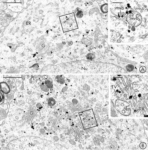
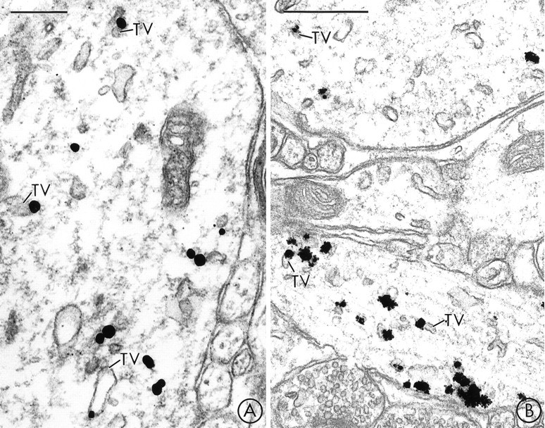
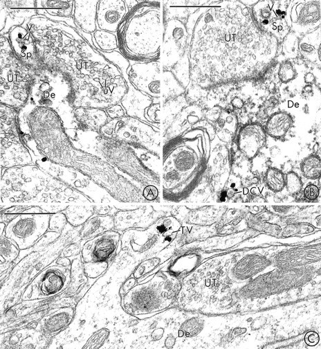
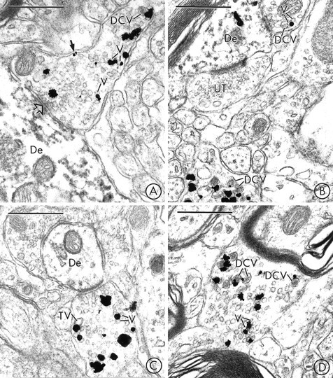
Midbrain dopaminergic neurons are known to release dopamine from somata and/or dendrites located in the substantia nigra (SN) and the ventral tegmental area (VTA). There is considerable controversy, however, about the subcellular sites for somatodendritic dopamine storage in these regions. In the present study, we used dual-labeling electron microscopic immunocytochemistry to localize the vesicular monoamine transporter-2 (VMAT2), a novel marker for sites of intracellular monoamine storage, within identified dopaminergic (tyrosine hydroxylase-containing) neurons in the rat SN and VTA. In dopaminergic perikarya, immunogold labeling for VMAT2 was localized to the Golgi apparatus, tubulovesicles that resembled smooth endoplasmic reticulum (SER), and the limiting membranes of multivesicular bodies. In dopaminergic dendrites, VMAT2 was extensively localized to tubulovesicles that resembled saccules of SER, and less frequently localized to isolated small synaptic vesicles (SSVs) or large dense-core vesicles (DCVs). In rare cases, VMAT2-immunoreactive SSVs were clustered within the cytoplasm of an SN or a VTA dendrite. Dopaminergic dendrites in the VTA contained a significantly higher number of immunogold particles for VMAT2 per unit than those in the SN. Together, these observations support the proposal that dopamine is stored in and may be released from dendritic SSVs and DCVs, but suggest that the SER is the major site of dopamine storage within midbrain dopaminergic neurons. In addition, they provide new evidence that dopaminergic dendrites in the VTA may have greater potential for reserpine-sensitive storage and release of dopamine than those in the SN.


Figures

Fig. 1.
Fig. 1.
VMAT2 is localized to saccules of Golgi and multivesicular bodies in neuronal perikarya in the SN and VTA.A, Immunogold particles for VMAT2 are seen in the region of the Golgi apparatus (G) of a labeled perikaryon in the SNC, but are not detected in the adjacent rough endoplasmic reticulum (ER). The inset shows the boxed regionat higher magnification. Immunogold labeling for VMAT2 is seen along the limiting membranes of two multivesicular bodies (mvb).B, Immunogold labeling for VMAT2 is localized to the Golgi apparatus (G) of a perikaryon in the VTA. Many of the gold particles contact lateral saccules of Golgi lamellae (curved arrows). The inset shows the boxed region at higher magnification. Gold particles are localized to saccules of Golgi (G) and associated vesicles and tubulovesicles (TV). Nu, Nucleus. Scale bars: A, B, 1 μm; inset in A, B, 0.2 μm.
Fig. 2.
Fig. 2.
In midbrain dendrites, prominent immunogold labeling for VMAT2 is localized to tubulovesicles that resemble smooth endoplasmic reticulum. A, B, A large dendrite in the SN (A) and two large dendrites in the VTA (B) contain immunogold labeling for VMAT2 that is primarily localized to large, electron-lucent tubulovesicles (TV) that resemble SER. Scale bars: A, 0.25 μm; B, 0.5 μm.
Fig. 3.
Fig. 3.
In SN dendrites, VMAT2 is localized to tubulovesicles of SER, and more rarely to SSVs or DCVs. A, In dually labeled tissue, gold particles for VMAT2 are seen within the cytoplasm of a medium-sized dendrite that also contains immunoperoxidase labeling for TH. The peroxidase reaction product is seen as an electron-dense precipitate throughout the cytoplasm, which is notably absent in dendrites processed only for VMAT2 (C). The immunogold particles are localized to tubulovesicles (TV) that are larger than the unlabeled synaptic vesicles (uV) seen in adjacent unlabeled axon terminals (UT). One of the unlabeled terminals (UT) forms a synaptic junction with the labeled dendrite (arrow). Scale bars: B, C, 0.5 μm. In larger dendrites, immunogold particles for VMAT2 are also localized to large tubulovesicles (TV), clusters of smaller SSVs, and an occasional DCV. Scale bars: 0.5 μm;inset, 0.2 μm.
Fig. 4.
Fig. 4.
In the VTA, VMAT2-labeled SSVs are sometimes clustered near dendrodendritic contacts. A, Immunogold labeling for VMAT2 and immunoperoxidase labeling for TH are seen in two large dendrites (De). Some of the immunogold particles are localized to a cluster of small synaptic vesicles (V) and larger electron-lucent tubulovesicles (TV) near a point of contact (straight arrow) between the two dendrites (De). A similar cluster of vesicles and tubulovesicles (curved arrow) contains no detectable immunogold labeling in the observed plane of section, but contained several immunogold particles for VMAT2 in a serial section (not shown). A VMAT2-labeled dense-core vesicle (DCV) is also seen in the “presynaptic” dendrite. B, In a section processed for single labeling, immunogold particles for VMAT2 are localized to a cluster of small synaptic vesicles (V) in a dendrite (De) near its site of contact (arrow) with an unlabeled dendrite (UDe). The labeled vesicles are similar in size to the unlabeled synaptic vesicles (uV) seen in an adjacent unlabeled terminal (UT). Scale bars:A, B, 0.5 μm.
Fig. 5.
Fig. 5.
In rare cases, VMAT2-labeled vesicles are localized to dendritic spines in the SNR and the VTA. A, B, Immunogold labeling for VMAT2 and immunoperoxidase labeling for TH are colocalized in two dendrites in the SNR. Immunogold particles are localized to small synaptic vesicles (V) in dendritic spines (Sp). These vesicles are similar in size to the unlabeled small synaptic vesicles (uV) seen in adjacent unlabeled axon terminals (UT).C, Gold particles for VMAT2 are localized to a tubulovesicular organelle (TV) in a dendritic spine in the VTA. The labeled tubulovesicle is larger than the synaptic vesicles seen in an adjacent unlabeled axon terminal (UT). De, Dendrite. Scale bars:A–C, 0.5 μm.
Fig. 6.
Fig. 6.
In the SN and VTA, VMAT2 is localized to SSVs and DCVs in axon terminals that usually lack detectable TH-immunoreactivity. A, Immunogold particles for VMAT2 are localized to small synaptic vesicles (V), an isolated DCV, and the plasma membrane (closed arrow) of an axon terminal in the SN that lacks detectable peroxidase reaction product for TH. The VMAT2-containing terminal forms a synaptic contact (open arrow) with a dendrite (De) that contains intense peroxidase reaction product for TH, but no detectable immunogold labeling for VMAT2. B, Immunogold particles for VMAT2 are seen in two axon terminals in the SN. The terminal at thetop contacts a dendrite (De) that is labeled with immunoperoxidase for TH and immunogold for VMAT2. The terminal at thebottom contains numerous VMAT2-labeled large dense-core vesicles (DCV). The VMAT2-immunoreactive terminals contain no detectable peroxidase labeling for TH. UT, Unlabeled terminal. C, Immunogold particles for VMAT2 are localized to small synaptic vesicles (V) and a larger electron-lucent tubulovesicular structure (TV) in an axon terminal in the VTA that lacks detectable peroxidase reaction product for TH. The VMAT2-labeled terminal contacts a dendrite (De) that is lightly immunoperoxidase-labeled for TH. D, An axon terminal in the VTA contains immunogold particles that are localized to the membranes of large dense-core vesicles (DCV) and small synaptic vesicles (V). The VMAT2-labeled terminal contains no detectable peroxidase reaction product for TH. Scale bars:A–D, 0.5 μm.

Similar articles

Cited by

References

    1. Ayala J. Transport and internal organization of membranes: vesicles, membrane networks and GTP-binding proteins. J Cell Sci. 1994;107:753–763. - PubMed
    1. Bannon MJ, Granneman JG, Kapatos G. The dopamine transporter: potential involvement in neuropsychiatric disorders. In: Bloom FE, Kupfer DJ, editors. Psychopharmacology: the fourth generation of progress. Raven; New York: 1995. pp. 179–188.
    1. Bernardini GL, Gu X, Viscardi E, German DC. Amphetamine-induced and spontaneous release of dopamine from A9 and A10 dendrites: an in vitro electrophysiological study in the mouse. J Neural Transm Gen Sect. 1991;84:183–193. - PubMed
    1. Björklund A, Lindvall O. Dopamine in dendrites of substantia nigra neurons: suggestions for a role in dendritic terminals. Brain Res. 1975;83:531–537. - PubMed
    1. Broadwell RD, Cataldo AM. The neuronal endoplasmic reticulum: its cytochemistry and contribution to the endomembrane system. I. Cell bodies and dendrites. J Histochem Cytochem. 1983;31:1077–1088. - PubMed

Publication types

LinkOut - more resources