. 1996 Jul 1;16(13):4186-94.
doi: 10.1523/JNEUROSCI.16-13-04186.1996.

Protection of retinal ganglion cells from natural and axotomy-induced cell death in neonatal transgenic mice overexpressing bcl-2

Affiliations

Protection of retinal ganglion cells from natural and axotomy-induced cell death in neonatal transgenic mice overexpressing bcl-2

L Bonfanti et al. J Neurosci. .

Abstract

Approximately half of the retinal ganglion cells (RGCs) present in the rodent retina at birth normally die during early development. Overexpression of the photo-oncogene bcl-2 recently has been shown to rescue some neuronal populations from natural cell death and from degeneration induced by axotomy of nerves within the peripheral nervous system. Here we study in vivo the role of the overexpression of bcl-2 in the natural cell death of RGCs and in the degenerative process induced in these cells by transection of the optic nerve. We find that in newborn bcl-2 transgenic mice, the number of RGCs undergoing natural cell death is considerably lower than in wild-type pups. Consistently, a vast majority (90%) of the ganglion cells found in the retina of neonatal transgenics are maintained in adulthood, whereas only 40% survive in wild-type mice. After transection of the optic nerve, the number of degenerating ganglion cells, determined by counting pyknotic nuclei or nuclei with fragmented DNA, is substantially reduced in transgenic mice. In wild-type animals, almost 50% of ganglion cells degenerate in the 24 hr after the lesion, whereas almost the entire ganglion cell population survives axotomy in transgenic mice. Therefore, overexpression of bcl-2 is effective in preventing degeneration of this neuronal population, raising the possibility that ganglion cells are dependent on the endogenous expression of bcl-2 for survival. The remarkable rescue capacity of bcl-2 overexpression in these neurons makes it an interesting model for studying natural cell death and responses to injury in the CNS.


Figures
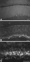
Fig. 1.
Fig. 1.
Immunostaining for the human Bcl-2 protein in radial sections of neonatal retina. A, Wild-type mouse retina showing no staining. Print has been underexposed to allow visualization of the structure. B, bcl-2 transgenic mouse retina, line NSE 73 a/b, showing high levels of human Bcl-2 protein in the ganglion cells (white arrows). C, High magnification of B. Apical dendrites of ganglion cells are visible (white arrows); nuclei are negative. Some differentiating cells in the inner nuclear layer (most probably amacrine cells) show weak immunoreactivity. Eventually all retinal neuronal classes except for photoreceptors will express the human Bcl-2 protein in this line of transgenics. Scale bars, 50 μm.
Fig. 2.
Fig. 2.
Natural cell death of retinal ganglion cells from wild-type and bcl-2 transgenic mice. Light micrographs of the ganglion cell layer of whole-mount retinas stained with cresyl violet at P1–2.A, Wild-type mouse; arrows point to pyknotic nuclei. B, bcl-2 transgenic mouse, at corresponding location and focal plane shown in A. No pyknotic profiles are visible in this field. Scale bar, 10 μm.
Fig. 3.
Fig. 3.
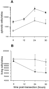
Pyknosis in the RGC layer. A, Time course of the appearance of pyknotic cells in the ganglion cell layer of wild-type (black triangles) and bcl-2 transgenic mice (open circles) after transection of the optic nerve.B, Time course of the reduction in the number of living cells in the ganglion cell layer of wild-type (black triangles) and bcl-2 transgenic mice (open circles) after the transection of the optic nerve.
Fig. 4.
Fig. 4.
Effects of optic nerve transection on RGCs in wild-type and bcl-2 animals at 24 hr postlesion. Light micrographs of the ganglion cell layer of whole-mount retinas stained with cresyl violet at P1-P2. A, Wild-type mouse, exhibiting numerous apoptotic cells; B, bcl-2 transgenic mouse, where only one apoptotic cell is visible (arrow). Scale bar, 10 μm.
Fig. 5.
Fig. 5.
In situ labeling of DNA fragmentation (TUNEL) in retinal sections 24 hr after optic nerve transection. Radial section of wild-type (A) and bcl-2 transgenic (B) retinas, where TUNEL-positive nuclei appear as white fluorescent profiles against a black background, particularly in the wild-type RGC layer (arrowheads). Scale bar, 50 μm.
Fig. 6.
Fig. 6.
Effects of optic nerve transection on DNA fragmentation of RGCs. TUNEL-positive cells counted in retinal sections of wild-type and bcl-2 transgenic mice 24 hr after section of the optic nerve. Bars represent the number of labeled cells for every 1000 cells of the ganglion cell layer.
Fig. 7.
Fig. 7.
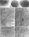
Morphology of wild-type and bcl-2 optic nerves 24 hr after transection. A, Section (1 μm thick) of intact, wild-type optic nerve, in the proximity of the posterior pole of the eye. Astrocytic processes form a regular plexus across the whole surface of the nerve. B, Proximal stump of the optic nerve of a wild-type mouse 24 hr after transection. Same location as inA. Note that astrocytic bodies appear isolated because their processes are no longer visible. The matrix shows white spaces.C, Proximal stump of bcl-2 optic nerve 24 hr after transection. Astrocytic desegregation is restricted to a limited area (arrowheads). The remaining part of the nerve appears normal. Scale bar: A–C, 200 μm. D, E, Electron micrographs of specimens shown in B and C, respectively. The morphology of the nerve of the wild-type mouse (D) is altered profoundly. Arrows point to a bundle of astrocytic filaments indicating a glial reaction. Theasterisk indicates the empty space left from degenerating fibers. These changes are much less prominent in the nerve of the bcl-2 transgenic mouse (E). a, Astrocytic nucleus. Scale bar: F, G, 1 μm. High-magnification micrographs of preparations shown in D and E, respectively. Single axons of both wild-type (F) and bcl-2 (G) optic nerves have undergone similar modifications. Fibers are swollen, and anomalous tubular and vesicular profiles fill their lumen (arrowheads). The normal ultrastructure of the fibers, with their complement of microtubules and intermediate filaments, is totally lost. Scale bar, 0.2 μm.

References

    1. Allsopp TE, Kiselov S, Wyatt S, Davies AM. Role of bcl-2 in the brain-derived neurotrophic factor survival response. Eur J Neurosci. 1995;7:1266–1272. - PubMed
    1. Allsopp TE, Wyatt S, Patterson HF, Davies AM. The proto-oncogene bcl-2 can selectively rescue neurotrophic factor-dependent neurons from apoptosis. Cell. 1993;73:295–307. - PubMed
    1. Beazley LD, Perry VH, Baker B, Darby EJ. An investigation into the role of ganglion cells in the regulation of division and death of other retinal cells. Dev Brain Res. 1987;33:169–184. - PubMed
    1. Berkelaar M, Clarke DB, Wang Y-C, Bray GM, Aguayo AJ. Axotomy results in delayed death and apoptosis of retinal ganglion cells in adult rats. J Neurosci. 1994;14:4368–4374. - PMC - PubMed
    1. Carmignoto G, Maffei L, Candeo P, Canella R, Comelli C. Effect of NGF on the survival of rat retinal ganglion cells following optic nerve section. J Neurosci. 1989;9:1263–1272. - PMC - PubMed

Publication types

Substances

LinkOut - more resources