. 1996 Aug 15;16(16):4846-60.
doi: 10.1523/JNEUROSCI.16-16-04846.1996.

Voltage-gated K+ channel beta subunits: expression and distribution of Kv beta 1 and Kv beta 2 in adult rat brain

Affiliations

Voltage-gated K+ channel beta subunits: expression and distribution of Kv beta 1 and Kv beta 2 in adult rat brain

K J Rhodes et al. J Neurosci. .

Abstract

Recent cloning of K+ channel beta subunits revealed that these cytoplasmic polypeptides can dramatically alter the kinetics of current inactivation and promote efficient glycosylation and surface expression of the channel-forming alpha subunits. Here, we examined the expression, distribution, and association of two of these beta subunits, Kv beta 1 and Kv beta 2, in adult rat brain. In situ hybridization using cRNA probes revealed that these beta-subunit genes are heterogeneously expressed, with high densities of Kv beta 1 mRNA in the striatum, CA1 subfield of the hippocampus, and cerebellar Purkinje cells, and high densities of Kv beta 2 mRNA in the cerebral cortex, cerebellum, and brainstem. Immunohistochemical staining using subunit-specific monoclonal and affinity-purified polyclonal antibodies revealed that the Kv beta 1 and Kv beta 2 polypeptides frequently co-localize and are concentrated in neuronal perikarya, dendrites, and terminal fields, and in the juxtaparanodal region of myelinated axons. Immunoblot and reciprocal co-immunoprecipitation analyses indicated that Kv beta 2 is the major beta subunit present in rat brain membranes, and that most K+ channel complexes containing Kv beta 1 also contain Kv beta 2. Taken together, these data suggest that Kv beta 2 is a component of almost all K+ channel complexes containing Kv 1 alpha subunits, and that individual channels may contain two or more biochemically and functionally distinct beta-subunit polypeptides.


Figures
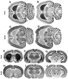
Fig. 1.
Fig. 1.
Expression of Kvβ1 and Kvβ2 mRNA in adult rat brain. Horizontal and coronal sections of rat brain were processed byin situ hybridization histochemistry to localize Kvβ1 (A, B, EG) and Kvβ2 (C, D, HJ) mRNA. Areas containing a high density of hybridization signal appear dark in these bright-field images. The autoradiograms inAD were exposed for 3 d, whereas those inEJ were exposed for 7 d. At the shorter exposure time, subtle differences in expression levels are more easily discernible. For example, there is a comparatively greater density of Kvβ1 mRNA in the CA1 subfield of the hippocampus as compared with the adjacent CA3 subfield (B). CA1, Hippocampal subfield; CB, cerebellum; CPu, caudate putamen;EC, entorhinal cortex; RN, red nucleus;ms, medial septal nuclei; AN, anterior thalamic nucleus; MD, mediodorsal thalamic nucleus.
Fig. 2.
Fig. 2.
Cellular localization of Kvβ1 and Kvβ2 mRNA expression. Emulsion autoradiograms were prepared to localize Kvβ1 and Kvβ2 mRNA within individual cells. Cells containing a high density of mRNA contain a correspondingly high density of silver grains, which appear as bright spots in these dark-field images. In posterior cingulate cortex (area 23), Kvβ1 mRNA (A) is highly expressed in many small cells in layers II and III and larger cells in layer V (arrows), whereas Kvβ2 (B) is expressed predominantly in small cells in layer II and larger cells in layer V (arrows). In the entorhinal cortex, Kvβ1 mRNA (C) is expressed in large and small cells in layers II, III, V, and VI, whereas Kvβ2 mRNA (D) is expressed at high density in layer II cells and with a lower density in cells in the remaining layers of this structure. In the caudate nucleus, there is a very high level of Kvβ1 expression (E) and a lower level of Kvβ2 expression (F) in virtually all cells (arrows). The bundles of myelinated axons that course through this structure do not contain hybridization signal (arrowheads). In the basal forebrain, Kvβ1 (G) and Kvβ2 (H) mRNAs are expressed in large cells (arrows) with the distribution and frequency characteristic of the large cholinergic neurons present in this region. The short arrows in G and H mark the midline of the brain.
Fig. 3.
Fig. 3.
Immunoblot analyses of the Kvβ1 and Kvβ2 β-subunit polypeptides in rat brain membranes and in transfected COS-1 cells. Crude rat brain membranes (RBM) (50 μg) and the detergent extracts of COS-1 cells transfected with Kvβ1/RBG4 (Kvβ1), Kvβ2/RBG4 (Kvβ2), or RBG4 alone (vector) were fractionated on a 12.5% SDS gel and transferred to nitrocellulose, and the resultant immunoblot probed with either rabbit anti-Kvβ1 polyclonal antibody at 1:50 (left) or mouse anti-Kvβ2 mAb K17/70 tissue culture supernatant neat (right). Signals were visualized using ECL (left, 30 min; right, 1 min). Numbers on leftrefer to mobility of prestained molecular weight standards.
Fig. 4.
Fig. 4.
Analysis of antibody specificity. Immunofluorescence staining of Kvβ1 and Kvβ2 β subunits expressed in COS-1 cells. COS-1 cells were transfected with Kvβ1/RBG4 (AD) or Kvβ2/RBG4 (EH) cDNAs. Transfected cells then were fixed, permeabilized, and incubated with rabbit anti-Kvβ1 polyclonal antibody at 1:100 (A, E), rabbit anti-Kvβ2 polyclonal antibody at 1:200 (B, F), mouse anti-Kvβ1 mAb K9/40 at 1:2 (C, G), or mouse anti-Kvβ2 mAb K17/70 at 1:2 (D,H). Cells then were incubated with Texas Red-conjugated anti-rabbit (A, B, E,F) or anti-mouse (C, D,G, H) secondary antibody.
Fig. 5.
Fig. 5.
Presence of Kvβ1 and Kvβ2 in rat brain K+ channel complexes. Samples of adult rat brain membranes (RBM) (60 μg) and aliquots of products of immunoprecipitation reactions from detergent extracts of 500 μg RBM with polyclonal antibodies specific for both Kvβ1 and Kvβ2 (pan-β, 1:100), Kvβ1 (anti-Kvβ1, 1:200), Kvβ2 (anti-Kvβ2, 1:500), or the Kv2.1 α subunit (anti-Kv2.1, 1:200) were size fractionated by 12.5% SDS-PAGE. Samples were transferred to nitrocellulose and probed with rabbit anti-Kvβ1 polyclonal antibody at 1:50 (left panel), mouse anti-Kvβ2 mAb K17/70 neat (middle panel), or rabbit anti-Kv1.2 polyclonal antibody at 1:100 (right panel). Bound antibody detected by ECL–autoradiography for 40 min (left panel) or 3 min (middle and right panels). Arrowspoint to mobility of the heavy-chain polypeptides of the rabbit immunoglobulins used in the immunoprecipitation reactions and of the respective K+ channel polypeptides;numbers at left of the panels denoteMr of prestained molecular weight standards. Bands at ∼50 kDa (left and right panels) are heavy chains of rabbit IgG used for immunoprecipitations, which react with anti-rabbit, but not anti-mouse, secondary antibody.
Fig. 6.
Fig. 6.
Immunohistochemical localization of Kvβ1 and Kvβ2 in the cerebral cortex and striatum. In parietal cortex, immunoreactivity for Kvβ1 (K9/40 mAb) (A) is concentrated in large pyramidal cells in layer V and in small interneurons (arrows) concentrated primarily in layers II and III. Immunoreactivity for Kvβ2 (affinity-purified polyclonal antibody) (C) is concentrated in the somas and apical dendrites of layer V pyramidal cells. The small interneurons that contain a high density of immunoreactivity for Kvβ1 contain a much lower density of immunoreactivity for Kvβ2. In the striatum, there is a very high density of immunoreactivity for Kvβ1 (B) and Kvβ2 (D) in the globus pallidus (GP) and a lower density in the caudate nucleus (CD). In the globus pallidus, immunoreactivity for Kvβ1 and Kvβ2 is primarily concentrated in terminal fields and is present in both the supra- and subcommissural segments. AC, Anterior commissure.
Fig. 7.
Fig. 7.
Immunohistochemical localization of Kvβ1 and Kvβ2 in the midbrain and cerebellum. In the midbrain, immunoreactivity for Kvβ1 (K9/40 mAb) (A) and Kvβ2 (affinity-purified polyclonal antibody) (C) is concentrated in terminal fields in the pars reticulata of the substantia nigra (SNr) and in large neurons in the red nucleus (RN). In the cerebellar cortex, immunoreactivity for Kvβ1 (B) and Kvβ2 (D) is concentrated in the cell bodies of Purkinje cells (P) and in terminal fields throughout the molecular layer. Immunoreactivity for Kvβ2 also is concentrated in the dendrites of Purkinje cells and in the axon terminals of basket cells, which form a characteristic synaptic plexus (arrows) that terminates on the initial segment of Purkinje cell axons.
Fig. 8.
Fig. 8.
Hypothetical model of β-subunit associations in rat brain. The total rat brain β-subunit pool contains at least three β-subunit isoforms, designated Kvβ1, Kvβ2, and KvβX in this Venn diagram. Immunoreactivity observed using Kvβ2-specific antibodies recapitulates virtually the entire pattern and extent of immunoreactivity revealed using the pan-β antibody (Rhodes et al., 1995), suggesting that Kvβ2 is present in virtually all β subunit-containing K+ channel complexes. In contrast, immunoreactivity for Kvβ1 is present in a small proportion of pan-β/Kvβ2-immunoreactive structures. Because the C-terminal epitope contained within the pan-β antibody is present in all cloned Kvβ subunits, it is likely that these β-subunit isoforms (designated KvβX) also will associate with a subset of the total brain β-subunit-containing complexes, i.e., in complexes that also contain Kvβ2.

References

    1. Arai M, Prystowsky MB, Cohen JA. Expression of the T-lymphocyte activation gene, F5, by mature neurons. J Neurosci Res. 1992;33:527–537. - PubMed
    1. Chouinard SW, Wilson GF, Schliomgen AK, Ganetsky B. A potassium channel β subunit related to the aldo-keto reductase superfamily is encoded by the Drosophila hyperkinetic locus. Proc Natl Acad Sci USA. 1995;92:6763–6767. - PMC - PubMed
    1. Cohen JA, Arai M, Luning Prak E, Brooks SA, Young LH, Prystowsky MB. Characterization of a novel mRNA expressed by neurons in mature brain. J Neurosci Res. 1992;31:273–284. - PubMed
    1. England SK, Uebele VN, Kodali J, Bennett PB, Tamkun MM. A novel K+ channel β-subunit (hKvβ1.3) is produced via alternative mRNA splicing. J Biol Chem. 1995a;270:28531–28534. - PubMed
    1. England SK, Uebele VN, Shear H, Kodali J, Bennett PB, Tamkun MM. Characterization of a voltage-gated K+ channel β subunit expressed in human heart. Proc Natl Acad Sci USA. 1995b;92:6309–6313. - PMC - PubMed

Publication types

LinkOut - more resources