Comparative Study
. 1996 Aug 15;16(16):4881-9.
doi: 10.1523/JNEUROSCI.16-16-04881.1996.

Effects of salicylate and lanthanides on outer hair cell motility and associated gating charge

Affiliations
Comparative Study

Effects of salicylate and lanthanides on outer hair cell motility and associated gating charge

S Kakehata et al. J Neurosci. .

Abstract

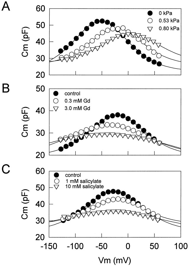
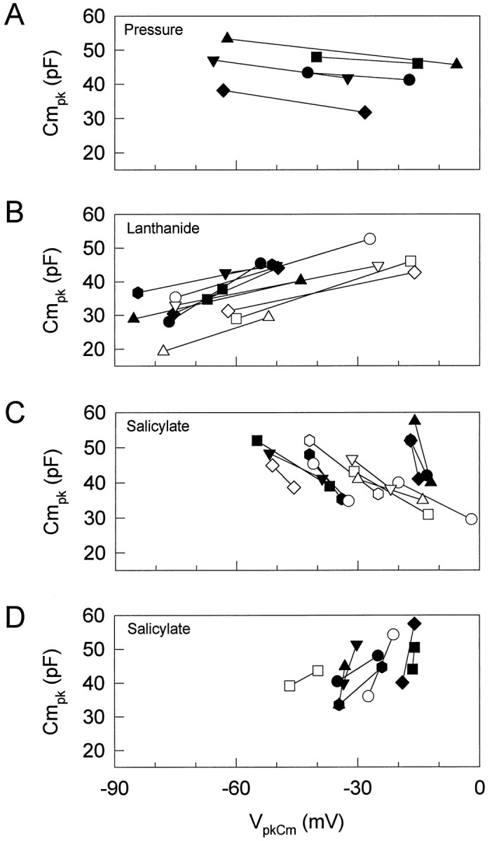
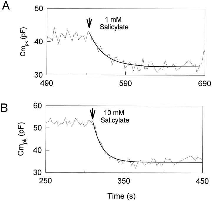
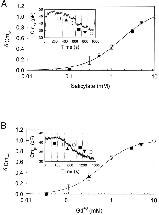
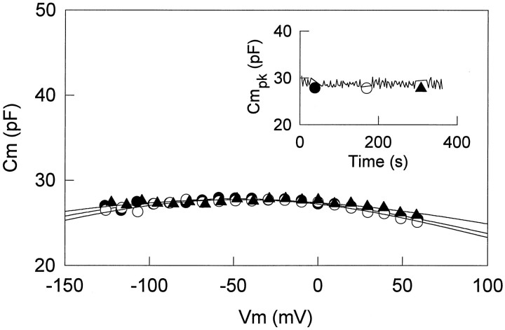
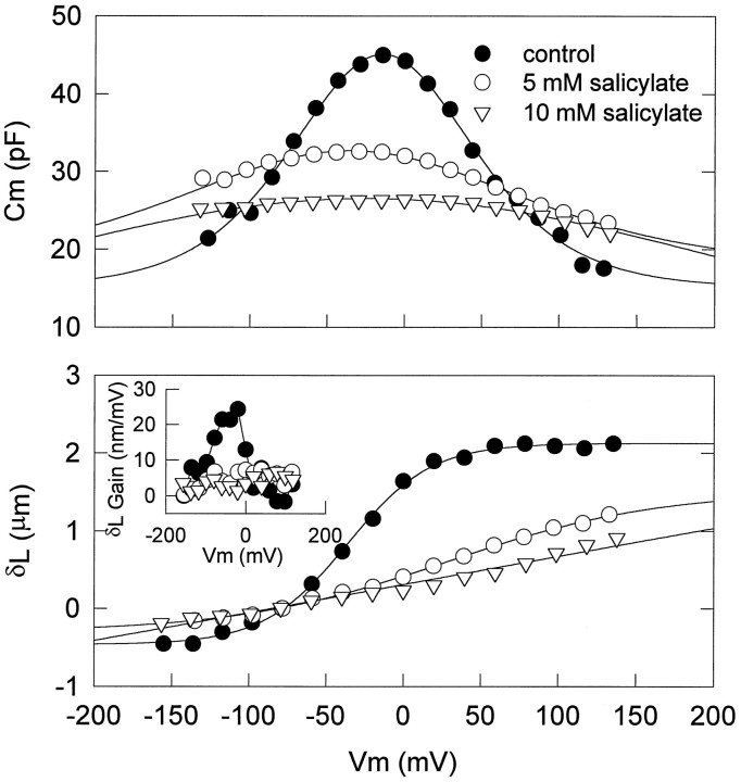
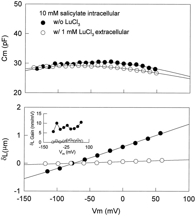
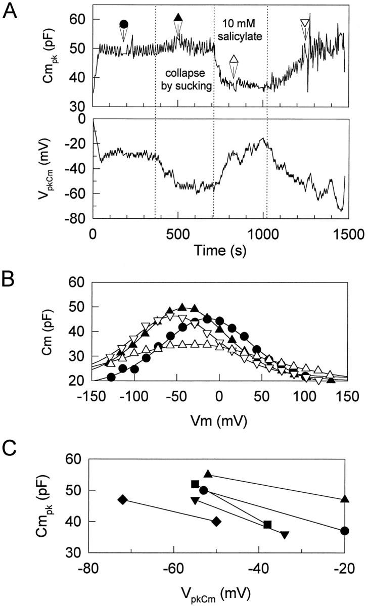
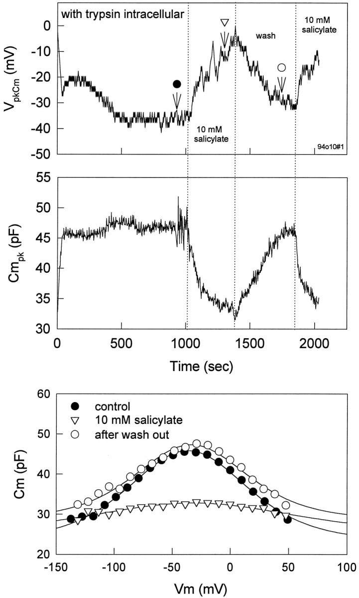
Salicylate, one of the most widely used drugs, is known to induce reversible tinnitus and hearing loss. Salicylate interferes with outer hair cells (OHCs), which are believed to underlie normal auditory frequency selectivity and sensitivity. In the present experiments, the effects of salicylate and lanthanides on OHC motility and nonlinear capacitance were investigated by using isolated guinea-pig OHCs while attempting to avoid inadvertent intracellular pressure change, which itself can affect OHC motility and capacitance. Either extracellularly or intracellularly applied salicylate reduced nonlinear peak capacitance (Cmpk) and shifted the voltage at peak capacitance to depolarized levels. Concentration-response curves for reduction in Cmpk by salicylate and GdCl3 revealed a half-maximal concentration and Hill coefficient of 1.6 mM and 1.0, and 0.6 mM and 1.2, respectively. In comparable groups of OHCs, the normal Cmpk values of which were near 40 pF, average Cmpk decreased to 28 and 36 pF for intracellularly and extracellularly applied salicylate, respectively. Salicylate reduced, but did not completely block, the voltage-induced length change. Extracellularly, but not intracellularly, applied lanthanide blocked voltage-induced movement and capacitance almost completely. After intracellular trypsin treatment, salicylate reduced voltage-dependent capacitance reversibly, suggesting that salicylate directly acts on the sensor/motor and not via effects on intracellular structures, such as the subsurface cisternae. The results are consistent with the hypothesis that the dissociated, charged form of salicylate directly interacts with the sensor/motor on the inner aspect of the OHC plasma, whereas lanthanides interact on the outer aspect.


Figures

Fig. 1.
Fig. 1.
Effects of intracellular pressure (A), lanthanides (B), and salicylate (C) onCm. Voltage-dependent capacitance was obtained by the voltage stair-step technique. A, AfterVpkCm stabilized after intracellular pressure dissipation (∼10 min), an increasing intracellular pressure caused a positive shift in the VpkCm with decreasing peak capacitance (Cmpk).Rs (series resistance), 1.9 MΩ (pipette tip, ∼1.5 μm); Rm (membrane resistance), 139 MΩ. The initial cell length was 75 μm; the cell length was reduced to 73% when intracellular pressure was increased to 0.8 kPa. B, In another cell, afterVpkCm stabilized, GdCl3 was applied extracellularly by a Y-tube delivery system. GdCl3 caused a negative shift inVpkCm with decreasingCmpk. Rs, 2.8 MΩ (pipette tip, ∼1 μm); Rm, 220 MΩ. The initial cell length was 50 μm; the cell length elongated to 107%. C, In another cell, with the use of a small-tipped pipette to maintain initial intracellular turgor pressure, extracellularly applied salicylate caused a positive shift inVpkCm with decreasingCmpk. Rs, 27.2 MΩ; Rm, 350 MΩ. The initial cell length was 65 μm; the cell length was reduced to 78%. All treatments were done after VpkCm stabilized.
Fig. 2.
Fig. 2.
Effects of intracellular pressure (A), lanthanides (B), and salicylate (C, D) on the relationship between VpkCm andCmpk. Each point shows the relationship between VpkCm andCmpk. Increasing pressure, extracellularly applied lanthanides, or salicylate reducesCmpk. A,Cmpk and VpkCmunder two pressure conditions, −0.41 ± 0.26 (mean ± SE) and 0.64 ± 0.23 kPa. Increasing pressure caused a positive shift in VpkCm. B,Cmpk and VpkCmbefore and during application of lanthanides (3 or 10 mm GdCl3, n = 7; 1 mm LuCl3,n = 3; 1 mmCeCl3, n = 1). Extracellularly applied lanthanides caused a negative shift inVpkCm with decreasingCmpk in all cells tested (n = 11). Reversibility was dependent on concentration. C,D, Cmpk andVpkCm before and during application of 10 mm salicylate. Extracellularly applied salicylate reduced Cmpk, although the direction of the shift in VpkCm was variable. The data are plotted in separate graphs for clarity. Of 21 cells tested, 13 showed a positive shift in VpkCm (C), and 8 cells showed a negative shift (D). Each symbol indicates a different cell. All treatments were done afterVpkCm stabilized.
Fig. 3.
Fig. 3.
Effects of extracellularly applied salicylate on OHC capacitance. The effective time course of extracellularly applied salicylate was examined by using a tracking procedure. A small-tipped pipette was used to permit any turgor pressure change that salicylate may cause. Salicylate was applied afterVpkCm and Cmpkreached steady state. A, Effects of 1 mm salicylate. Peak capacitance is shown as a function of time. The reduction of Cmpk is well fit by using a single exponential curve fit. Tau is 19 sec.Rs, 27.1 MΩ;Rm, 255 MΩ. B, Effects of 10 mm salicylate in a different cell. Tau is 12 sec.Rs, 26.6 MΩ;Rm, 292 MΩ.
Fig. 4.
Fig. 4.
Concentration–response curve for reduction inCmpk by salicylate and GdCl3. A, The concentration–response curve for salicylate. The inset shows the effect on theCmpk by various concentrations of salicylate in a representative example. Rs, 5.5 MΩ; Rm, 450 MΩ. Pipette pressure was kept at 0 kPa. Reductions of Cmpkinduced by various concentrations were normalized to the reduction induced by 10 mm salicylate. Each point is the mean ± SE of five cells. B, The concentration–response curve for GdCl3. Theinset shows the effect on theCmpk by various concentrations of GdCl3 in a representative example.Rs, 6.5 MΩ;Rm, 400 MΩ. Pipette pressure was kept at 0.11 kPa. Reductions of Cmpk induced by various concentrations were normalized to the reduction induced by 10 mm GdCl3. Each point is the mean ± SE of five cells determined at the end of each perfusion.
Fig. 5.
Fig. 5.
Effects of intracellularly applied salicylate on OHC capacitance. The OHC was dialyzed with a pipette solution containing 10 mm salicylate. The insetshows peak capacitance as a function of time, measured with the tracking procedure (solid line) or stair-step protocol (symbols at 30, 160, and 300 sec).Rs, 6.01 MΩ;Rm, 270 MΩ. Capacitance drops to low levels immediately after obtaining whole-cell configuration. This figure is representative of 20 cells tested.
Fig. 6.
Fig. 6.
Effects of intracellularly applied salicylate on OHC capacitance and movement. Voltage-dependent capacitance and voltage-induced length change were measured 3–4 min after whole-cell configuration in three different cells. As the concentration of salicylate increased, voltage-dependent capacitance and nonlinearity of voltage-induced length change were reduced correspondingly. Length changes were induced by 20 mV steps from −150 to +150 mV at aVhold of −80 mV.Rs, 3.38 MΩ andRm, 66.5 MΩ (closedcircles); Rs, 3.68 MΩ andRm, 34.7 MΩ (opencircles); Rs, 6.57 MΩ andRm, 240 MΩ (opentriangles). Fits (solid lines) for capacitance indicate VpkCm,Qmax, and z of −14.3 mV, 4.74 pC, and 0.650 (open circles); −38.2 mV, 5.09 pC, and 0.317 (closed circles); −28.7 mV, 36.54 pC, and 0.111 (open triangles). Fits for the mechanical data indicate Vhold and z of −36.4 mV and 0.955 (closed circles), 2.89 mV and 0.376 (open circles), and 6.43 mV and 0.039 (opentriangles). The inset shows the first derivative of the mechanical responses, indicating the gain of the mechanical response with different concentrations of salicylate. See text for details.
Fig. 7.
Fig. 7.
Comparison of effects between salicylate and lanthanide on OHC capacitance and movement. Voltage-dependent capacitance and voltage-induced length change were measured before and during extracellularly applied LuCl3 (1 mm) in an OHC with intracellularly applied salicylate (10 mm). Length changes were induced by 20 mV steps from −20 to +60 mV at aVhold of −80 mV.Rs, 4.75 MΩ andRm, 89.2 MΩ (closedcircles); Rs, 4.39 MΩ andRm, 261 MΩ (opencircles). Fits (solid lines) for capacitance indicate VpkCm,Qmax, and z of −42.3 mV, 9.152 pC, and 0.205 (open circles); −71.3 mV, 7.35 pC, and 0.191 (closed circles). Fitting such depressed capacitance functions may not be reliable because of the limited voltage range that can be applied. The mechanical data could not be fit reliably with a two-state Boltzmann. Note that lanthanides, although changing the capacitance function minimally, effectively block mechanical responses. The inset shows mechanical gain of the cell under each condition.
Fig. 8.
Fig. 8.
Effects of salicylate on the limited value ofVpkCm. Intracellular pressure of the OHC was reduced directly through the patch pipette to collapse the cell.A, The cell was maintained in collapse after the point indicated by the first dotted line. As expected, collapsing the cell caused Cmpk to increase and VpkCm to shift to a limiting negative value (Kakehata and Santos-Sacchi, 1995). At the second dottedline, 10 mm salicylate was applied extracellularly. VpkCm shifted in the positive direction with a decrease in Cmpk. Washout at the third dotted line caused recovery.B, Capacitance function determined by the stair-step protocol for the same cell. Fits (solid lines) to the capacitance data indicate VpkCm,Qmax, and z of −14.3 mV, 4.74 pC, and 0.650 (closed circles, control); −41.0 mV, 4.09 pC, and 0.743 (closed triangles, after collapse); −32.3 mV, 3.86 pC, and 0.418 (open triangles, during application of salicylate); −53.6 mV, 3.54 pC, and 0.728 (closed triangles, after wash). C, Data from five cells showing consistent positive shift ofVpkCm and decrease inCmpk in collapsed cells after 10 mm salicylate treatment.
Fig. 9.
Fig. 9.
Effects of salicylate onVpkCm and Cmpkof a trypsin-treated cell. Trypsin (300 g/ml) was included in the patch pipette. After the cell became fully spherical at ∼13 min, 10 mm salicylate was applied extracellularly. Voltage-dependent capacitance was measured before, during, and after application of salicylate. Salicylate reduced voltage-dependent capacitance reversibly, despite permanent disruption of the subsurface cisternae. Fits (solid lines) for capacitance indicateVpkCm, Qmax, and z of −36.6 mV, 3.04 pC, and 0.757 (closedcircles); −32.0 mV, 1.69 pC, and 0.450 (opentriangles). The fit to open triangles is unreliable because of the shallowness of the function.

References

    1. Ashmore JF. A fast motile response in guinea-pig outer hair cells: the cellular basis of the cochlear amplifier. J Physiol (Lond) 1987;388:323–347. - PMC - PubMed
    1. Ashmore JF. Transducer motor coupling in cochlear outer hair cells. In: Kemp D, Wilson JP, editors. Mechanics of hearing. Plenum; New York: 1989. pp. 107–113.
    1. Ashmore JF. Mammalian hearing and the cellular mechanism of the cochlear amplifier. In: Corey DP, Roper SD, editors. Sensory transduction. Rockefeller UP; New York: 1992. pp. 395–412. - PubMed
    1. Brownell WE, Bader CR, Bertrand D, de Ribaupierre Y. Evoked mechanical responses of isolated cochlear outer hair cells. Science. 1985;227:194–196. - PubMed
    1. Carlyon RP, Butt M. Effects of aspirin on human auditory filters. Hear Res. 1993;66:233–244. - PubMed

Publication types

LinkOut - more resources