. 1996 Aug 15;16(16):5049-59.
doi: 10.1523/JNEUROSCI.16-16-05049.1996.

Distinct properties of neuronal and astrocytic endopeptidase 3.4.24.16: a study on differentiation, subcellular distribution, and secretion processes

Affiliations

Distinct properties of neuronal and astrocytic endopeptidase 3.4.24.16: a study on differentiation, subcellular distribution, and secretion processes

B Vincent et al. J Neurosci. .

Abstract

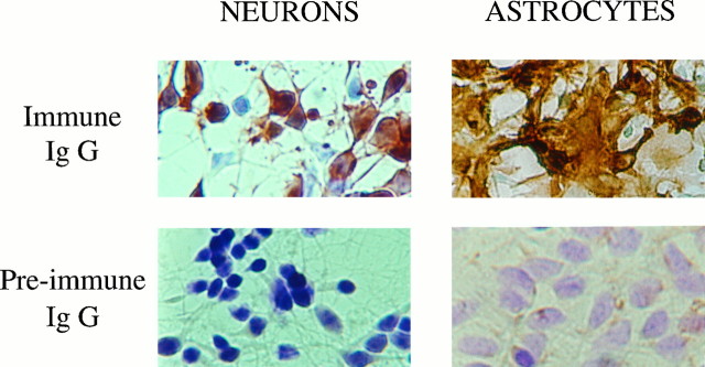
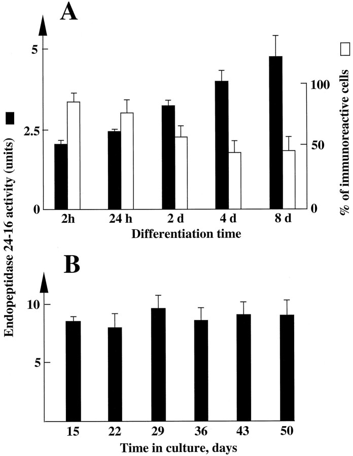
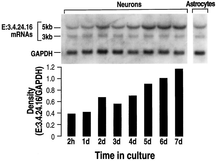
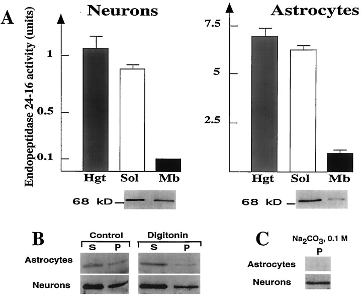
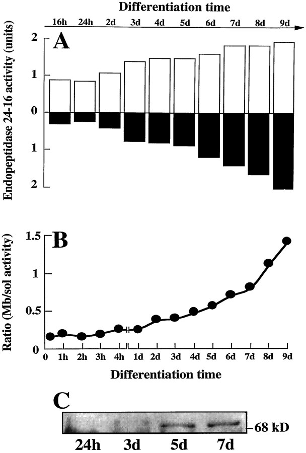
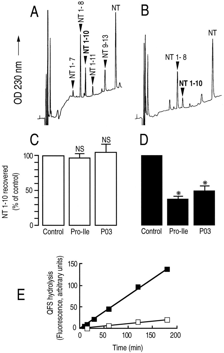
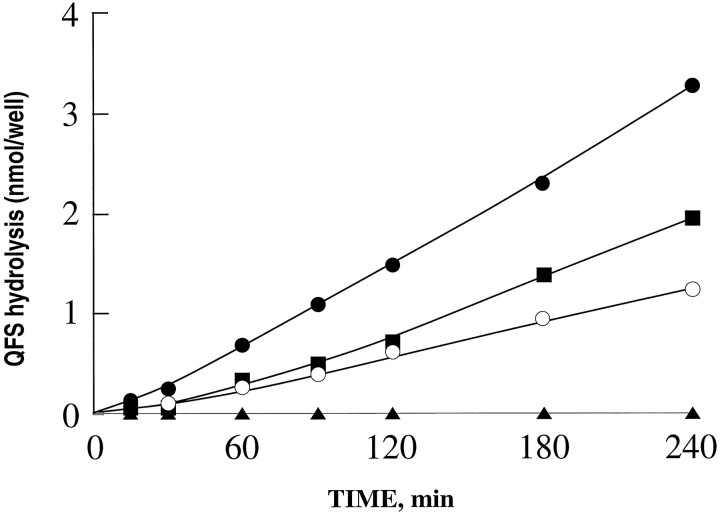
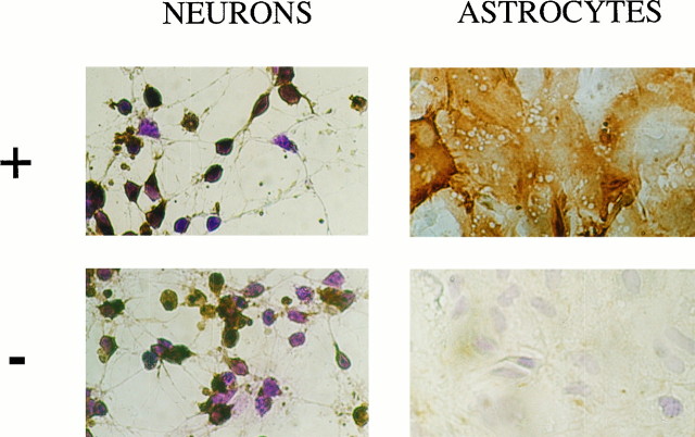
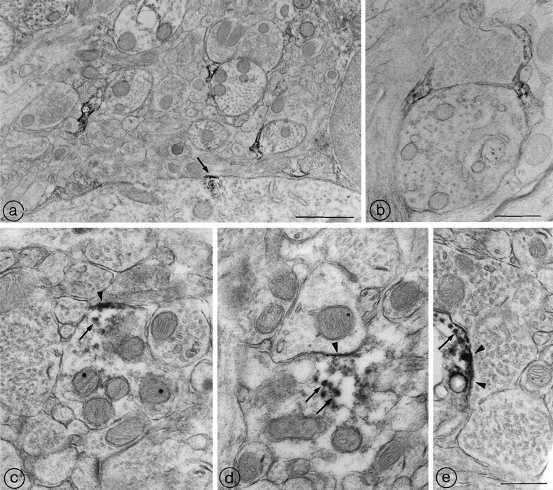
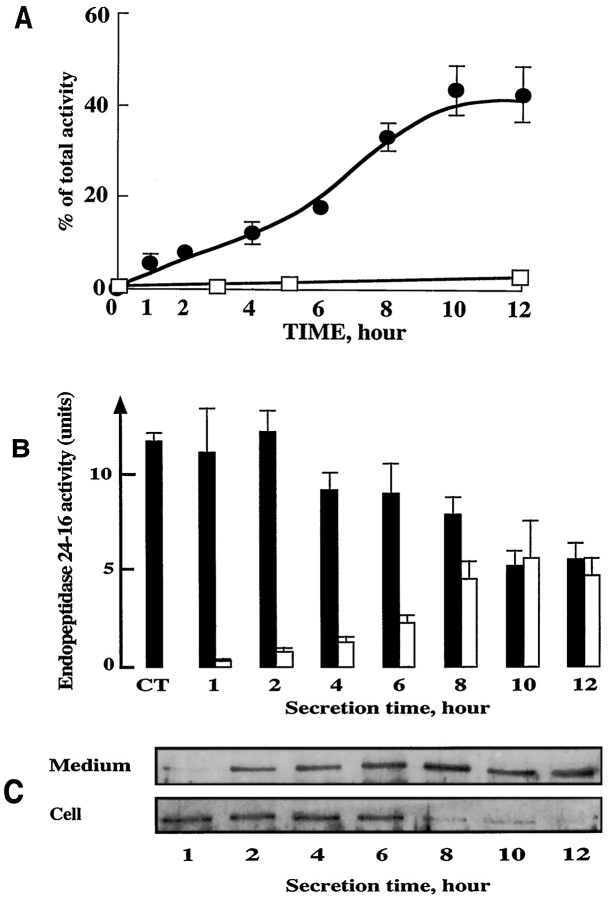
Endopeptidase 3.4.24.16 belongs to the zinc-containing metalloprotease family and likely participates in the physiological inactivation of neurotensin. The peptidase displays distinct features in pure primary cultured neurons and astrocytes. Neuronal maturation leads to a decrease in the proportion of endopeptidase 3.4.24.16-bearing neurons and to a concomitant increase in endopeptidase 3.4.24.16 activity and mRNA content. By contrast, there is no change with time in endopeptidase 3.4.24.16 activity or content in astrocytes. Primary cultured neurons exhibit both soluble and membrane-associated endopeptidase 3.4.24.16 activity. The latter behaves as an ectopeptidase on intact plated neurons and resists treatments with 0.2% digitonin and Na2CO3. Further evidence for an association of the enzyme with plasma membranes was provided by cryoprotection experiments and electron microscopic analysis. The membrane-associated form of endopeptidase 3.4.24.16 increased during neuronal differentiation and appears to be mainly responsible for the overall augmentation of endopeptidase 3.4.24.16 activity observed during neuronal maturation. Unlike neurons, astrocytes only contain soluble endopeptidase 3.4.24.16. Astrocytes secrete the enzyme through monensin, brefeldin A, and forskolin-independent mechanisms. This indicates that endopeptidase 3.4.24.16 is not released by classical regulated or constitutive secreting processes. However, secretion is blocked at 4 degrees C and by 8 bromo cAMP and is enhanced at 42 degrees C, two properties reminiscent of that of other secreted proteins lacking a classical signal peptide. By contrast, neurons appear unable to secrete endopeptidase 3.4.24.16.


Figures

Fig. 1.
Fig. 1.
Immunolabeling of endopeptidase 3.4.24.16 in primary cultures of neurons and astrocytes. Neurons and astrocytes were cultured for 4 and 15 d, respectively, in the conditions described in Materials and Methods. After fixation and cryoprotection, cells were incubated overnight at 4°C with the IgG-purified fractions of the immune (top panels) or preimmune (bottompanels) rabbit antiserum developed against rat brain endopeptidase 3.4.24.16. After exposure to a goat anti-rabbit IgG coupled to peroxidase, endopeptidase 3.4.24.16-bearing cells were revealed with diaminobenzidine (brown cells) as described, and immunonegative cells still reacted with cresyl violet (blue cells). Photographs were taken with Kodacolor 100 film at 200× magnification.
Fig. 2.
Fig. 2.
Endopeptidase 3.4.24.16 activity in whole homogenate of primary cultured neurons and astrocytes: effect of time in culture. Neurons and astrocytes were primary-cultured as described in Materials and Methods. At the indicated times, dishes were rinsed twice with PBS, cells were scraped in 5 mm Tris-HCl, pH 7.5, and homogenized with a syringe. Endopeptidase 3.4.24.16 activity (black bars) was monitored in neuron (A) and astrocyte (B) homogenates (50 and 25 μl, respectively) by means of the QFS assay and quantified as described in Materials and Methods. 1 unit = 1 nmol of QFS hydrolysed/hr/25 or 50 μl. Values are the mean ± SEM of six independent determinations performed with six different cultures. The white bars in A indicate the percent of neurons expressing endopeptidase 3.4.24.16 during neuronalin vitro differentiation and were taken from Chabry et al. (1990).
Fig. 3.
Fig. 3.
Northern blot analysis of endopeptidase 3.4.24.16 mRNA during in vitro neuronal differentiation. Total mRNAs (20 μg) were isolated as described in Materials and Methods from neurons cultured for the indicated times and from 15-d-old-plated astrocytes. RNAs were electrophoresed, blotted on a nitrocellulose sheet, and hybridized with the32P-labeled PCR fragment derived from the λ7a clone encoding rat brain endopeptidase 3.4.24.16 and the GAPDH probe (see Materials and Methods). RNA molecular weights are indicated on the left. Autoradiograms (top) were analyzed by densitometry. Bars indicate the mean of two independent quantifications performed with two distinct cell cultures and correspond to the 5 kb E:3.4.24.16 versus the GAPDH mRNA density recovered in the same sample. Ratio values did not differ by >0.005.
Fig. 4.
Fig. 4.
Effect of digitonin and Na2CO3 on endopeptidase 3.4.24.16 immunoreactivity in neurons and astrocytes. A, Homogenates, soluble and membrane-associated fractions of 4-d-old neurons and 15-d-old astrocytes, were prepared as described in Materials and Methods and assayed for their Pro-Ile-sensitive QFS-hydrolysing activity (1 unit = 1 nmol of QFS hydrolysed/hr). Values represent the mean ± SEM of six determinations performed with independent cultures. Bottom panel corresponding to Western blot analysis of proteins (10 μg) in soluble and membrane-associated fractions of neurons and astrocytes shows a single immunoreactive band around 75 kDa. B, Cells were prepared as described in Materials and Methods and incubated in the absence (Control) or in the presence of 0.2% Digitonin. Aliquots were then centrifuged (4°C, 35 min, 150,000 × g), and 5–15 μg of protein of supernatants (S) and pellets (P) were electrophoresed and analyzed by Western blot. C, Membrane-associated fractions of primary cultured neurons and astrocytes (5 and 50 μg of protein, respectively) were treated with 0.1 mNa2CO3 and centrifuged, and resulting pellets (P) were analyzed by Western blot.
Fig. 5.
Fig. 5.
Soluble and membrane-associated endopeptidase 3.4.24.16 activity during differentiation of primary cultured neurons.A, Primary cultured neurons were scraped at the indicated differentiation times in 5 mm Tris-HCl, pH 7.5, and subcellular fractions were prepared as described in Materials and Methods. Both soluble (white bars) and membrane-associated (black bars) fractions were tested for their QFS-hydrolysing activities as described in Materials and Methods.B illustrates the ratio between QFS-hydrolysing activity in membrane-associated versus soluble fractions according to differentiation time. C, Five micrograms of membrane-associated fractions taken at 1, 3, 5, and 7 d of culture were dried, submitted to an 8% acrylamide gel, and analyzed by Western blot with the anti-E 3.4.24.16 IgG fraction as described in Materials and Methods.
Fig. 6.
Fig. 6.
Endopeptidase 3.4.24.16 activity on plated neurons. Neurotensin (10 nmol, 10 μm) was incubated with 15-d-old cultured astrocytes (A) and with 4-d-old cultured neurons (B) for 2 and 3 hr, respectively, as described in Materials and Methods, in the absence (A, B) or in the presence (C, D) of Pro-Ile (10 mm) or phosphodiepryl 03 (100 nm). One hundred microliters of acidified medium were submitted to HPLC analysis. Arrows indicate the elution time of synthetic neurotensin fragments. Bars (C, D) represent the NT (1–10) recovered and are expressed as the percent of NT (1–10) recovered in the absence of inhibitor (Control). Values are the mean ± SEM of three to nine independent determinations. *p < 0.0001. NS, Nonstatistically significant. E, The Pro-Ile-sensitive QFS-hydrolysing activity detectable on plated neurons (▪) or astrocytes (□) was monitored as described in Materials and Methods.
Fig. 7.
Fig. 7.
QFS hydrolysis by plated neurons during in vitro differentiation. Neuronal cultures corresponding to 2 hr (▴), 1 d (○), 2 d (▪), or 4 d (•) of differentiation were incubated for the indicated times with QFS (50 nmol, 50 μm) in 1 ml of PBS-1% glucose, pH 7.4, as described in Materials and Methods, in the absence or in the presence of Pro-Ile (10 mm). At the end of the incubations, 100 μl of supernatants were removed and acidified, and endopeptidase 3.4.24.16 activity was fluorimetrically monitored as described in Materials and Methods. Curves represent kinetics of the Pro-Ile-sensitive QFS-hydrolysing activity and correspond to the mean of three independent determinations.
Fig. 8.
Fig. 8.
Effect of cryoprotection on endopeptidase 3.4.24.16 immunoreactivity in primary cultured neurons and astrocytes. Four-day-old plated neurons and 15-d-old plated astrocytes were fixed with glutaraldehyde, cryoprotected (+) or not (−) with NaK2 buffer containing 30% sucrose, and processed for immunochemical detection of endopeptidase 3.4.24.16 as described in Materials and Methods. Photographs were taken with Kodacolor 100 film at 200× magnification.
Fig. 9.
Fig. 9.
Ultrastructural distribution of endopeptidase 3.4.24.16 in the midbrain tegmentum of the adult rat. a, Three densely immunoreactive astrocytic leaflets are visible in this field of the midbrain tegmentum. Each of them is apposed to a cross-sectioned dendritic profile. At the bottom of the field, a neuronal soma exhibits a discrete patch of endopeptidase 3.4.24.16 immunoreactivity. The immunoreactivity is restricted to a short segment of the plasma membrane (arrow) and underlying cytoplasm. Scale bar, 1 μm. b, Two immunoreactive astrocytic leaflets seal off a synaptic junction between an axon terminal and an unlabeled dendritic shaft. Note that the reaction product pervades the entire glial cytoplasm. Scale bar, 0.5 μm.c, d, e, Endopeptidase 3.4.24.16-immunoreactive dendrites. In all three of these labeled dendritic shafts, the reaction product is concentrated along restricted zones of the plasmalemma (arrowheads), as well as within microtubules and/or vesicular organelles (arrows). Whereas the subplasmalemmal labeling in c and d is clearly extrasynaptic, the reaction product in e may be masking a postsynaptic specialization. Scale bar, 0.5 μm.
Fig. 10.
Fig. 10.
Secretion of endopeptidase 3.4.24.16 by cultured astrocytes. A, Time course of endopeptidase 3.4.24.16 recovery in the medium of 15-d-old cultured astrocytes (•) and 4-d-old cultured neurons (□). Incubations were performed in PBS+/1% glucose, pH 7.4. At the indicated times, medium was taken off and enzymatic activity was fluorimetrically measured with QFS as detailed in Materials and Methods. Values are expressed as the percentage of whole-cell activity detectable att = 0 and are the mean ± SEM of four independent experiments. B illustrates the medium (white bars) and cellular (black bars) endopeptidase 3.4.24.16 activity in astrocytes at each time of the secretion kinetics. Values are mean ± SEM of three independent experiments. C, Western blot analysis of endopeptidase 3.4.24.16 protein content in media and cells during astrocyte-secretion experiments. Five micrograms of proteins were loaded onto an 8% SDS-PAGE and immunoblotted as described in Materials and Methods.

Similar articles

Cited by

References

    1. Barelli H, Vincent JP, Checler F. Peripheral inactivation of neurotensin: isolation and characterization of a metallopeptidase from rat ileum. Eur J Biochem. 1988;175:481–489. - PubMed
    1. Barelli H, Dive V, Yiotakis A, Vincent JP, Checler F. Potent inhibition of endopeptidase 24.16 and endopeptidase 24.15 by the phosphonamide peptide N -(phenylethylphosphonyl)-Gly-l-Pro-l-aminohexanoic acid. Biochem J. 1992;287:621–625. - PMC - PubMed
    1. Barelli H, Vincent JP, Checler F. Rat kidney endopeptidase 24.16: purification, physico-chemical characteristics and specificity towards opiates, tachykinins and neurotensin-related peptides. Eur J Biochem. 1993;211:79–90. - PubMed
    1. Barelli H, Fox-Threlkeld JET, Dive V, Daniel EE, Vincent JP, Checler F. Role of endopeptidase 3.4.24.16 in the catabolism of neurotensin, in vivo , in the vascularly perfused dog ileum. Br J Pharmacol. 1994;112:127–132. - PMC - PubMed
    1. Chabry J, Checler F, Vincent JP, Mazella J. Colocalization of neurotensin receptors and of the neurotensin-degrading enzyme endopeptidase 24–16 in primary cultures of neurons. J Neurosci. 1990;10:3916–3921. - PMC - PubMed

Publication types

LinkOut - more resources