. 1996 Aug 15;16(16):5060-72.
doi: 10.1523/JNEUROSCI.16-16-05060.1996.

Optical recordings of the effects of cholinergic ligands on neurons in the ganglion cell layer of mammalian retina

Affiliations

Optical recordings of the effects of cholinergic ligands on neurons in the ganglion cell layer of mammalian retina

W H Baldridge. J Neurosci. .

Abstract

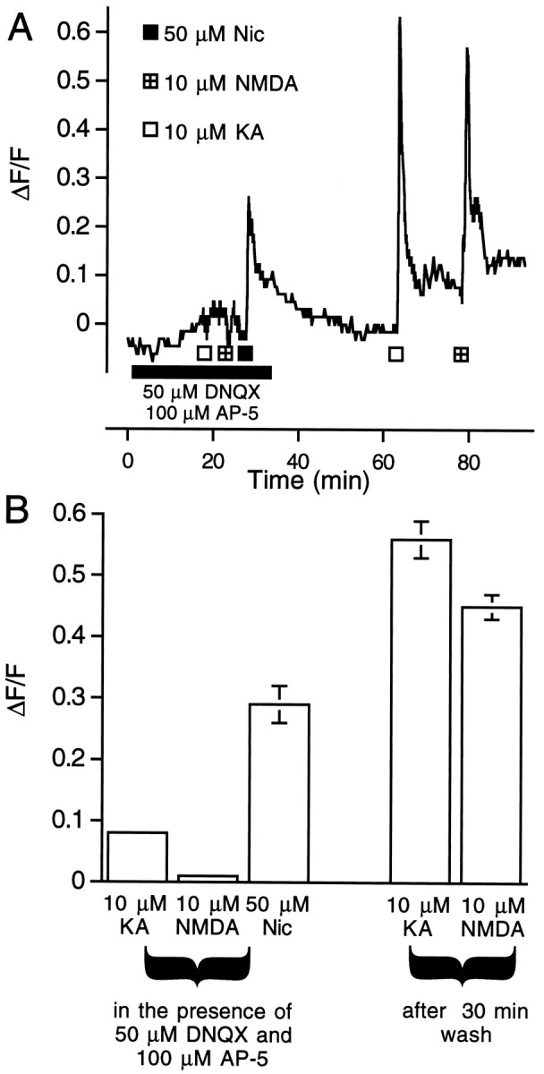
Cholinergic regulation of the activity of rabbit retinal ganglion cells and displaced amacrine cells was investigated using optical recording of changes in intracellular free calcium ([Ca2+]i). Labeling of neurons in the mature retina was achieved by injecting calcium green-1 dextran (CaGD) into the isolated retina. Nicotine increased ganglion cell [Ca2+]i, affecting every loaded cell in some preparations; the pharmacology of nicotine was consistent with an action at neuronal nicotinic receptors, and specifically it was kappa-(neuronal-)bungarotoxin-sensitive but alpha-bungarotoxin-insensitive. Muscarine also raised [Ca2+]i, but it was less potent than nicotine, affecting only a subpopulation of ganglion cells, with an M1-like muscarinic receptor pharmacology. Neither the nicotine- nor muscarine-induced increases of ganglion cell [Ca2+]i were blocked by the glutamate receptor antagonists 6,7-dinitroquinoxaline-2,3-dione and aminophosphonopentanoic acid. Therefore, the effects of cholinergic agonists on ganglion cell [Ca2+]i were not attributable to an indirect effect mediated by glutamatergic bipolar cells. The effects of nicotine and muscarine were abolished in calcium-free solution, indicating that the responses depend on calcium influx. Displaced (Cb) cholinergic amacrine cells were also loaded with CaGD and were identified by selective labeling with the nuclear dye 4',6-diamidino-2-phenyl-indole. Cb amacrine cells did not respond to either nicotine or muscarine, but responded vigorously to the glutamate receptor agonist kainic acid. There is anatomical evidence indicating that cholinergic amacrine cells make synaptic contact with each other, but the present results do not support the hypothesis that communication between these cells is cholinergic.


Figures

Fig. 1.
Fig. 1.
Video and pseudocolor images of the GCL from three pieces of rabbit retina (A–D, E–G, H–J) labeled with CaGD. A, Video image of CaGD-labeled ganglion cells imaged under 485 nm excitation. Some bundles of labeled ganglion cell axons are also visible. B, Pseudocolor representation of the response of the cells in A to 50 μmnicotine. Not every cell responded, but those that did showed responses of up to ΔF/F = 1.00, but more typically ΔF/F ∼ 0.50. The increase of CaGD fluorescence illustrated here and in all subsequent images indicates an increase of [Ca2+]i.C, Responses of cells from the same piece of retina to 50 μm muscarine. Compared with nicotine, fewer cells responded and the responses were weaker. D, Responses of cells from the same retina to 10 μmkainic acid. Virtually every CaGD-labeled cell responded, with some reaching ΔF/F ∼ 1.25. E, Video image of a piece of retina labeled with CaGD. F, Pseudocolor imaging illustrating the responses of the cells in E to 50 μm muscarine. Although several cells responded, the responses were weak. G, Response of the same cells to 50 μm nicotine. In this piece of retina, virtually every cell responded to nicotine with ΔF/F ∼ 0.50. H, Video image of a piece of retina stained with the nuclear stain DAPI and imaged under 380 nm excitation. Four nuclei were brightly labeled (red circles) and correspond to displaced (Cb) cholinergic amacrine cells. The other cells, with dimly stained nuclei, were characterized as presumptive ganglion cells (see Materials and Methods). These cells were also labeled with CaGD (not shown). I, Pseudocolor imaging of the same piece of retina as in H illustrating the response to 50 μm nicotine. Although several ganglion cells responded, the cholinergic amacrine cells did not.J, Both ganglion and amacrine cells (red circles) responded to 10 μm kainic acid. Scale bar in the last illustration of each set is 50 μm.
Fig. 2.
Fig. 2.
Cholinergic agonists and kainic acid increase ganglion cell [Ca2+]i.A, Optical recordings from a ganglion cell illustrating the dose-dependent increase of CaGD fluorescence (ΔF/F), indicating an increase of [Ca2+]i in response to increasing concentrations of nicotine. B, Mean responses to increasing concentrations of nicotine (squares), the nicotinic agonist DMPP (triangles), and muscarine (circles) from all ganglion cells studied. C, Optical recording from a ganglion cell illustrating the dose-dependent response to increasing concentrations of kainic acid. D, Mean responses to increasing concentrations of kainic acid from all ganglion cells studied (squares). For comparison, the effect of the same dose of kainic acid on the firing rate of a ganglion cell has also been plotted (open circles). The ganglion cell firing rate data were taken from Massey and Miller (1988). In all cases, error bars are SEM.
Fig. 3.
Fig. 3.
The effect of nicotinic receptor antagonists on the nicotine-induced rise of ganglion cell [Ca2+]i. A, The effect of 50 μm nicotine (Nic) was blocked by 25 μm (+)-tubocurarine (Tbc) and 100 μm hexamethonium (Hex; B), and in both cases the response to nicotine recovered after washing. C, Neither 1 nor 50 μm nicotine were blocked by 2.5 μm α-bungarotoxin (α-Bgt).D, Ten micromolar methyllycaconitine (Mla) did not block 50 μm nicotine, and 1 μm methyllycaconitine failed to block the response to 1 μm nicotine. E, Summary of the results from all ganglion cell studies expressed as mean ΔF/F. Error bars are SEM.
Fig. 4.
Fig. 4.
The effect of nicotine on ganglion cell [Ca2+]i is blocked by κ-bungarotoxin. A, After a 40 min incubation in 100 nm κ-bungarotoxin (κ-Bgt), the effect of 1 μm nicotine (Nic), but not 50 μm nicotine, on ganglion cell [Ca2+]i was blocked.B, In some cells, the block of the response to 1 μm nicotine was reversed by washing for 1 hr.C, Mean ΔF/F of all the ganglion cells studied in response to 1 and 50 μmnicotine, shortly and 1 hr after incubation in 100 nm κ-bungarotoxin. Although some cells recovered after 1 hr wash as illustrated in B, on average the block produced by 100 nm κ-bungarotoxin was irreversible. Error bars are SEM.
Fig. 5.
Fig. 5.
The effect of nicotine on ganglion cell [Ca2+]i is not blocked by glutamate receptor antagonists. A, During incubation with a mixture of non-NMDA (50 μm DNQX) and NMDA (100 μm AP-5) receptor antagonists, the ganglion cell responses to 10 μm kainic acid (KA) and 10 μmNMDA were blocked. However, the effect of 50 μm nicotine (Nic) was not blocked by these antagonists. After wash, the response to both kainic acid and NMDA recovered. B, Summary of the results from all ganglion cell studies expressed as mean ΔF/F. Error bars are SEM.
Fig. 6.
Fig. 6.
The effect of muscarine on ganglion cell [Ca2+]i has an M1-like muscarinic receptor pharmacology. The effect of 50–100 μm muscarine (Musc) on ganglion cell [Ca2+]i was blocked by 1 μm atropine (Atr; A) and 100 μm pirenzepine (Pirenz;B), an antagonist with M1 > M2 muscarinic receptor affinity. The block produced by both antagonists was reversed by washing. C, The effect of muscarine was not blocked by 100 μm gallamine (Gall), an M2 muscarinic receptor antagonist, or the mixture of the glutamate antagonists DNQX (50 μm) and AP-5 (100 μm) (D). E, Summary of the results from all ganglion cell studies expressed as mean ΔF/F. Error bars are SEM.
Fig. 7.
Fig. 7.
The effect of nicotine and muscarine on ganglion cell [Ca2+]i depends on extracellular calcium. When Ames solution was made nominally calcium-free by the addition of 5 mm EGTA, the response of ganglion cells to nicotine (Nic; A) and muscarine (Musc; B) was abolished. After subsequent washing, the response to both agonists recovered.C, The mean ΔF/F from all cells studied in response to nicotine and muscarine, during and after superfusion with calcium-free Ames medium. Error bars are SEM.
Fig. 8.
Fig. 8.
Nicotine does not increase [Ca2+]i in cholinergic amacrine cells. As illustrated in Figure 1H, displaced (Cb) cholinergic amacrine cells were identified in the GCL of the rabbit retina by their characteristic bright DAPI-labeled nucleus. Cells without this feature were identified as presumptive ganglion cells (see Materials and Methods).A, Optical recordings from two retinas illustrating the effects of nicotine (Nic) and kainic acid (KA) on these cells, and ganglion cells from the same pieces of retina. In both retinas, cholinergic amacrine cells failed to respond to 50 μm nicotine, but did respond to 10 μm kainic acid. In comparison, presumptive ganglion cells responded to both 50 μm nicotine and 10 μmkainic acid. B, The mean ΔF/Fof all cholinergic amacrine and ganglion cells studied in response to nicotine and kainic acid. Error bars are SEM.

References

    1. Adams DJ, Nutter TJ. Calcium permeability and modulation of nicotinic acetylcholine receptor-channels in rat parasympathetic neurons. J Physiol (Paris) 1992;86:67–76. - PubMed
    1. Ariel M, Daw NW. Effects of cholinergic drugs on receptive field properties of rabbit retinal ganglion cells. J Physiol (Lond) 1982;324:135–160. - PMC - PubMed
    1. Aizenman E, Forsch MP, Lipton SA. Responses mediated by excitatory amino acid receptors in solitary retinal ganglion cells from rat. J Physiol (Lond) 1988;396:75–91. - PMC - PubMed
    1. Aizenman E, Loring RH, Lipton SA. Blockade of nicotinic responses in rat retinal ganglion cells by neuronal bungarotoxin. Brain Res. 1990;517:209–214. - PubMed
    1. Ames A, III, Nesbett FB. In vitro retina as an experimental model of the central nervous system. J Neurochem. 1981;37:867–877. - PubMed

Publication types

LinkOut - more resources