. 1996 Aug 15;16(16):5073-81.
doi: 10.1523/JNEUROSCI.16-16-05073.1996.

Hippocampal astrocytes in situ respond to glutamate released from synaptic terminals

Affiliations

Hippocampal astrocytes in situ respond to glutamate released from synaptic terminals

J T Porter et al. J Neurosci. .

Abstract

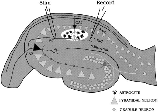
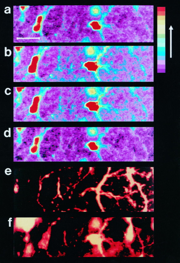
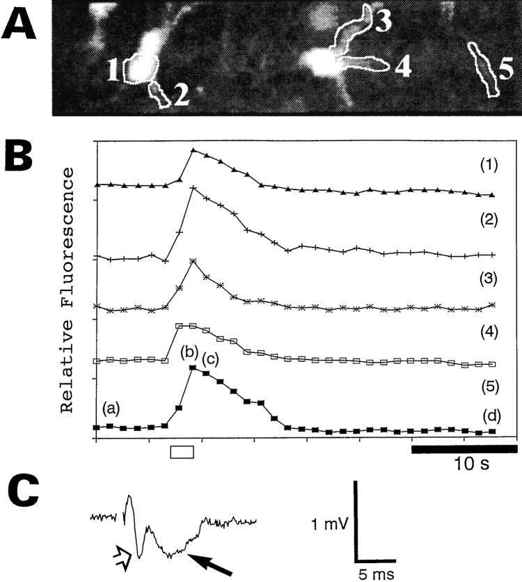
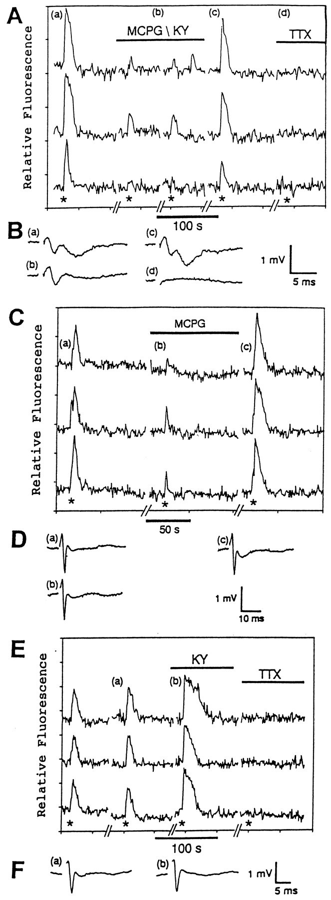
A long-standing question in neurobiology is whether astrocytes respond to the neuronal release of neurotransmitters in vivo. To address this question, acutely isolated hippocampal slices were loaded with the calcium-sensitive dye Calcium Green-1 and the responses of the astrocytes to electrical stimulation of the Schaffer collaterals were monitored by confocal microscopy. To confirm that the responsive cells were astrocytes, the slices were immunostained for the astrocytic marker glial fibrillary acidic protein. Stimulation of the Schaffer collaterals (50 Hz, 2 sec) resulted in increases in the concentration of intracellular calcium ([Ca2+]i) in the astrocytes located in the stratum radiatum of CA1. The astrocytic responses were blocked by the sodium channel blocker tetrodotoxin, the voltage-dependent calcium channel blocker omega-conotoxin-MVIIC, and the selective metabotropic glutamate receptor antagonist alpha-methyl-4-carboxyphenylglycine (MCPG). These results suggest that the astrocytic responses were induced by stimulation of metabotropic glutamate receptors on the astrocytes by neuronally released glutamate. The astrocytic responses to neuronal stimulation were enhanced in the presence of the K+ channel antagonist 4-aminopyridine (4-AP). Inhibition of the astrocytic responses in the presence of 4-AP required the presence of both MCPG and the ionotropic glutamate receptor antagonist kynurenic acid. These results suggest that higher levels of neuronal activity result in stimulation of both metabotropic and ionotropic glutamate receptors on the astrocytes. Overall, the results indicate that hippocampal astrocytes in situ are able to respond to the neuronal release of the neurotransmitter glutamate with increases in [Ca2+]i.


Figures

Fig. 1.
Fig. 1.
This diagram illustrates the relative positions of the stimulating (Stim) and recording (Record) electrodes and the studied astrocytes within the hippocampal slice. The Schaffer collaterals (SC) were electrically stimulated, and the changes in [Ca2+]iwere monitored in the astrocytes in the stratum radiatum of CA1 (white area) with a confocal microscope. The recording electrode was used to record the field potentials in response to single test pulses.
Fig. 2.
Fig. 2.
Electrical stimulation of Schaffer collaterals induces increases in [Ca2+]i in the hippocampal astrocytes in situ. The pseudocolorized confocal images of hippocampal astrocytes in situ loaded with Calcium Green-1 illustrate the relative fluorescence of the astrocytes before stimulation (a), at the end of the stimulation (200 μA, 50 Hz, 2 sec) (b, c), and 28 sec after the stimulation (d). The color bar on the right indicates that the relative [Ca2+]iincreases as the colors change from violet to red. Scale bar, 20 μm. The cells studied in ad were identified as astrocytes by subsequent immunostaining for the astrocytic marker GFAP (e). The astrocytic nuclei were stained with propidium iodide (f).
Fig. 3.
Fig. 3.
Graphical representation of the experiment shown in Figure 2. A, This image is the black and white equivalent of image b in Figure 2. The relative fluorescence of the indicated regions (15) of the astrocytes was monitored. B, This graph illustrates the relative changes in calcium for the regions labeled in A. The lettersad correspond to imagesad of Figure 2. The position of the letters indicates where in the time sequence the image was taken. Theopen bar at the bottom indicates when the stimulation (200 μA, 50 Hz, 2 sec) was applied to the Schaffer collaterals. The traces in this graph and all subsequent graphs were arbitrarily shifted along the ordinate axis for clarity. C, This field potential was induced by a single test pulse of 200 μA just before the experiment. The presynaptic and postsynaptic portions of the field potential are indicated by the open arrow and the filled arrow, respectively.
Fig. 4.
Fig. 4.
The astrocytic responses to stimulation of the Schaffer collaterals are enhanced by the K+channel antagonist 4-AP and are inhibited by the Na+ channel antagonist TTX and the voltage-dependent Ca2+ channel antagonist MVIIC.A, This graph illustrates the responses of three different astrocytes in situ to electrical stimulation of the Schaffer collaterals (200 μA, 50 Hz, 2 sec) in the presence or absence of 100 μm 4-AP and 1 μm TTX. The asterisks indicate when the electrical stimulations were given. The letters ac indicate the approximate times when the field potentials shown in B were recorded.B, The field potentials (ac) indicate the relative level of neuronal activity in the absence of 4-AP and TTX (a), in the presence of 4-AP (b), and in the presence of 4-AP and TTX (c). C, This graph demonstrates that the responses of three different hippocampal astrocytes in situ to Schaffer collateral stimulation (400 μA, 50 Hz, 2 sec; asterisks) in the presence of 100 μm 4-AP are blocked by 5 μm MVIIC. The letters a andb indicate when the field potentials shown in Dwere recorded. D, The field potentials indicate the relative level of neuronal activity in the presence of 4-AP before (a) and after (b) MVIIC treatment.
Fig. 5.
Fig. 5.
In the absence of 4-AP, the astrocytic responses are attributable to the stimulation of metabotropic glutamate receptors. A, This graph demonstrates the effect of the selective metabotropic glutamate receptor antagonist MCPG (1 mm) and the ionotropic glutamate receptor antagonists KY (3 mm) and TTX (1 μm) on the responses of three different hippocampal astrocytes in situ to Schaffer collateral stimulation (200 μA, 50 Hz, 2 sec; asterisks). The lettersad indicate when the field potentials inB were measured. B, The field potentials indicate the relative levels of neuronal activity before (a), during (b), and after (c) treatment with MCPG and KY. TTX treatment blocked the field potential (d). C, This graph illustrates the effect of MCPG (1 mm) on the responses of three different astrocytes in situ to Schaffer collateral stimulation (200 μA, 50 Hz, 2 sec;asterisks). The letters ac indicate when the field potentials in D were recorded. D, These field potentials were recorded before (a), during (b), and after (c) treatment with MCPG.E, This graph illustrates the effect of KY (3 mm) and TTX (1 μm) on the responses of three different hippocampal astrocytes in situto stimulation of the Schaffer collaterals (300 μA, 50 Hz, 2 sec;asterisks). The letters a and bindicate when the field potentials shown in F were measured.F, These field potentials were recorded before (a) and during (b) the KY treatment.
Fig. 6.
Fig. 6.
In the presence of 4-AP, the astrocytic responses are attributable to stimulation of both metabotropic and ionotropic glutamate receptors. A, This graph illustrates the effect of MCPG (1 mm) and KY (3 mm) on the responses of three different hippocampal astrocytes in situ to Schaffer collateral stimulation (400 μA, 50 Hz, 2 sec; asterisks) in the presence of 4-AP (100 μm). The lettersac indicate when the field potentials inB were measured. B, The field potentials were measured before (a), during (b), and after (c) treatment with MCPG and KY. C, This graph demonstrates the effect of MCPG (1 mm) and TTX (1 μm) on the responses of three different hippocampal astrocytes in situ to Schaffer collateral stimulation (200 μA, 50 Hz, 2 sec; asterisks) in the presence of 4-AP (100 μm). The lettersac indicate when the field potentials inD were measured. D, The field potentials were recorded in the absence of 4-AP (a), in the presence of 4-AP (b), and in the presence of 4-AP and MCPG (c).E, This graph illustrates the effect of KY (3 mm) on the responses of three different hippocampal astrocytes in situ to Schaffer collateral stimulation (200 μA, 50 Hz, 2 sec; asterisks) in the presence of 4-AP (100 μm). The lettersad indicate when the field potentials inF were recorded. F, The field potentials were recorded in the absence of 4-AP (a), in the presence of 4-AP (b), in the presence of 4-AP and KY (c), and after washout of KY (d).

References

    1. Aoki C, Pickel VM. Ultrastructural relations between β-adrenergic receptors and catecholaminergic neurons. Brain Res Bull. 1992;29:257–263. - PubMed
    1. Aoki C, Joh TH, Pickel VM. Ultrastructural localization of β-adrenergic receptor-like immunoreactivity in the cortex and neostriatum of rat brain. Brain Res. 1987;437:264–282. - PubMed
    1. Bashir ZI, Bortolotto ZA, Davies CH, Berretta N, Irving AJ, Seal AJ, Henley JM, Jane DE, Watkins JC, Collingridge GL. Induction of LTP in the hippocampus needs synaptic activation of glutamate metabotropic receptors. Nature. 1993;363:347–350. - PubMed
    1. Buckle PJ, Haas HL. Enhancement of synaptic transmission by 4-aminopyridine in hippocampal slices of the rat. J Physiol (Lond) 1982;326:109–122. - PMC - PubMed
    1. Buijs RM, van Vulpen EHS, Geffard M. Ultrastructural localization of GABA in the supraoptic nucleus and neural lobe. Neuroscience. 1987;20:347–355. - PubMed

Publication types

LinkOut - more resources