. 1996 Aug 15;16(16):5141-53.
doi: 10.1523/JNEUROSCI.16-16-05141.1996.

Actions of substance P on rat neostriatal neurons in vitro

Affiliations

Actions of substance P on rat neostriatal neurons in vitro

T Aosaki et al. J Neurosci. .

Abstract

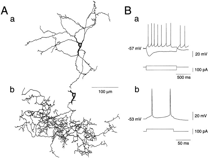
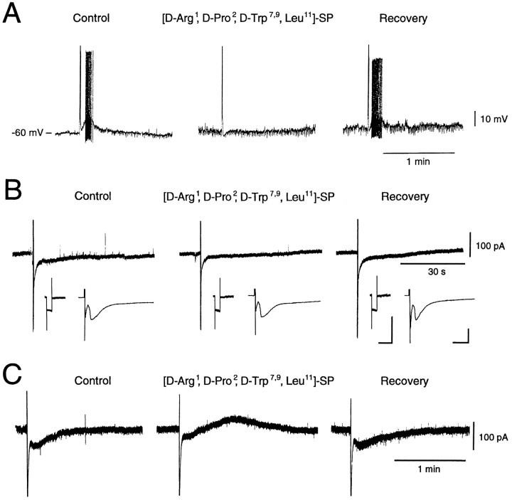
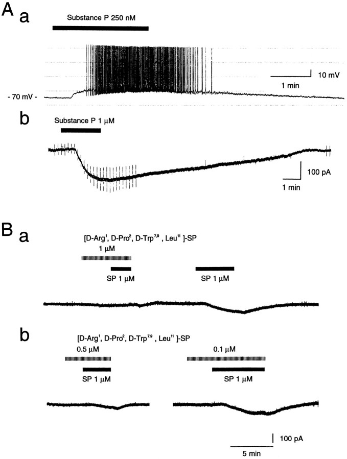
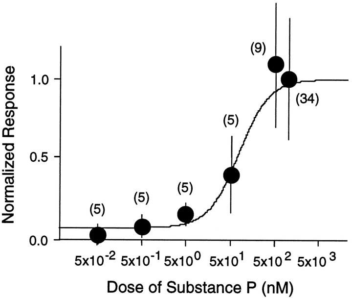
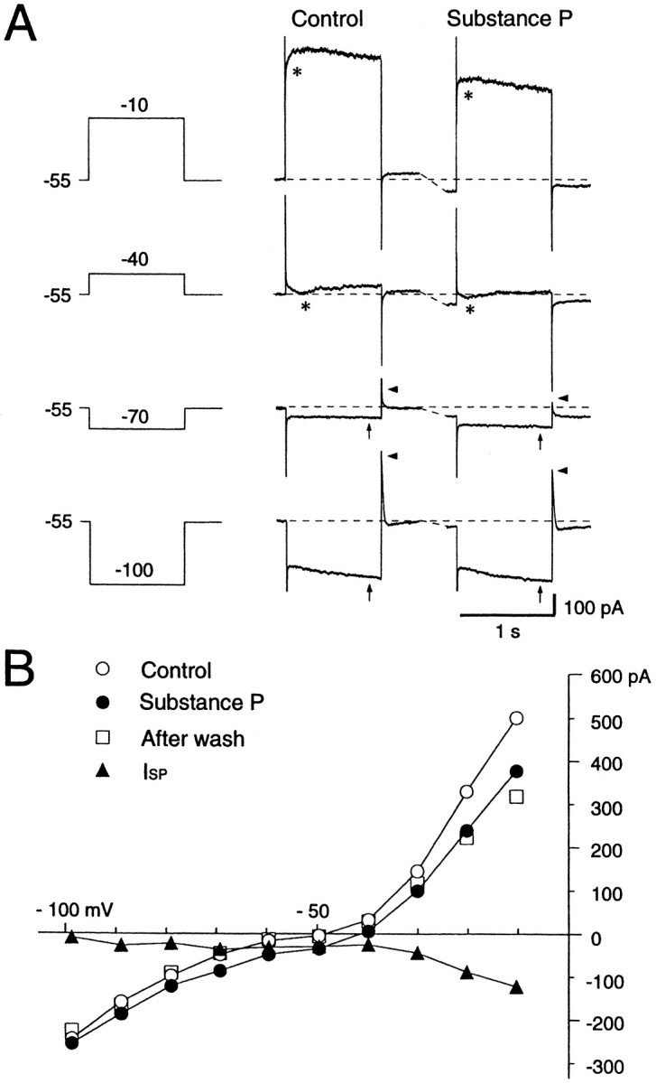
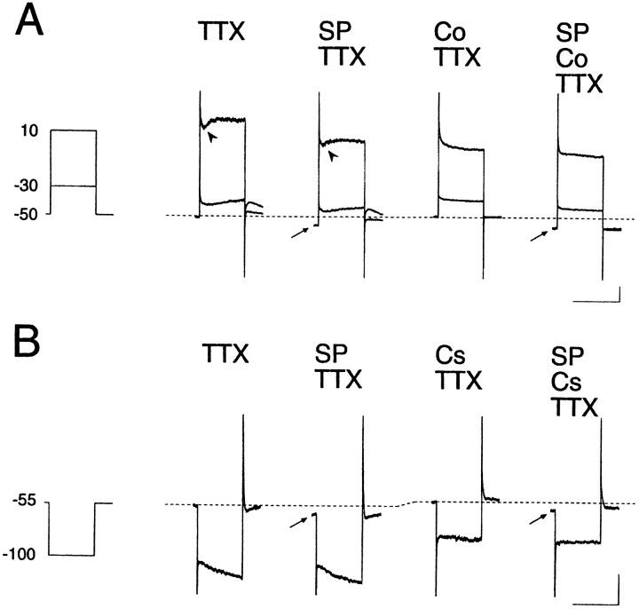
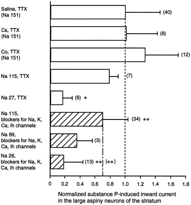
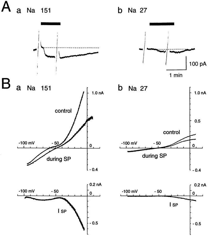
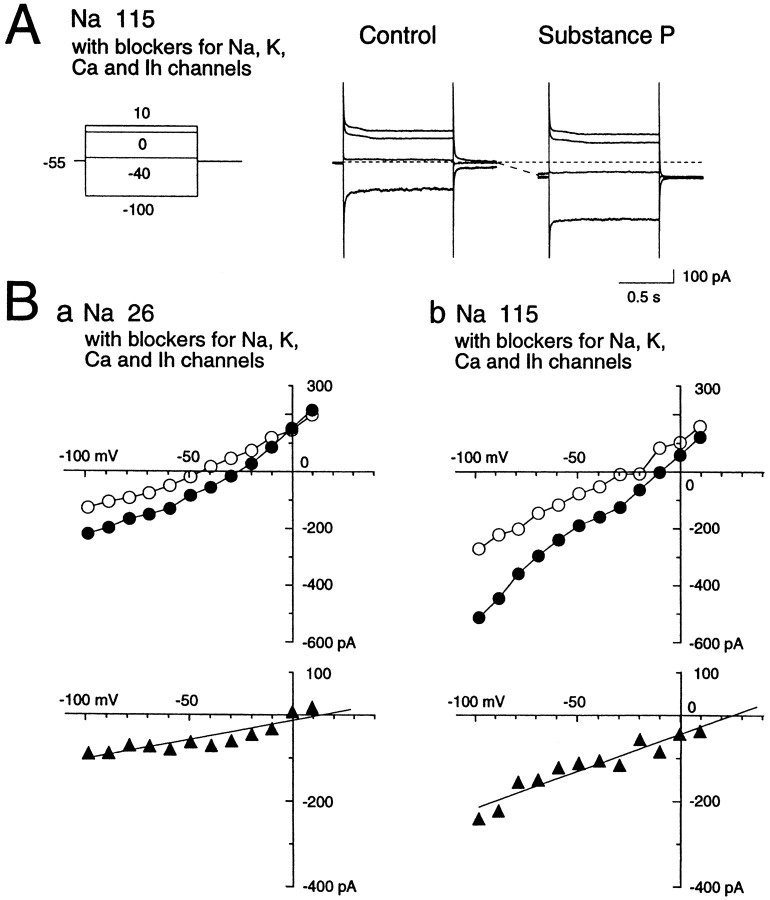
Actions of substance P (SP) on the neostriatal neurons in in vitro rat slice preparations were studied via whole-cell patch-clamp recording. Almost all large aspiny neurons (cholinergic cells) and half of the low-threshold spike (LTS) cells (somatostatin/ NOS-positive cells) showed depolarization or an inward shift of the holding currents in response to bath-applied SP in a dose-dependent manner. In contrast, no responses were observed in fast-spiking (FS) cells (parvalbumin-positive cells) and medium spiny cells. Spike discharges followed by slow EPSPs/EPSCs were evoked by intrastriatal electrical stimulation in the large aspiny neurons. Pretreatment with [D-Arg1, D-Pro2, D-Trp7,9, Leu11]-SP, an antagonist of the SP receptor, reversibly suppressed the induction of the slow EPSPs/EPSCs and unmasked slow IPSCs. The SP-induced inward current, although almost unchanged even after the blockade of Ih channels and voltage-dependent Na+, Ca2+, and K+ channels, changed its amplitude according to the Na+ concentration used in both the large aspiny neurons and LTS cells. Thus, the cation current could account for virtually all of the inward current at resting levels in both neurons. These results suggest that the firing of afferent neurons such as striatonigral medium spiny neurons, one of the possible sources of SP, would increase the firing probability of the two types of interneurons of the neostriatum by SP-receptor-mediated opening of tetrodotoxin-insensitive cation channels.


Figures

Fig. 1.
Fig. 1.
Reconstruction of a large aspiny neuron of the rat neostriatum that was stained with biocytin during whole-cell recording (A). Dendrites (a) and an axon (b) are shown separately. B, Membrane properties of the same cell in response to constant-current pulses applied intracellularly. The resting membrane potentials (approximately −60 mV), input resistances (∼430 MΩ), as well as the long-duration, large-amplitude afterhyperpolarization and the prominent sag during hyperpolarizing current pulses all fit well with the physiological properties of large aspiny neurons (long-lasting afterhyperpolarization, LA cells).
Fig. 2.
Fig. 2.
Responses of large aspiny neurons to intrastriatal stimulation. A, Intrastriatal stimulation (10 pulses of 0.5 msec duration, 20 Hz) elicited a slow EPSP with action potentials in a large aspiny neuron in the current-clamp mode. Pretreatment with [d-Arg1,d-Pro2,d-Trp7,9,Leu11]-SP (10 μm), an antagonist for the NK1 receptor, suppressed this completely and reversibly. B, Intrastriatal repetitive stimulation elicited a slow EPSP in the voltage-clamp mode (holding potential, −60 mV), and [d-Arg1,d-Pro2,d-Trp7,9,Leu11]-SP (10 μm) suppressed this reversibly.Inset: Left, Membrane currents elicited by hyperpolarizing step pulses of 5 mV. Calibration: 1 sec (horizontal), 100 pA (vertical). Note that membrane conductance did not change throughout the experiment. Right, Fast excitatory synaptic currents elicited by single test pulses of the same intensity (∼250 A) as that of repetitive pulses for the slow EPSC. Calibration: 10 msec (horizontal), 100 pA (vertical). Intensity of the stimulating pulses was adjusted to evoke fast synaptic currents but suppress spike firing in this cell. Note that the fast synaptic currents did not change their amplitude during the treatment. C, Slow postsynaptic responses consist of excitatory and inhibitory components. Treatment with [d-Arg1,d-Pro2,d-Trp7,9,Leu11]-SP (20 μm) unmasked an inhibitory component, and removal of the antagonist resulted in complete recovery.
Fig. 3.
Fig. 3.
Effects of substance P on large aspiny neurons.A, Substance P evoked depolarization in the current-clamp mode (a) and elicited an inward current in the voltage-clamp mode (−60 mV) (b). TTX (0.3 μm) was also applied in b. Note that membrane conductance increased after application of SP. Depolarizing and hyperpolarizing voltage pulses (±10 mV, 100 msec) were applied before and during SP application. B, Pretreatment with an SP antagonist suppressed the SP-induced inward current in a dose-dependent manner. The recordings in a and b are for the same cell.
Fig. 4.
Fig. 4.
Dose–response curve for substance P-induced inward current evoked at a membrane potential of −60 mV in the large aspiny neurons of the neostriatum. Each point andvertical bar represents mean ± SD. Numbersin parentheses refer to numbers of test cells.
Fig. 5.
Fig. 5.
Effects of SP on a voltage-clamped large aspiny neuron bathed in normal saline containing TTX (0.3 μm). A, Current responses to step pulses (−100, −70, −40, and −10 mV) before (Control) and during the SP application. A prominent sag (arrows) at the hyperpolarizing pulse of −100 mV and a fast outward transient (arrowheads) on cessation of hyperpolarization are present. Depolarizing step pulses more positive than −40 mV evoked an inward current (asterisks). SP evoked an inward shift of the holding current. B, Steady-stateIV curves before (○), during exposure to (•), and after washout (□) of SP. The curves were constructed from the measurements of current level attained at the end of each 1 sec hyperpolarizing or depolarizing voltage step before, during, and after SP application. The IV curve forISP was obtained by subtraction of the control from the SP values (▴). Slope conductance decreased in the suprathreshold region, increased at resting membrane potential levels, and decreased again at potentials more negative than −70 mV during SP application.
Fig. 6.
Fig. 6.
Ca2+ channels and hyperpolarization-activated cation channels (Ih) do not contribute to the SP-induced inward current. In A, Ca2+ was replaced by Co2+ (2.4 mm) in the external solution. The rapidly decaying current was suppressed by SP application (arrowheads). In B, Cs+ (2 mm) was added to the test solution. TTX was contained in the external solution at 0.3 μm in A and B. The amplitude of the inward shift of the holding potential during SP application was unchanged either in Co2+-containing or in Cs+-containing solutions (arrows). Calibration in A and B: 1 sec (horizontal), 100 pA (vertical).
Fig. 9.
Fig. 9.
SP-induced inward shift of the holding current at the resting potential in solutions of differing ionic composition. The inward shift is illustrated as the ratio to the shift recorded in the control saline solution containing TTX (0.3 μm) (mean ± SD = −81.2 ± 36.7 pA). SDs are shown withbars. Numbers in parentheses refer to numbers of test cells. The external solutions tested were: saline (Na+ 151 mm) with TTX, saline with Cs+ (2 mm) and TTX, saline with Co2+ (2 mm) and TTX, low-Na+ (115 mm) solution with TTX, low-Na+ (27 mm) solution with TTX, and solutions for study of the nonselective cation channel (hatched bars), which included TEA (30 mm), 4-AP (10 mm), Mg2+ (3.6 mm), no Ca2+, Cs+ (2 mm), and TTX. Occasionally, nifedipine (5 μm) was added. The internal solution contained Cs-methanesulfonate in the study of the nonselective cation channel. Comparisons were made with the Student’s t test against the same group of neurons tested in the control saline solution (*p < 0.05, significant; **p < 0.01, very significant) or in the solutions containing blockers for Na+, K+, Ca2+, and Ih channels ({**}p < 0.01, very significant).
Fig. 7.
Fig. 7.
Effects of SP (1 μm) in a low (27 mm)- or control (151 mm)-Na+ solution. NaCl was replaced by choline-Cl in the low-Na+ solution. Ramp voltage pulses (from −100 to 10 mV, 40 sec) were applied before and during the application of SP. TTX (0.3 m) was applied throughout the experiment. A, The SP-induced current was substantially decreased in the low-Na+solution. B, IV curves before and during SP application obtained in the normal (a)- and low (b)-Na solutions. The SP-induced current (ISP) curve was constructed by subtracting the IV curve obtained before SP application from the curve obtained during the peptide application. Note the dramatic reduction of ISP in a low-Na+ solution.
Fig. 8.
Fig. 8.
Effects of SP (1 μm) on nonselective cation channels in a low (26 mm)- or normal (115 mm)-Na solution. NaCl was replaced by choline-Cl. In this experiment, K+,Ih, Na+, and Ca2+ channel conductances were all suppressed by the addition of TEA (30 mm), 4-AP (5 mm), Cs+ (2 mm), Mg2+ (3.6 mm), or TTX (0.3 m) to the external solution, and Cs-methanesulfonate (120 mm) to the internal solution. CaCl2 was omitted from the external solution. Note the substantially smaller SP-induced current in the low-Na+ solution than in the normal-Na+ solution. ○, Control; •, substance P responses; ▴, ISP.
Fig. 10.
Fig. 10.
Actions of SP on identified LTS cells in the neostriatum. A, Morphology of an LTS cell. Dendrites (a) and an axon (b) are reconstructed.c, A burst of action potentials, a characteristic of the LTS cells, seen immediately after breaking a patch membrane in the normal saline solution. Calibration bars: 40 mV, 400 pA, 500 msec (fromtop to bottom). B, Responses to SP in an identified LTS cell in an external solution containing TEA, 4-AP, Mg2+, no Ca2+, Cs+, and TTX with Cs-methanesulfonate in the pipette. Holding potential, −55 mV. a, SP at 1 μm evoked an inward current in the LTS cell.b, IV curve before and during SP application. The same cell as in a.

References

    1. Adams PR, Brown DA, Jones SW. Substance P inhibits the M-current in bullfrog sympathetic neurones. Eur J Pharmacol. 1983;79:330–333. - PMC - PubMed
    1. Akins PT, Surmeier DJ, Kitai ST. Muscarinic modulation of a transient K+ conductance in rat neostriatal neurons. Nature. 1990;344:240–242. - PubMed
    1. Anderson JJ, Chase TN, Engber TM. Substance P increases release of acetylcholine in the dorsal striatum of freely moving rats. Brain Res. 1993;623:189–194. - PubMed
    1. Aosaki T, Kawaguchi Y. Actions of neuropeptides on large aspiny neurons of rat neostriatum in vitro. Soc Neurosci Abstr. 1995;21:913.
    1. Aosaki T, Graybiel AM, Kimura M. Effects of the nigrostriatal dopamine system on acquired neural responses in the striatum of behaving monkeys. Science. 1994a;265:412–415. - PubMed

Publication types