. 1996 Aug 15;16(16):5205-15.
doi: 10.1523/JNEUROSCI.16-16-05205.1996.

Differential sensitivity of human visual cortex to faces, letterstrings, and textures: a functional magnetic resonance imaging study

Affiliations

Differential sensitivity of human visual cortex to faces, letterstrings, and textures: a functional magnetic resonance imaging study

A Puce et al. J Neurosci. .

Abstract

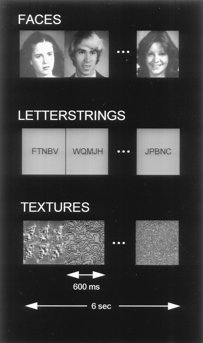
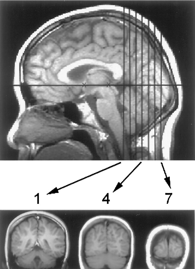
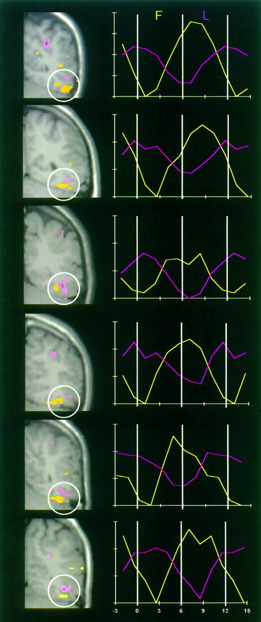
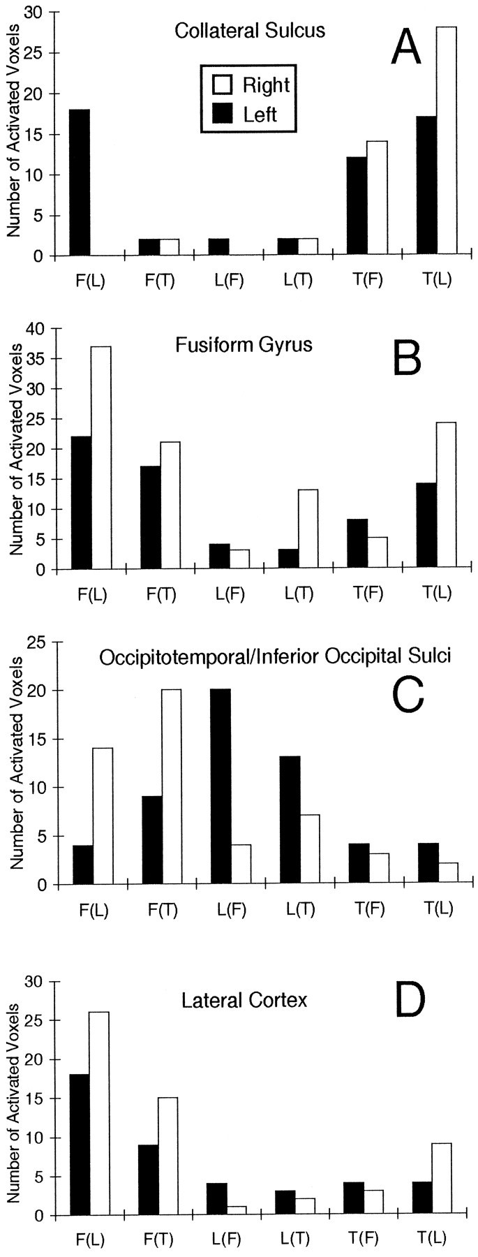
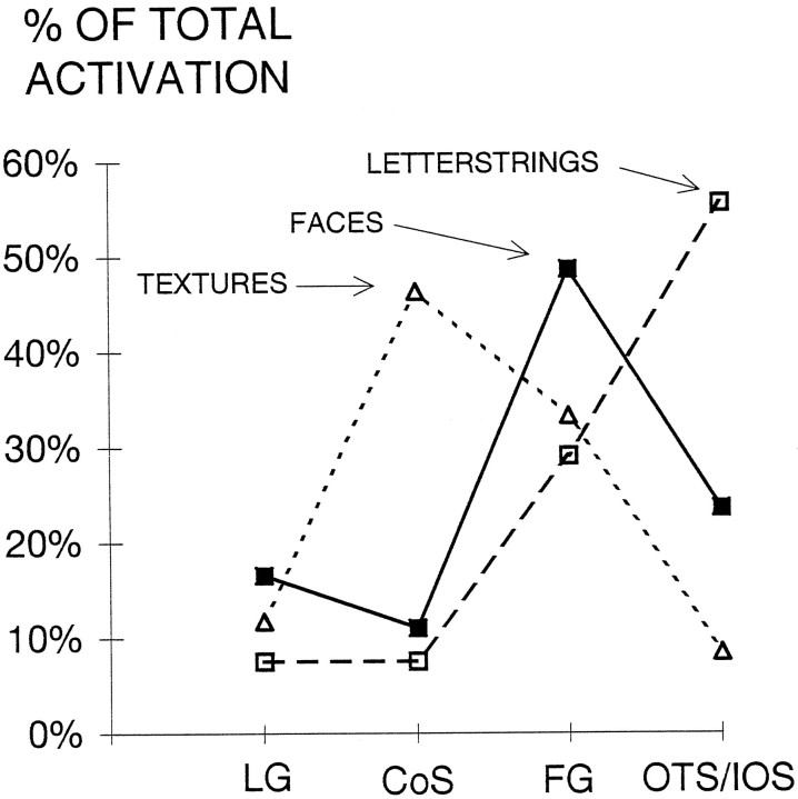
Twelve normal subjects viewed alternating sequences of unfamiliar faces, unpronounceable nonword letterstrings, and textures while echoplanar functional magnetic resonance images were acquired in seven slices extending from the posterior margin of the splenium to near the occipital pole. These stimuli were chosen to elicit initial category-specific processing in extrastriate cortex while minimizing semantic processing. Overall, faces evoked more activation than did letterstrings. Comparing hemispheres, faces evoked greater activation in the right than the left hemisphere, whereas letterstrings evoked greater activation in the left than the right hemisphere. Faces primarily activated the fusiform gyrus bilaterally, and also activated the right occipitotemporal and inferior occipital sulci and a region of lateral cortex centered in the middle temporal gyrus. Letterstrings primarily activated the left occipitotemporal and inferior occipital sulci. Textures primarily activated portions of the collateral sulcus. In the left hemisphere, 9 of the 12 subjects showed a characteristic pattern in which faces activated a discrete region of the lateral fusiform gyrus, whereas letterstrings activated a nearby region of cortex within the occipitotemporal and inferior occipital sulci. These results suggest that different regions of ventral extrastriate cortex are specialized for processing the perceptual features of faces and letterstrings, and that these regions are intermediate between earlier processing in striate and peristriate cortex, and later lexical, semantic, and associative processing in downstream cortical regions.


Figures

Fig. 1.
Fig. 1.
Examples of stimuli (unfamiliar faces, unpronounceable nonwords, and textures) used in this study. In experiment 1, faces were alternated with letterstrings. In experiment 2, textures were alternated with faces or letterstrings in separate imaging runs. Ten stimuli in each category were presented in each 6.0 sec epoch, followed by 10 stimuli of another category.
Fig. 2.
Fig. 2.
Midline sagittal T1-weighted image of the brain showing seven coronal slices. The center of the first slice was located at the posterior margin of the splenium (indicated by thefirst vertical black line). Examples of slices 1, 4, and 7 are shown below the sagittal image. The subject’s head was positioned using the canthomeatal line. In most subjects, this corresponded to the line between the anterior and posterior commissures (shown as black Xs). Average anterio-posterior locations of slices 1–7 in Talairach coordinates were, respectively,y = −42, −49, −56, −63, −70, −77, and −84. The temporal lobe anterior to slice 1 could not be imaged reliably because of susceptibility artifact from the ear canals.
Fig. 3.
Fig. 3.
Activation in a subject (slice 4) in experiment 1. A, Activated voxels identified by split-t test for faces (white) and letterstrings (black) are shown superimposed on a T1-weighted coronal anatomical image. In this and subsequent figures, the right side of the brain is represented on the left side of the image. In the left hemisphere, a region at the border of the temporal and occipital lobes (white circle) was activated by faces in the fusiform gyrus and by letterstrings in the occipitotemporal sulcus. B, Activated voxels for faces (white) and letterstrings (black) identified by the frequency analysis are superimposed on the anatomical image. C, Time course of signal change for the voxels activated by faces (whitevoxels in circle) from B for both task orders. Percent signal change (ΔS/S) is shown on the y-axis. Vertical bars indicate onsets of the 14 successive stimulus cycles. The F–L and L–F conditions are represented by the solid and dotted lines, respectively. D, Time course of signal change for the voxels activated by letterstrings (blackvoxels in circle) from B for both task orders. E, Power spectra corresponding to the activation time course shown in C. Note the peak at the stimulus alternation frequency (0.083 Hz). Power (in arbitrary units) is shown on the y-axis. F, Power spectra corresponding toD. G, Power at the stimulus alternation frequency as a function of phase (in bins of 20°) for the activation time course shown in C. Note that the change in stimulus order produces a phase shift of 180°. H, Power at the stimulus alternation frequency as a function of phase for the activation time course shown in D.
Fig. 4.
Fig. 4.
Average t test data of 12 subjects in the F–L condition overlaid on five averaged anatomical images.A is the most anterior and E the most posterior slice. Activation by faces is indicated by theyellowred color scale, and activation by letterstrings is indicated by the pinkpurplecolor scale.
Fig. 5.
Fig. 5.
Activation data from the ventral left hemisphere (white circle) in six subjects. In the leftcolumn, activated voxels identified by split-t test analysis of the F–L condition are superimposed on corresponding anatomical images. Faces activated a region of the lateral fusiform gyrus (yellow), whereas letterstrings activated a region of the occipitotemporal sulcus (pink). The rightcolumn shows the corresponding activation time course averaged over all alternation cycles for faces (yellow lines) and letterstrings (pink lines). They-axis shows percent signal change with vertical ticks of 0.5%. Time (in sec) is shown on the x-axis.
Fig. 6.
Fig. 6.
Histograms of activated voxels (identified by frequency analysis) in regions of left and right cortices.A, Collateral sulcus. B, Fusiform gyrus.C, Combined occipitotemporal and inferior occipital sulci.D, Combined middle temporal and occipital gyri, and superior temporal and lateral occipital sulci.
Fig. 7.
Fig. 7.
Activation in ventral extrastriate regions for each stimulus category, expressed as a percentage of total activation by that stimulus category. CoS, Collateral sulcus;FG, fusiform gyrus; LG, lingual gyrus;OTS/IOS, occipitotemporal and inferior occipital sulci.

References

    1. Albright TD. Cortical processing of visual motion. In: Miles FA, Wallman J, editors. Visual motion and its role in the stabilization of gaze. Elsevier; New York: 1993. pp. 177–201. - PubMed
    1. Allison T, Begleiter A, McCarthy G, Roessler E, Nobre AC, Spencer DD. Electrophysiological studies of color processing in human visual cortex. Electroencephalogr Clin Neurophysiol. 1993;88:343–355. - PubMed
    1. Allison T, Ginter H, McCarthy G, Nobre A, Puce A, Luby M, Spencer DD. Face recognition in human extrastriate cortex. J Neurophysiol. 1994a;71:821–825. - PubMed
    1. Allison T, McCarthy G, Nobre A, Puce A, Belger A. Human extrastriate visual cortex and the perception of faces, words, numbers, and colors. Cereb Cortex. 1994b;5:544–554. - PubMed
    1. Allison T, McCarthy G, Belger A, Puce A, Luby M, Kim J, Spencer DD, Bentin S (1995) What is a face? Electrophysiological responsiveness of human extrastriate visual cortex to human and animal faces, face components, and complex objects. Proc Cognit Neurosci Soc, San Francisco, p 49.

Publication types