Comparative Study
. 1996 Sep 1;16(17):5393-404.
doi: 10.1523/JNEUROSCI.16-17-05393.1996.

Mechanisms of H+ and Na+ changes induced by glutamate, kainate, and D-aspartate in rat hippocampal astrocytes

Affiliations
Comparative Study

Mechanisms of H+ and Na+ changes induced by glutamate, kainate, and D-aspartate in rat hippocampal astrocytes

C R Rose et al. J Neurosci. .

Abstract

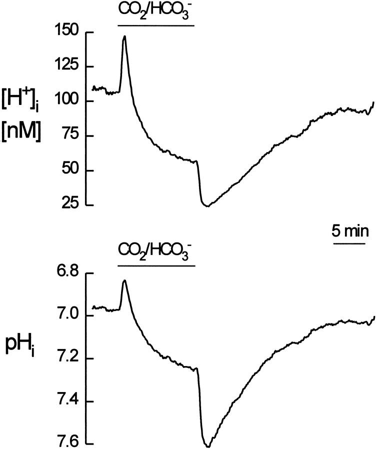
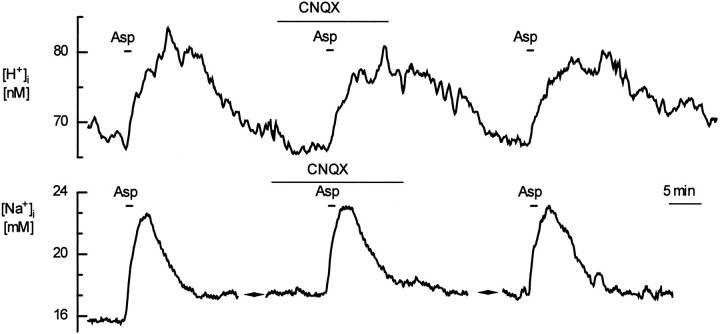
The excitatory transmitter glutamate (Glu), and its analogs kainate (KA), and D-aspartate (D-Asp) produce significant pH changes in glial cells. Transmitter-induced pH changes in glial cells, generating changes in extracellular pH, may represent a special form of neuronal-glial interaction. We investigated the mechanisms underlying these changes in intracellular H+ concentration ([H+]i) in cultured rat hippocampal astrocytes and studied their correlation with increases in intracellular Na+ concentration ([Na+]i), using fluorescence ratio imaging with 2',7'-bis(carboxyethyl)-5,6-carboxyfluorescein (BCECF) or sodium-binding benzofuran isophthalate (SBFI). Glu, KA, or D-Asp evoked increases in [Na+]i; Glu or D-Asp produced parallel acidifications. KA, in contrast, evoked biphasic changes in [H+]i, alkaline followed by acid shifts, which were unaltered after Ca2+ removal and persisted in 0 CI(-)-saline, but were greatly reduced in CO2/HCO3(-)-free or Na(+)-free saline, or during 4,4'-diisothiocyanato-stilbene-2,2'-disulphonic acid (DIDS) application. The non-NMDA receptor antagonist 6-cyano-7-nitroquinoxaline-2,3-dione (CNQX) blocked KA-evoked changes in [H+]i and [Na+]i, indicating that they were receptor-ionophore mediated. In contrast, CNQX increased the [H+]i change and decreased the [Na+]i change induced by Glu. D-Asp, which is transported but does not act at Glu receptors, induced [H+]i and [Na+]i changes that were virtually unaltered by CNQX. Our study indicates that [Na+]i increases are not primarily responsible for Glu- or KA-induced acidifications in astrocytes. Instead, intracellular acidifications evoked by Glu or D-Asp are mainly caused by transmembrane movement of acid equivalents associated with Glu/Asp-uptake into astrocytes. KA-evoked biphasic [H+]i changes, in contrast, are probably attributable to transmembrane ion movements mediated by inward, followed by outward, electrogenic Na+/HCO3- cotransport, reflecting KA-induced biphasic membrane potential changes.


Figures

Fig. 1.
Fig. 1.
Model showing possible interrelationships between Glu-induced ion fluxes and [H+]i regulation in hippocampal astrocytes. Glu andd-Asp are substrates for the electrogenic Glu transporter (A, hatched area), which exchanges extracellular Glu ord-Asp and 2 Na+ for intracellular K+ and OH. This causes cellular depolarization and acidification. Activation of ionotropic non-NMDA receptors by Glu or KA (indicated by arrowhead) depolarizes the cells by influx of Na+, and sometimes Ca2+, and efflux of K+ through cation channels (A). Various secondary changes in [H+]i could be caused by these actions of Glu. Additional intracellular acidification could originate secondary to increase in intracellular Ca2+ activating a Ca2+/H+ pump in the plasma membrane (B), and/or because of the increase in [Na+]i attenuating cellular acid secretion by Na+/H+ exchange and Na+-dependent Cl/HCO3 exchange (C). Cellular depolarization could cause intracellular alkalinization by stimulating inwardly directed electrogenic Na+/HCO3 cotransport (C); a late hyperpolarization follows a period of elevated [Na+]i attributable to Na+, K+-ATPase activity (D), and this could activate outwardly directed Na+/HCO3 cotransport resulting in intracellular acidification.
Fig. 2.
Fig. 2.
Relationship between pHi and intracellular H+ concentration. The distortion caused by presenting changes in intracellular H+ concentration ([H+]i) as changes in pHi is illustrated here. A hippocampal astrocyte was switched from a HEPES-buffered, CO2/HCO3-free saline to a CO2/HCO3-buffered saline and back. The changes in [H+]i are shown along with the changes in pHi. In the pHitracing, the absolute magnitude of the alkaline shift appears larger than the acid shift, whereas the [H+]i trace reveals just the opposite. To avoid these distortions, all subsequent data are presented as [H+]i.
Fig. 3.
Fig. 3.
[H+]i changes evoked by Glu, KA, or d-Asp in CO2/HCO3-containing or CO2/HCO3-free solution.A illustrates the typical changes in intracellular H+ concentration (Δ[H+]i) caused by bath application of Glu, KA, or d-Asp in standard, CO2/HCO3-buffered saline. Substances were applied for 1 min (indicated bybars) at a concentration of 1 mm. These recordings were from three different cells. To facilitate comparison, baseline [H+]i was set to 0 in all cells.B, Record showing changes [H+]i caused by 1 min bath application (indicated by bars) of KA or Glu (1 mm) in HEPES-buffered, CO2/HCO3-free saline, and CO2/HCO3-buffered saline.
Fig. 4.
Fig. 4.
Concentration dependence of kainate-induced [H+]i changes. A, The biphasic KA-induced responses in [H+]iincreased with KA concentration. All applications were for 2 min periods; KA was applied at 0.1, 0.5, and 1 mm(indicated by bars). B, Graphic summary of the amplitudes of biphasic alkaline-acid transients elicited by 2 min bath applications of KA at 0.1, 0.5, 1, and 2 mm in CO2/HCO3-buffered saline. Shown are the mean values of 49 cells; bars indicate SD.
Fig. 5.
Fig. 5.
Ca2+- and Cl-dependence of kainate-induced [H+]i changes. A, Alkaline-acid transients elicited by 1 mm KA applications for 1 min (indicated by bars) in standard CO2/HCO3-buffered saline (2 mm Ca2+) and after removal of extracellular Ca2+ (0 [Ca2+]e, solution contained 0.5 mm EGTA). The KA response was unchanged in the absence of [Ca2+]e.B, Alkaline-acid transients elicited by 1 mmKA application for 1 min (indicated by bars) in standard CO2/HCO3-buffered saline (containing 122.75 mm Cl) and after replacement of extracellular Cl by gluconic acid (0 [Cl]e). Removing Cl caused an alkaline shift probably because of reverse Cl/HCO3 exchange. Note that the KA response was preserved qualitatively in the absence of [Cl]e.
Fig. 6.
Fig. 6.
Na+ dependence and influence of DIDS on kainate-induced [H+]i changes.A, Alkaline-acid transients elicited by 1 mmKA application for 2 min (indicated by bars) in standard CO2/HCO3-buffered saline and after replacement of extracellular Na+ by NMDG and choline (0 [Na+]e). Removal of Na+ caused a marked acid shift, because it blocks or reverses acid-exporting mechanisms. The KA-induced changes in [H+]i were reversibly blocked in the absence of Na+. B, Alkaline-acid transients elicited by 1 mm KA application for 1 min (indicated bybars) are shown in standard CO2/HCO3-buffered saline and during application of the anion transport blocker DIDS (0.5 mm). DIDS caused a partially reversible acid shift, because it blocks acid- exporting mechanisms. The KA response was largely blocked by DIDS.
Fig. 7.
Fig. 7.
Neuronal [H+]i changes induced by KA. KA application (1 mm for 1 min) (indicated by bar) rapidly increased [H+]i in a cultured hippocampal neuron in CO2/HCO3-buffered saline. Unlike the situation in astrocytes, KA never evoked alkaline shifts in neurons.
Fig. 8.
Fig. 8.
Comparison between glutamate- and kainate-induced [Na+]i and [H+]itransients. A, Recordings showing changes in intracellular Na+ and H+ concentrations (Δ[Na+]i, Δ[H+]i) induced by application of Glu (1 mm for 1 min) (indicated by bars) in CO2/HCO3-buffered saline (solid lines) and CO2/HCO3-free saline (dashed lines). Although Glu-induced [H+]i changes were greatly altered when switching between CO2/HCO3-free and CO2/HCO3-containing solution, very small alterations were seen in the induced changes in [Na+]i. B, Recordings showing KA-induced [Na+]i and [H+]i changes. KA (1 mm) was applied for 1 min as indicated by the bars. Again, the significant changes in KA-induced [H+]i transients caused by switching from CO2/HCO3-containing solution to CO2/HCO3-free solution were not associated with significant changes in the [Na+]i transients. A,B, Na+ and H+ measurements were obtained from different cells.
Fig. 10.
Fig. 10.
CNQX reduced the increase in [Na+]i induced by Glu, but not the increase in [H+]i. Glu-induced [H+]i and [Na+]itransients are shown in standard CO2/HCO3-buffered saline and during application of the ionotropic, non-NMDA receptor blocker CNQX (25 μm) (solid bars). Glu (1 mm) was applied for 1 min (indicated by short bars). The recordings were obtained from two different cells, and two segments were deleted from the lower trace(arrows) to enable better comparison between the traces.
Fig. 9.
Fig. 9.
CNQX blocked kainate-induced [H+]i and [Na+]itransients. KA-induced [H+]i and [Na+]i transients are shown in standard CO2/HCO3-buffered saline and during application of the ionotropic, non-NMDA receptor blocker CNQX (25 μm) (solid bars). KA (1 mm) was applied for 1 min (upper trace) or 2 min (lower trace, indicated by short bars). The recordings were obtained from two different cells; note the different time scales. CNQX completely blocked both the KA-induced [H+]i and [Na+]i transients.
Fig. 11.
Fig. 11.
CNQX had only minor effects on aspartate-induced [H+]i and [Na+]itransients. [H+]i and [Na+]i transients were induced byd-Asp (Asp) in standard CO2/HCO3-buffered saline and during application of CNQX (25 μm) (solid bars). d-Asp (1 mm) was applied for 1 min (short solid bars). The recordings were obtained from two different cells, and two segments were deleted from thelower trace (arrowheads) to enable better comparison between the traces.
Fig. 12.
Fig. 12.
Proposed voltage shifts and transmembrane Na+/HCO3 movements in astrocytes during kainate application in CO2/HCO3-buffered saline.A, Model of KA-induced changes in membrane potential, [Na+]i, and Na+/HCO3-cotransporter reversal potential in astrocytes. KA causes depolarization and increases [Na+]i (1). This is followed by Na+ pump stimulation leading to hyperpolarization and normalization of [Na+]i(2). The reversal potential of Na+/HCO3 cotransport is altered by the changes in [Na+]i, illustrated by the hypothetical curves to the right. The upper trace shows the presumed changes in membrane potential (Em) resulting from KA application (1 mm for 1 min) (indicated bybars) (see also Bowman and Kimelberg, 1984; Kettenmann and Schachner, 1985; Backus et al., 1989). The middle trace shows the averaged KA-induced [Na+]i change of six representative cells. The lower trace shows changes in the reversal potential of Na+/HCO3 cotransport (Erev), calculated from the average [Na+]i change in the middle trace (see Discussion). B, Proposed mechanism of the alkaline-acid transients seen in astrocytes resulting from KA application (1 mm for 1 min) (indicated bybars). The upper trace shows changes in the Erev (solid line). Superimposed on the Erev trace is the presumed change in Em induced by KA (dashed line) (see above). During the fast, KA-induced membrane depolarization,Em is more positive thanErev, favoring influx of Na+/HCO3 and, therefore, intracellular alkalinization. During repolarization and hyperpolarization of the membrane, Em is more negative than Erev, because of the relatively slower recovery of [Na+]i, favoring efflux of Na+ and HCO3 and intracellular acidification. The lower trace shows the KA-induced biphasic alkaline-acid shift in [H+]i averaged from six representative cells.

References

    1. Amato A, Ballerini L, Attwell D. Intracellular pH changes produced by glutamate uptake in rat hippocampal slices. J Neurophysiol. 1994;72:1686–1696. - PubMed
    1. Aronson PS. Kinetic properties of the plasma membrane Na+-H+ exchanger. Annu Rev Physiol. 1985;47:545–560. - PubMed
    1. Backus KH, Kettenmann H, Schachner M. Pharmacological characterization of the glutamate receptor in cultured astrocytes. J Neurosci Res. 1989;22:274–282. - PubMed
    1. Ballanyi K, Kettenmann H. Intracellular Na+activity in cultured mouse oligodendrocytes. J Neurosci Res. 1990;26:455–460. - PubMed
    1. Ballanyi K, Schlue WR, Dorner R. Glutamate and kainate increase intracellular sodium activity in leech neuropile glial cells. Glia. 1989;2:51–54. - PubMed

Publication types

MeSH terms

LinkOut - more resources