. 1996 Sep 1;16(17):5466-77.
doi: 10.1523/JNEUROSCI.16-17-05466.1996.

Regenerative proliferation in organ cultures of the avian cochlea: identification of the initial progenitors and determination of the latency of the proliferative response

Affiliations

Regenerative proliferation in organ cultures of the avian cochlea: identification of the initial progenitors and determination of the latency of the proliferative response

M E Warchol et al. J Neurosci. .

Abstract

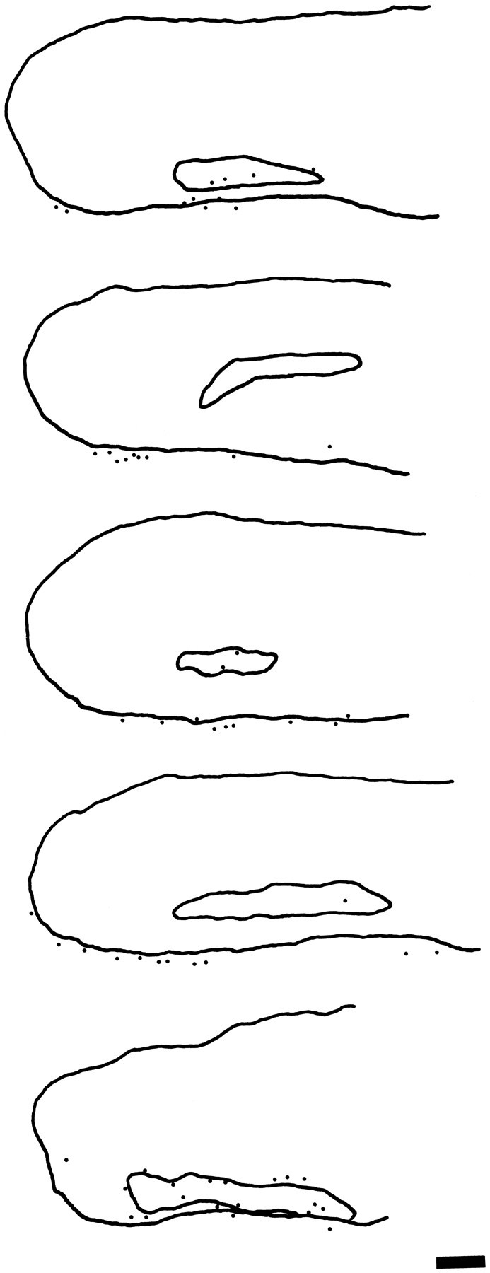
Sensory hair cells in the cochleae of birds are regenerated after the death of preexisting hair cells caused by acoustic over-stimulation or administration of ototoxic drugs. Regeneration involves renewed proliferation of cells in an epithelium that is otherwise mitotically quiescent. To determine the identity of the first cells that proliferate in response to the death of hair cells and to measure the latency of this proliferative response, we have studied hair-cell regeneration in organ culture. Cochleae from hatchling chicks were placed in culture, and hair cells were killed individually by a laser microbeam. The culture medium was then replaced with a medium that contained a labeled DNA precursor. The treated cochleae were incubated in the labeling media for different time periods before being fixed and processed for the visualization of proliferating cells. The first cells to initiate DNA replication in response to the death of hair cells were supporting cells within the cochlear sensory epithelium. All of the labeled supporting cells were located within 200 microns of the hair-cell lesions. These cells first entered S-phase approximately 16 hr after the death of hair cells. The results indicate that supporting cells are the precursors of regenerated hair cells and suggest that regenerative proliferation of supporting cells is triggered by signals that act locally within the damaged epithelium.


Figures

Fig. 1.
Fig. 1.
Photomicrograph showing a portion of the inferior region of a living chick cochlea after 24 hr in culture in a Rose chamber. The specimen was viewed on an inverted microscope using a 100× objective lens. Hair cells and supporting cells in the cultured explants retain their normal morphology and can be visualized easily for laser ablation (see text). The nuclei of the hyaline cells are visible near the lower left corner. Scale bar, 20 μm.
Fig. 2.
Fig. 2.
Cells in living cultures of the chick cochlea can be individually lesioned using a laser microbeam. A, Cuticular plates of hair cells in the distal/inferior portion of a living chick cochlea after 24 hr in culture. B, Photo of the same region 5 min after seven hair cells were killed individually using a pulsed laser microbeam. Arrows indicate the borders of the lesion. Note that the hair cells are completely missing from the cellular mosaic of the sensory epithelium, but that other cells appear unaffected. Scale bar, 20 μm.
Fig. 3.
Fig. 3.
Patterns of hair-cell lesions used in the experiments. All hair-cell lesions were confined to the distal-most 1200 μm of the cochlear sensory epithelium. A, One pattern of hair-cell lesion consisted of 100 hair cells that were arranged in a 10 × 10 hair cell square and located in the mid-inferior region of the sensory epithelium. The cells were killed individually with the laser microbeam (see text). B, The second type of lesion was composed of 250 hair cells, in a 5 × 50 hair cell array that originated near the distal tip of the epithelium. Experimental cochleae received one or the other of these lesions.
Fig. 4.
Fig. 4.
A, Autoradiograph showing a section from a chick cochlea 8 hr after the laser ablation of hair cells. Hair cells are missing from a region near the center of the epithelium.B, Higher-power photomicrograph of the lesioned region in the same specimen. Normal-appearing supporting cells are present within the lesion. Note also the presence of extruded hair cells directly above the lumen of the lesioned portion of the sensory epithelium. Scale bars: A, 50 μm; B, 10 μm.
Fig. 5.
Fig. 5.
Autoradiographs of sections from chick cochleae that were incubated in medium that contained [3H]thymidine for 16 hr after hair-cell lesions. Labeled supporting cells (arrows) were present within (A) and directly adjacent to (B) the lesioned areas. No lesion-related labeling of hyaline cells was present in these specimens. Scale bar, 20 μm.
Fig. 6.
Fig. 6.
Labeled cells in whole mounts of cultured chick cochleae that were incubated in medium that contained the mitotic tracer BrdU for 16 hr after hair-cell lesions. The lower dashed lines in each photograph indicate the inferior borders of the sensory epithelia (the line separating border cells from hyaline cells). The other dashed lines indicate the borders of the hair-cell lesions, which were determined by microscopic examination of the whole-mount specimens. Labeled supporting cells were present in the lesioned regions of the sensory epithelia (arrows). Scale bar, 50 μm.
Fig. 7.
Fig. 7.
Plots showing the distribution of labeled supporting cells and labeled hyaline cells along the length of the sensory epithelia of two cultured chick cochleae after incubation in [3H]thymidine for 16 hr after the laser ablation of hair cells. Labeled cells in the autoradiographic sections were counted every 15 μm over a total distance of 600 μm. In both plots, thex-axis denotes longitudinal distance along the cochlea, beginning ∼200 μm from the distal tip and proceeding proximally. Labeled supporting cells were present only near the lesion sites. In contrast, labeled hyaline cells were commonly present outside the sensory region of the cochlea, but were not concentrated near the sites of the hair-cell lesions.
Fig. 8.
Fig. 8.
Autoradiographs of sections of chick cochleae that were incubated in medium that contained [3H]thymidine for 24 hr after the lesioning of hair cells. Labeled supporting cells are indicated by arrows. The pattern of mitotic labeling that was present after 24 hr incubations was similar to that present after the 16 hr incubations. Labeled supporting cells were present within and near the hair-cell lesions. Scale bar, 20 μm.
Fig. 9.
Fig. 9.
Photographs of whole mounts of cochleae that were cultured in medium that contained BrdU for 24 hr after the lesioning of hair cells. The inferior borders of the sensory epithelia are oriented toward the bottom of both photographs and are indicated by thelower dashed lines. The upper dashed lines indicate the borders of the hair-cell lesions. Labeled supporting cells are present in the sensory epithelia of both specimens (arrows). Labeled cells are also present in the region outside of the inferior boundary of the sensory epithelia (A). Scale bar, 50 μm.
Fig. 10.
Fig. 10.
Plots showing the distribution of labeled supporting cells and labeled hyaline cells along the length of the sensory epithelia in two cultured chick cochleae after incubation in [3H]thymidine for 24 hr after the laser ablation of hair cells. Labeled cells in autoradiographic sections were counted every 15 μm over a total distance of 600 μm. In both plots, thex-axis denotes longitudinal distance along the cochleae, beginning ∼200 μm from the distal tip and proceeding proximally. The general patterns of cell labeling were similar to those observed after 16 hr incubations (Fig. 8), although labeled cells were more numerous. Labeled supporting cells were concentrated near the lesion sites, whereas labeled hyaline cells were distributed along the entire inferior edge.
Fig. 11.
Fig. 11.
Camera lucida drawings of whole mounts of cultured cochleae that were incubated in medium that contained BrdU for 16 hr after the lesioning of hair cells. In all cases, the cochleae are drawn with their inferior borders oriented downward. The outlines of the lesioned areas within the sensory epithelia are also indicated. Labeled supporting cells were present either within or near the hair-cell lesions. Labeled hyaline cells (see text) were also present in these specimens, but were not concentrated near the lesioned areas. Scale bar, 100 μm.
Fig. 12.
Fig. 12.
Camera lucida drawings of whole mounts of cochleae that were cultured in media that contained BrdU for 24 hr after the lesioning of hair cells. The cochleae are oriented with their inferior borders downward, and the boundaries of the hair cells lesions are traced within the outlines of the sensory epithelia. The patterns of mitotic labeling after 24 hr incubations were similar to those present after 16 hr incubations (Fig. 11). Labeled supporting cells were present within the lesions and at short distances from the lesions. Although labeled hyaline cells were present in all specimens, they were not concentrated near the lesion sites. Scale bar, 100 μm.
Fig. 13.
Fig. 13.
Plot showing the numbers and distribution of BrdU-labeled supporting cells and hyaline cells in 10 unlesioned cochleae. Cochlear cultures were prepared as described in the text but did not receive hair-cell lesions. The mitotic tracer BrdU was added to the medium for the final 24 hr of culture. After fixation and histochemical processing, counts of BrdU-labeled supporting cells and hyaline cells were made from 10 adjoining 100 μm regions of the cochleae, beginning at the distal tip and extending 1000 μm toward the proximal end. Proliferating supporting cells were rare in unlesioned cochleae, except near the distal tip. In contrast, the numbers of proliferating hyaline cells were approximately uniform along the length of the unlesioned specimens. Mean ± SEM.

Similar articles

Cited by

References

    1. Balak KJ, Corwin JT, Jones JE. Regenerated hair cells can originate from supporting cell progeny: evidence from phototoxicity and laser ablation experiments in the lateral line system. J Neurosci. 1990;10:2505–2512. - PMC - PubMed
    1. Berns MW, Aist J, Edwards J, Strahs K, Girton J, McNeill P, Rattner JB, Kitzes M, Hammer-Wilson M, Liaw L-H, Siemens A, Koonce M, Peterson S, Brenner S, Burt J, Walter R, Bryant PJ, van Dyk D, Coulombe J, Cahill T, Berns GS. Laser microsurgery in cell and developmental biology. Science. 1981;213:505–513. - PubMed
    1. Bhave SA, Stone JS, Rubel EW, Coltrera MD. Cell cycle progression in gentamicin-damaged avian cochleas. J Neurosci. 1995;15:4618–4628. - PMC - PubMed
    1. Brooks RF. Regulation of the fibroblast cell cycle by serum. Nature. 1976;260:248–250. - PubMed
    1. Bunting EC, Cotanche DA, Girod DA. The role of hyaline cell migration in cochlear hair cell regeneration in chick basilar papilla following severe noise damage. Assoc Res Otolaryngol Abstr. 1996;19:15.

Publication types

LinkOut - more resources