

Anatomical demonstration of ocular dominance columns in striate cortex of the squirrel monkey
- PMID: 8757263
- PMCID: PMC6578890
- DOI: 10.1523/JNEUROSCI.16-17-05510.1996
Anatomical demonstration of ocular dominance columns in striate cortex of the squirrel monkey
Abstract
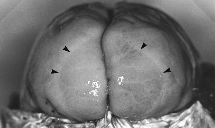
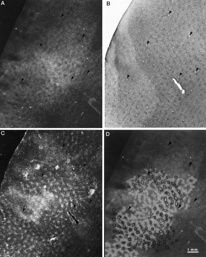
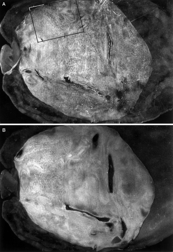
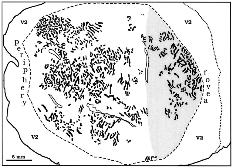
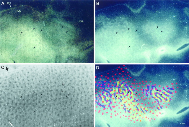
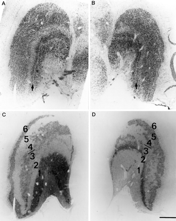
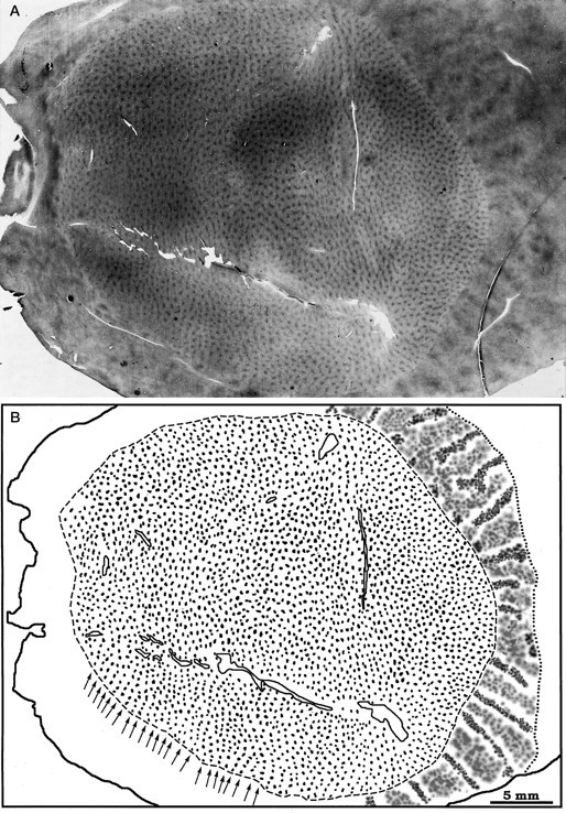
The squirrel monkey is the only primate reported to lack ocular dominance columns. Nothing anomalous about the visual capacity of squirrel monkeys has been found to explain their missing columns, leading to the suggestion that ocular dominance columns might be "an epiphenomenon, not serving any purpose" (Livingstone et al., 1995). Puzzled by the apparent lack of ocular dominance columns in squirrel monkeys, we made eye injections with transneuronal tracers in four normal squirrel monkeys. An irregular mosaic of columns, averaging 225 microns in width, was found throughout striate cortex. They were double-labeled by placing wheat germ agglutinin-horseradish peroxidase into the left eye and [3H]proline into the right eye. The tracers labeled opposite sets of interdigitating columns, proving they represent ocular dominance columns. The columns were much clearer in layer IVc alpha (magno-receiving) than IVc beta (parvo-receiving). In the lateral geniculate body, the parvo laminae showed extensive mixing of ocular inputs, suggesting that increased label spillover contributes to the blurred columns in layer IVc beta. The cytochrome oxidase (CO) patches were organized into distinct rows, but they bore no consistent relationship to the ocular dominance columns. These experiments indicate that ocular dominance columns are less well segregated in squirrel monkeys than macaques, but they are present. This fact is pertinent to a recent study reporting that ocular dominance columns are absent in normal squirrel monkeys, but induced to form by strabismus (Livingstone, 1996).
Figures
Similar articles
-
Ocular dominance columns: enigmas and challenges.Neuroscientist. 2009 Feb;15(1):62-77. doi: 10.1177/1073858408327806. Neuroscientist. 2009. PMID: 19218231 Free PMC article. Review.
-
Anatomical consequences of long-term monocular eyelid closure on lateral geniculate nucleus and striate cortex in squirrel monkey.J Comp Neurol. 1984 Jul 20;227(1):1-13. doi: 10.1002/cne.902270103. J Comp Neurol. 1984. PMID: 6088593
-
An adult-like pattern of ocular dominance columns in striate cortex of newborn monkeys prior to visual experience.J Neurosci. 1996 Mar 1;16(5):1791-807. doi: 10.1523/JNEUROSCI.16-05-01791.1996. J Neurosci. 1996. PMID: 8774447 Free PMC article.
-
Ocular dominance columns in strabismus.Vis Neurosci. 2006 Sep-Oct;23(5):795-805. doi: 10.1017/S0952523806230116. Vis Neurosci. 2006. PMID: 17020634
-
Ocular integration in the human visual cortex.Can J Ophthalmol. 2006 Oct;41(5):584-93. doi: 10.1016/S0008-4182(06)80027-X. Can J Ophthalmol. 2006. PMID: 17016529 Review.
Cited by
-
Overall patterns of eye-specific retino-geniculo-cortical projections to layers III, IV, and VI in primary visual cortex of the greater galago (Otolemur crassicudatus), and correlation with cytochrome oxidase blobs.Vis Neurosci. 2022 Nov 2;39:E007. doi: 10.1017/S0952523822000062. Vis Neurosci. 2022. PMID: 36321413 Free PMC article.
-
A neurotrophic model of the development of the retinogeniculocortical pathway induced by spontaneous retinal waves.J Neurosci. 1999 Sep 15;19(18):7951-70. doi: 10.1523/JNEUROSCI.19-18-07951.1999. J Neurosci. 1999. PMID: 10479696 Free PMC article.
-
Oriented axon projections in primary visual cortex of the monkey.J Neurosci. 2001 Jun 15;21(12):4416-26. doi: 10.1523/JNEUROSCI.21-12-04416.2001. J Neurosci. 2001. PMID: 11404428 Free PMC article.
-
Ocular dominance columns: enigmas and challenges.Neuroscientist. 2009 Feb;15(1):62-77. doi: 10.1177/1073858408327806. Neuroscientist. 2009. PMID: 19218231 Free PMC article. Review.
-
Development of ocular dominance columns across rodents and other species: revisiting the concept of critical period plasticity.Front Neural Circuits. 2024 Jul 5;18:1402700. doi: 10.3389/fncir.2024.1402700. eCollection 2024. Front Neural Circuits. 2024. PMID: 39036421 Free PMC article. Review.
References
-
- Boyd J, Matsubara J. Laminar and columnar patterns of geniculocortical projections in the cat: relationship to cytochrome oxidase. J Comp Neurol. 1996;365:659–682. - PubMed
-
- Casagrande VA, Harting JK. Transneuronal transport of tritiated fucose and proline in the visual pathways of tree shrew Tupaia glis . Brain Res. 1975;96:367–372. - PubMed
-
- Connolly M, Van Essen DC. The representation of the visual field in parvicellular and magnocellular layers of the lateral geniculate nucleus in the macaque monkey. J Comp Neurol. 1984;226:544–564. - PubMed
Publication types
MeSH terms
Substances
Grants and funding
LinkOut - more resources
Full Text Sources