. 1996 Aug 1;16(15):4596-603.
doi: 10.1523/JNEUROSCI.16-15-04596.1996.

Involvement of a phorbol ester-insensitive protein kinase C in the alpha2-adrenergic inhibition of voltage-gated calcium current in chick sympathetic neurons

Affiliations

Involvement of a phorbol ester-insensitive protein kinase C in the alpha2-adrenergic inhibition of voltage-gated calcium current in chick sympathetic neurons

S Boehm et al. J Neurosci. .

Abstract

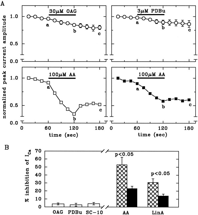
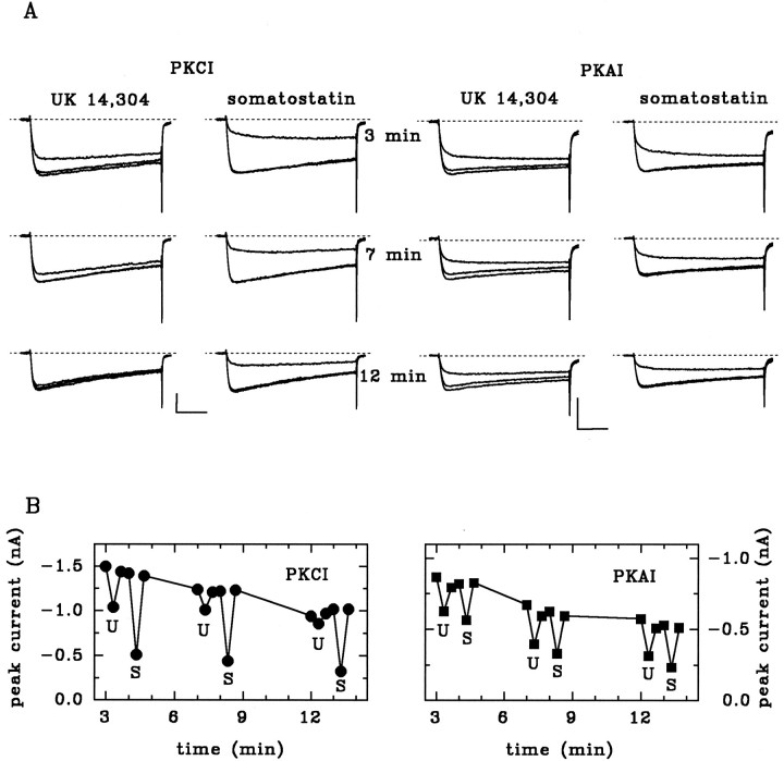
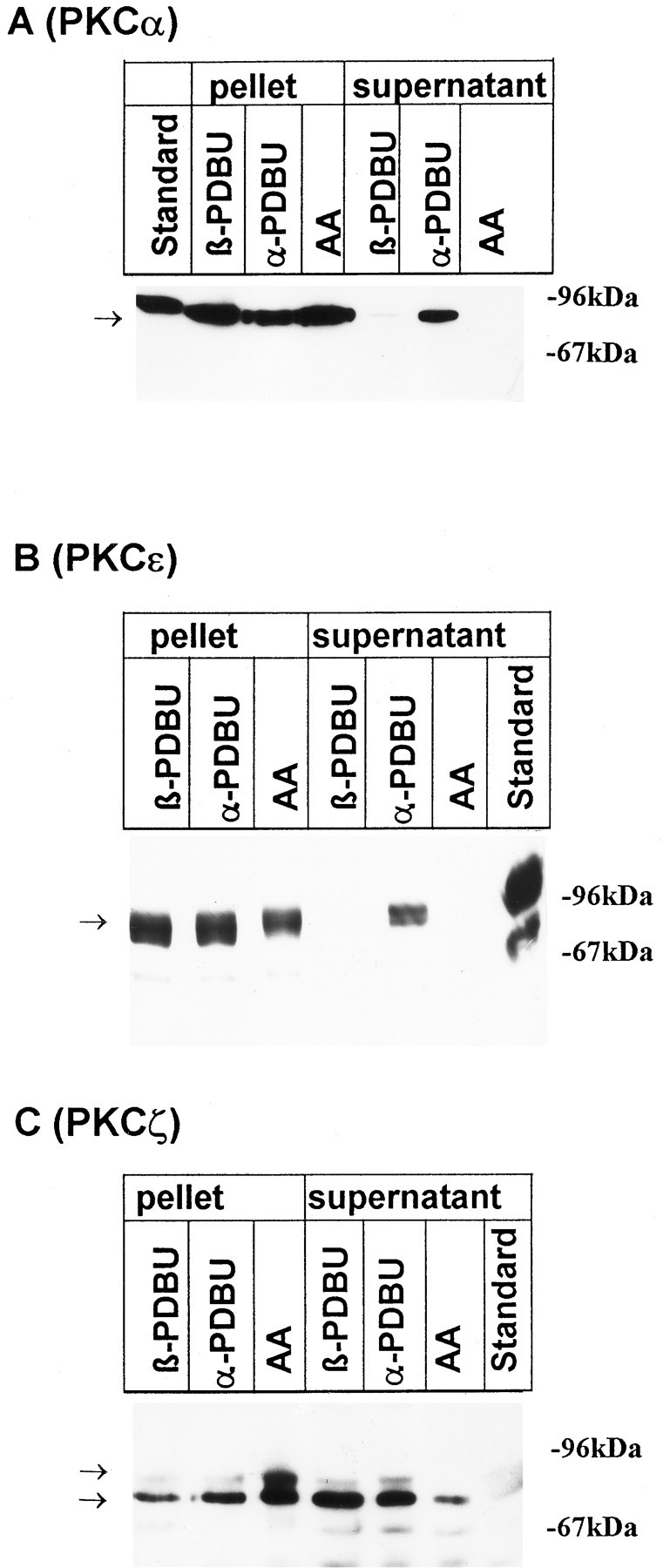
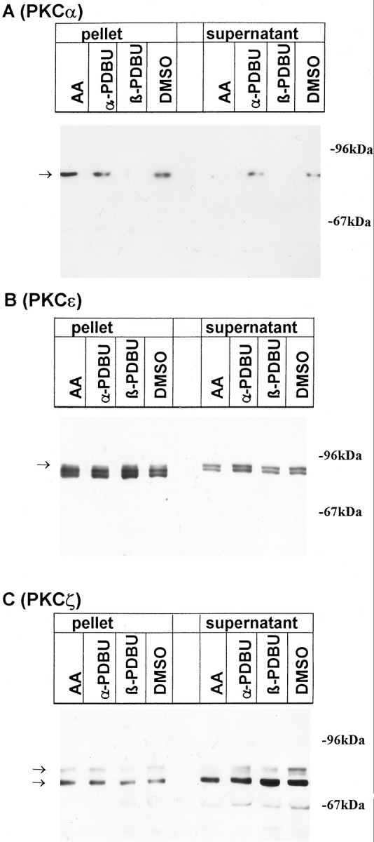
alpha2-Adrenoceptors regulate the efficacy at the sympathoeffector junction by means of a feedback inhibition of transmitter release. In chick sympathetic neurons, the mechanism involves an inhibition of N-type calcium channels, and we now present evidence that this effect involves an atypical, phorbol ester-insensitive protein kinase C (PKC). The inhibition of voltage-gated Ca2+ currents by the specific alpha2-adrenergic agonist UK 14,304 was significantly attenuated when the PKC inhibitors PKC(19-36), staurosporine, or calphostin C were included in the internal solution used to fill the patch pipettes, or if staurosporine or calphostin C were applied extracellularly; however, phorbol esters as classical activators of PKC or oleoylacetylglycerol did not mimic the effect of UK 14,304, and chronic exposure to 4-beta-phorbol dibutyrate (PDBu) did not attenuate it, ever though PKCalpha and -epsilon isozymes were translocated to plasma membranes by PDBu. The atypical isozyme PKCzeta was translocated by 100 micrometer AA and this effect was attenuated when PKC(19-36) was added to the patch pipette solution. Our observations indicate that classical, new, and atypical PKC isozymes are present in chick sympathetic neurons and that an atypical, phorbol ester-insensitive PKC is involved in the inhibition of voltage-activated calcium currents by alpha2-adrenoceptor activation.


Figures

Fig. 3.
Fig. 3.
AA and LinA, but not phorbol esters or OAG, inhibit Ca2+ currents. A, Time course of peak current amplitudes (normalized to the first amplitude) and effects of OAG, PDBu, and AA. These drugs were applied to cells dialyzed for at least 10 min with standard pipette solution (open circles), with 200 U/ml SOD (open squares), or with SOD plus 10 μm of the peptide inhibitor PKCI(19–36) (filled squares). n = 5–6.B, Inhibition of Ca2+ currents by 30 μm OAG, 3 μm PDBu, 10 μm SC-10, 100 μm AA, or 100 μm LinA, calculated as % inhibition = 100 − (200 b/a + c), wherea, b, and c are the current amplitudes measured after 60, 120, and 180 sec, respectively, as indicated inA. Neurons were dialyzed for at least 10 min with standard pipette solution (open bars), with SOD (hatched bars), or with SOD plus 10 μm of the peptide inhibitor PKCI(19–36) (filled bars), which significantly attenuates the effects of AA and LinA. Levels of significance for the difference between corresponding bars are indicated. n = 5–6.
Fig. 1.
Fig. 1.
The PKC inhibitor PKCI(19–36) prevents the α2-adrenoceptor-mediated but not the somatostatin-mediated inhibition of Ca2+currents. A, Calcium currents were induced from a holding potential of −80 mV by depolarizing pulses to 0 mV. UK (10 μm) or somatostatin (1 μm) were applied to the same cells 3, 7, and 12 min after establishing the whole-cell condition. The recording pipette contained 10 μm peptide inhibitor against protein kinase C [PKCI(19–36)] (left panel) or 10 μm peptide inhibitor against protein kinase A [PKI(6–22)amide] (right panel). Traces show currents before, during, and after application of agonists. Calibration: 0.5 nA, 30 msec. B, Time-plots of recordings from the cells shown above in A. Filled symbols indicate test pulses in the absence or presence of 10 μm UK 14,304 (U) or 1 μm somatostatin (S).
Fig. 2.
Fig. 2.
α2-Adrenoceptor-mediated inhibition of Ca2+ currents: summary of effects of PKC inhibitors. A, Inhibition of Ca2+ currents by 10 μm UK 14,304 (left) or 1 μm somatostatin (right) in cells dialyzed for at least 10 min with 0.1% DMSO, 1 μm calphostin C, or 1 μm staurosporine. Traces show currents before, during, and after the application of agonists. Calibration: 0.5 nA for DMSO, 0.25 nA for staurosporine, 0.1 nA for calphostin C; 30 msec. B, Inhibition of peak Ca2+ current amplitudes (ICa) by 10 μm UK 14,304 under control conditions (ctl) or with 10 μm PKCI(19–36), a peptide inhibitor of protein kinase C (PKCI); 10 μmglu27-PKCI(19–36), a noninhibitory analog peptide (PKCnI); 10 μmPKAI(6–22)amide, a peptide inhibitor of PKA (PKAI); 0.1% DMSO (DMSO); 1 μm calphostin C (calph); 1 μm staurosporine (stau); or 1 μm PDBu added to the pipette solution. Alternatively, cells pretreated for 24 hr with 10 μm β-phorbol-12,13-dibutyrate (24 hr PDBu) were tested with 10 μm UK 14,304 using regular pipette solution. Currents were recorded 10 min or later after breaking the cell membrane. **, p < 0.01 vs control; ##, p < 0.01 vs DMSO; n indicated in the bars.
Fig. 4.
Fig. 4.
Translocation of PKCα, ε, and ζ isozymes by phorbol esters and AA. The distribution of protein kinase C isoforms was investigated by immunoblots after a 10 min incubation of cultures with 100 μmAA, with the inactive phorbol ester αPDBU, or with the active phorbol ester βPDBU, both at 1 μm. Cells were lysed as described in Materials and Methods; aliquots of the pellet and supernatant corresponding to 2 × 105 cells were applied to SDS-polyacrylamide gels. Nitrocellulose blots were probed with peptide antisera specific for PKCα (A), PKCε (B), and PKCζ (C). Standard: 10 μg protein from rat neocortical homogenate. Arrows indicate the position of bands that were suppressed when antibodies had been preincubated with the corresponding immunogenic peptide (not shown). Data are representative of three additional experiments performed on different preparations.
Fig. 5.
Fig. 5.
Downregulation of classical, but not atypical, PKC isozymes by phorbol esters. The distribution of protein kinase C isoforms was analyzed by immunoblots after a 24 hr treatment of cultures with 100 μmAA, with 1 μm of the inactive and active phorbol esters αPDBU and βPBDU, respectively, or with the vehicle 0.1% DMSO. Cells were lysed as described in Materials and Methods; aliquots of the pellet and supernatant corresponding to 2 × 105 cells were applied to SDS-polyacrylamide gels. Nitrocellulose blots were probed with peptide antisera specific for PKCα (A), PKCε (B), and PKCζ (C). Arrows indicate the position of bands that were blocked when antibodies had been preincubated with the corresponding immunogenic peptide (not shown). Data are representative of three additional experiments performed on different preparations.

References

    1. Asaoka Y, Nakamura S-I, Yoshida K, Nishizuka Y. Protein kinase C, calcium and phospholipid degradation. Trends Biochem Sci. 1992;17:414–417. - PubMed
    1. Axelrod J. Receptor-mediated activation of phospholipase A2 and arachidonic acid release in signal transduction. Biochem Soc Trans. 1990;18:503–507. - PubMed
    1. Axelrod J, Burch RM, Jelsema CL. Receptor-mediated activation of phospholipase A2 via GTP-binding proteins: arachidonic acid and its metabolites as second messengers. Trends Neurosci. 1988;11:117–123. - PubMed
    1. Beech DJ, Bernheim L, Hille B. Pertussis toxin and voltage dependence distinguish multiple pathways modulating calcium channels of rat sympathetic neurons. Neuron. 1992;8:97–106. - PubMed
    1. Bley KR, Tsien RW. Inhibition of Ca2+ and K+ channels in sympathetic neurons by neuropeptides and other ganglionic transmitters. Neuron. 1990;2:379–391. - PubMed

MeSH terms

LinkOut - more resources