. 1996 Aug 1;16(15):4733-41.
doi: 10.1523/JNEUROSCI.16-15-04733.1996.

Chemically mediated cross-excitation in rat dorsal root ganglia

Affiliations

Chemically mediated cross-excitation in rat dorsal root ganglia

R Amir et al. J Neurosci. .

Abstract

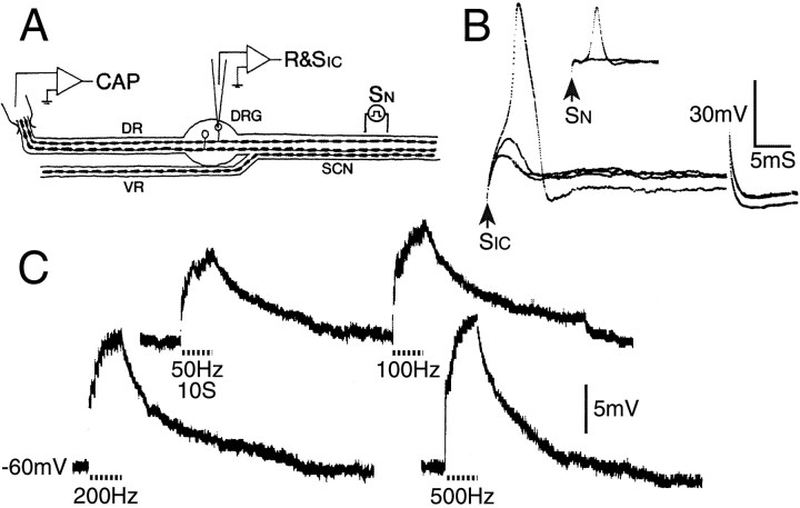
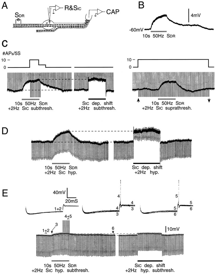
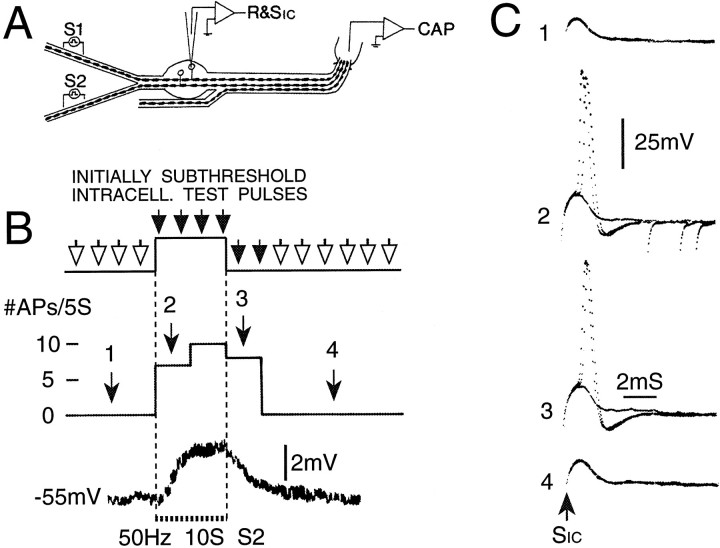
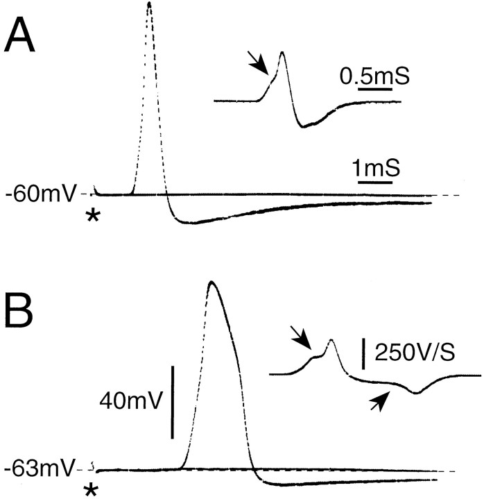
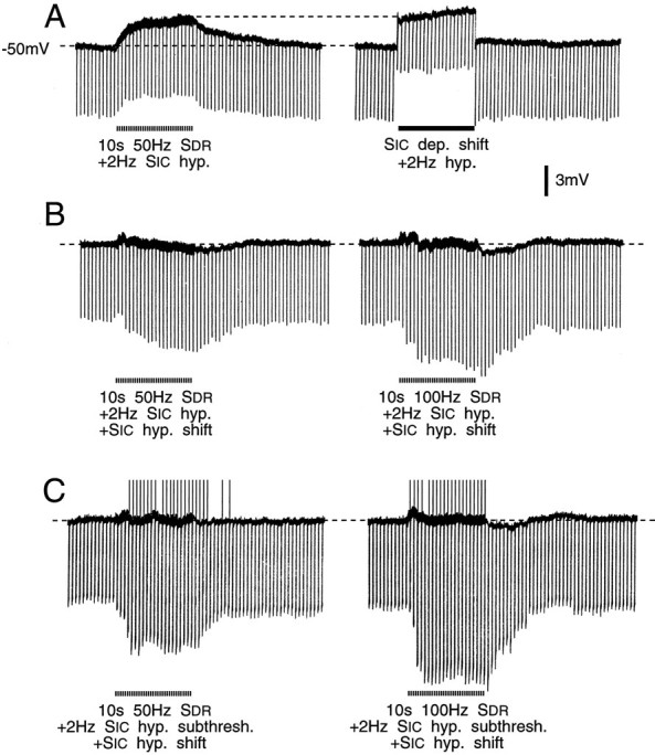
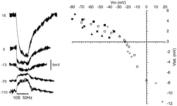
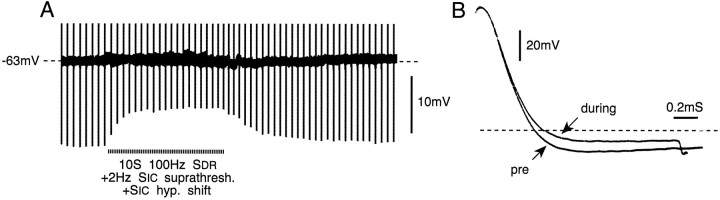
Primary afferent neurons in mammalian dorsal root ganglia (DRGs) are anatomically isolated from one another and are not synaptically interconnected. As such, they are classically thought to function as independent sensory communication elements. However, it has recently been shown that most DRG neurons are transiently depolarized when axons of neighboring neurons of the same ganglion are stimulated repetitively. Here we further characterize this functional coupling. In electrophysiological recordings made from excised rat DRGs, we found that DRG "cross-depolarization" is excitatory in that it is accompanied by an increase in the probability of spiking in response to otherwise subthreshold test pulses delivered intracellularly. Cross-depolarization contributes to this mutual cross-excitation. However, at least as important a contribution comes from a net increase in the neurons' input resistance (Rin) triggered by the stimulation of neighboring neurons. This change in Rin occurs even when cross-depolarization is absent or is balanced out. The amplitude of cross-depolaration was found to be voltage-dependent, with a reversal potential at approximately -23mV. Reversibility and the change in Rin both indicated that activity of neighboring neurons causes a membrane conductance change that is chemically mediated. Thus, far from being isolated, most DRG neurons participate in ongoing mutual interactions in which neuronal excitability is continuously modulated by afferent spike activity. This intraganglionic dialog appears to be mediated, at least in part, by an activity-dependent diffusable substance(s) released from neuronal somata and/or adjacent axons, and detected by neighboring cell somata and/or axons.


Figures

Fig. 2.
Fig. 2.
Cross-depolarization in DRG neurons. A, Sketch of one of the alternative experimental protocols (see Materials and Methods). B, Intracellular voltage (R&SIC) recording from an AINF neuron in response to sub- and suprathreshold pulses delivered through the intracellular micropipette (SIC, 35 msec pulse) and the sciatic nerve (SN, 0.1 msec pulse). The SN -evoked spike is small because it did not invade the soma (same calibration for both trials). C, Cross-depolarization in this cell to 10 sec SNtetani (dashed line) using stimuli subthreshold for the axon of the impaled neuron, and frequency as indicated.
Fig. 5.
Fig. 5.
Increased firing probability during cross-depolarization is associated with increasedRin. A, B, A third experimental protocol, and an example of cross-depolarization evoked by DR conditioning (SDR) subthreshold for the axon of the impaled neuron. C, Left, Initially subthreshold test pulses were delivered at 2 Hz through the intracellular electrode (SIC, vertical voltage deviations). Firing probability increased during conditioning (note spikes during the peak of the cross-depolarization), but was unaffected when an equivalent depolarizing shift was imposed by passing current through the recording electrode. C, Right, Probing with just suprathreshold test pulses (betweenarrows) yielded no sign of excitability suppression. Upward deflections before and after this time are just subthreshold test pulses. D, Hyperpolarizing constant-current test pulses reveal slightly increased Rin during DR conditioning, but much decreased Rin during an equivalent imposed depolarizing shift. E, Conditioning also increased the probability of anodal break responses (using higher amplitude hyperpolarizing test pulses than in D). An imposed depolarizing shift of similar amplitude had no effect on firing probability. Note the different change inRin during cross-depolarization versus imposed depolarization. Sample voltage traces from time windows 1–6 are shown.
Fig. 3.
Fig. 3.
Firing probability is increased during cross-depolarization. A, A second experimental protocol. The DR was split, and conditioning tetani were delivered to the half not containing the axon of the impaled neuron (S2).B, (Initially) subthreshold test pulses delivered at 2 Hz (SIC) yielded firing probability of 0 action potentials/5 sec epoch (#APs/5S). During conditioning, firing probability increased, reaching the maximum possible of 10 APs/5 sec. C, Evoked responses during time windows 1–4 inB. Each trace shows three consecutive sweeps.
Fig. 1.
Fig. 1.
A0 and AINF spikes. A0(A) and AINF (B) spikes (direct and differentiated record) after axonal stimulation (*) just below and just above threshold (single sweeps). Note the inflection on the rising phase (downward pointing arrows) and on the falling phase (B only, upward pointing arrow). CV = 8.6 m/sec for A, 4.3 m/sec for B.
Fig. 4.
Fig. 4.
Cross-excitation. A, Increased firing probability during cross-depolarization in a cell that fired bursts in response to intracellular stimulus pulses (10 msec, 2 Hz). The experimental protocol was as in Figure 5A. Conditioning, with resulting cross-depolarization, caused an increase in firing probability (bursts/5 sec). Spike bursts during time windows 1–4 (3 or 4 superimposed traces each) are shown below. Note that spikes in A and in subsequent figures are truncated.B, Conditioning tetani may produce cross-excitation even without triggering cross-depolarization. The experimental protocol was as in Figure 3. Conditioning did not trigger cross-depolarization in this AINF neuron (top), but nonetheless produced cross-excitation (bottom). Firing probability, initially set at 5/20, increased to 13/20 during the conditioning tetanus (p < 0.05). Downward deflections are shock artifacts, and upward deflections are spikes (note the prolonged postspike AHPs).
Fig. 6.
Fig. 6.
Balancing out cross-depolarization did not abolish cross-excitation. A, Hyperpolarizing test pulses (1 nA, 20 msec, 2 Hz) were applied during cross-depolarization (left trace) and indicated ∼10% increase inRin from a control level of 7 MΩ. Identical test pulses revealed a 25% decrease inRin when depolarization of similar amplitude was imposed intracellularly (right trace).B, Cross-depolarization was balanced out with intracellular hyperpolarizing current. This revealed a 35% increase inRin during conditioning at 50 Hz (left) and a 45% increase during conditioning at 100 Hz (right). C, Intracellular hyperpolarizing test pulses initially 25% subthreshold for anodal break spikes (1.3 nA, 85 msec, 2 Hz) evoked such spikes during 50 Hz (left) and 100 Hz (right) conditioning tetani (upward deflections, spikes truncated). Cross-depolarization was quenched during these trials.Rin increased ∼55% and 75%, respectively.
Fig. 7.
Fig. 7.
Reversal of cross-depolarization.Right, Peak amplitude of voltage shift evoked by 10 sec, 50 Hz conditioning tetani (Vtet., ordinate) as a function of membrane holding potential (Vm). Symbols identify five neurons, two of which reversed. Traces on theleft are from cell (○). Resting potentials = −55 mV (), −54 mV (▴), −61 mV(+), −53 mV (✖), −56mV (▪).
Fig. 8.
Fig. 8.
Spike AHP decreases during conditioning tetani (10 sec, 100 Hz), with kinetics resembling those of cross-depolarization. In the record shown, cross-depolarization was quenched. Traces inB show spike shape before (pre) andduring conditioning. Note delay in spike falling phase during conditioning. The horizontal dashed line indicates the resting membrane potential.

References

    1. Bean BP, Williams CA, Ceelen PW. ATP-activated channels in rat and bullfrog sensory neurons: current-voltage relation and single-channel behavior. J Neurosci. 1990;10:11–19. - PMC - PubMed
    1. Belmonte C, Gallego R. Membrane properties of cat sensory neurones with chemoreceptor and baroreceptor endings. J Physiol (Lond) 1983;342:603–614. - PMC - PubMed
    1. Brown JE, Muller KJ, Murray G. Reversal potential for an electrophysiological event generated by conductance changes: mathematical analysis. Science. 1971;174:318. - PubMed
    1. Brunet PC, Jirounek P. Long-range intercellular signalling in glial cells of the peripheral nerve. NeuroReport. 1994;5:635–638. - PubMed
    1. Callahan MJ, Korn SJ. Permeation of Na+ through a delayed rectifier K+ channel in chick dorsal root ganglion neurons. J Gen Physiol. 1994;104:747–772. - PMC - PubMed

Publication types