. 1996 Aug 1;16(15):4749-56.
doi: 10.1523/JNEUROSCI.16-15-04749.1996.

Compartmental localization of a metabotropic glutamate receptor (mGluR7): two different active sites at a retinal synapse

Affiliations

Compartmental localization of a metabotropic glutamate receptor (mGluR7): two different active sites at a retinal synapse

J H Brandstätter et al. J Neurosci. .

Abstract

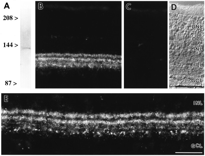
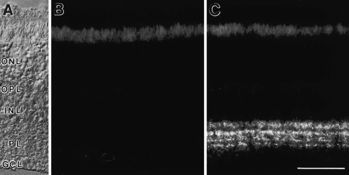
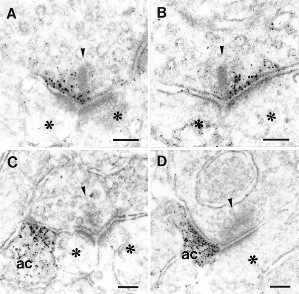
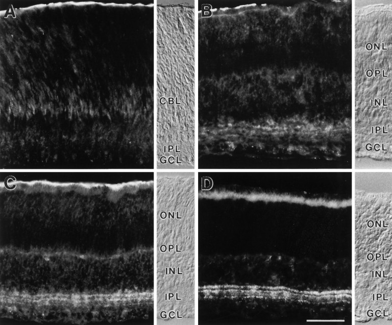
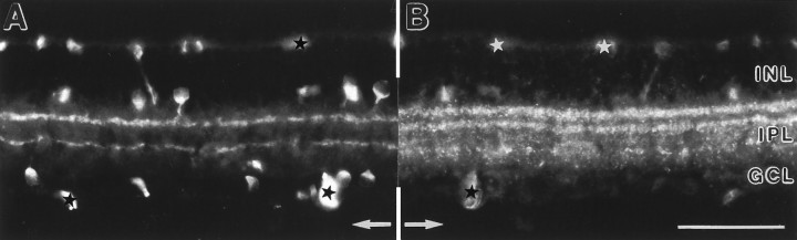
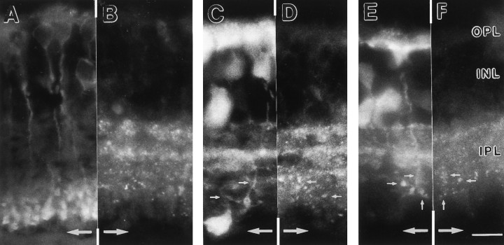
The distribution of the metabotropic glutamate receptor 7 (mGluR7) was studied in the rat retina using a specific antiserum. Punctate immunofluorescence that corresponded to synaptic localization was present exclusively in the inner plexiform layer. Double-labeling experiments suggested that mGluR7 is expressed at the synaptic terminals of certain cone bipolar cells. Electron microscopy showed that mGluR7 was present both presynaptically, as an autoreceptor in cone bipolar cell ribbon synapses, and postsynaptically in amacrine cells. There are usually two postsynaptic processes at a bipolar cell ribbon synapse; however, the presynaptic aggregation of mGluR7 was restricted to one half of the active zone and therefore was opposed to only one of the postsynaptic processes. This selective localization of mGluR7 could differentially regulate the glutamate release from the ribbon synapse, thus leading to a differential activation of the postsynaptic neurons.


Figures

Fig. 1.
Fig. 1.
Specificity of the antiserum against mGluR7.A, Western blot of rat retina membrane proteins (80 μg/lane) showed a single band at ∼120 kDa. B, MGluR7 immunoreactivity in vertical cryostat sections of rat retina was confined to the IPL. C, Preadsorption of anti-mGluR7 antiserum with the immunogen resulted in complete loss of specific immunoreactivity. D, The retinal layers are shown with Nomarski optics. E, Higher-power view showing the punctate staining for mGluR7. ONL, Outer nuclear layer;OPL, outer plexiform layer; INL, inner nuclear layer; IPL, inner plexiform layer; GCL, ganglion cell layer. Scale bar (shown in D): B–D, 50 μm; E, 25 μm.
Fig. 2.
Fig. 2.
Retina of mGluR7 knock-out mouse. A, The retinal layers are shown with Nomarski optics (abbreviations as in Fig. 1). B, Absence of specific mGluR7 immunoreactivity in vertical cryostat sections of the retina of mGluR7 knock-out mouse compared with (C) the wild-type staining pattern. Unspecific staining of photoreceptor inner segments can be seen in the outer retina. Scale bar (shown in C): 50 μm.
Fig. 3.
Fig. 3.
High-power electron micrographs showing the pre- and postsynaptic localization of mGluR7. A, B, Presynaptic localization of mGluR7 in an (A) OFF-cone and (B) ON-cone bipolar cell. Note that the receptor-labeling is present along only one part of the active zone, left or rightof the presynaptic ribbon. C, D, Postsynaptic localization of mGluR7 to an (C) OFF-cone and (D) ON-cone bipolar cell. Note that the receptor-labeling is present in only one of the postsynaptic neurons, the amacrine cell (ac), as identified by the presence of vesicles. Presynaptic ribbons are marked with arrowheads, postsynaptic neurons withasterisks. Scale bars, 0.1 μm.
Fig. 4.
Fig. 4.
Vertical sections of rat retinas showing the postnatal development of mGluR7 immunoreactivity. The retinal layers are shown with Nomarski optics accompanying each micrograph (abbreviations as in Fig. 1). CBL, Cytoblast layer.A, At postnatal day 5 (P5), no mGluR7-specific staining is found. B, A first, diffuse staining of cell somata in the INL and GCL and of processes in the IPL is seen at P7. C, Between P7 and P10, staining of somata decreases and preferential labeling of processes in the IPL increases. D, MGluR7 expression reaches the adult labeling pattern at around P16. Unspecific staining of photoreceptor inner segments and the pigment epithelium can be seen in the outer retina. Scale bar (shown in D): 25 μm.
Fig. 5.
Fig. 5.
Vertical section of rat retina double-labeled for mGluR7 and ChAT. The two bands of cholinergic amacrine cell processes labeled with an antibody against ChAT (A) are not congruent with the mGluR7-immunoreactive bands in the IPL (B). The micrographs are printed as mirror images and are aligned along a common midline, as indicated by the arrows. Corresponding points therefore are found at equal distances from the midline. Unspecifically stained, larger blood vessels are marked by stars. Abbreviations as in Figure 1. Scale bar, 50 μm.
Fig. 6.
Fig. 6.
Vertical sections of rat retinas that were double-stained with the antiserum against mGluR7 and antibodies that recognize distinct cell types. The micrographs are printed as mirror images and cut and aligned along a common border. Identical points of the sections therefore are found at equal distances from the midline (large arrows). A, Rod bipolar cells are stained with an antibody against PKCα. The lack of symmetry across the midline indicates no colocalization with the mGluR7 staining inB. In C and E, calbindin-immunopositive bands in the IPL and labeled ON-cone bipolar cells can be seen. Their terminals are colocalized with mGluR7 immunoreactive puncta in D and F (small arrows). Additionally, the outermost calbindin-immunoreactive band (C, E) is congruent with one of the mGluR7-immunoreactive bands (D, F). Abbreviations as in Figure 1. Scale bar (shown in F): 10 μm.

References

    1. Allbritton NL, Meyer T, Stryer L. Range of messenger action of calcium ion and inositol 1,4,5-triphosphate. Science. 1992;258:1812–1815. - PubMed
    1. Baude A, Nusser Z, Roberts JDB, Mulvihill E, McIlhinney RAJ, Somogyi P. The metabotropic glutamate receptor (mGluR1α) is concentrated at perisynaptic membrane of neuronal subpopulations as detected by immunogold reaction. Neuron. 1993;11:771–787. - PubMed
    1. Brandstätter JH, Hartveit E, Sassoè-Pognetto M, Wässle H. Expression of NMDA and high-affinity kainate receptor subunit mRNAs in the adult rat retina. Eur J Neurosci. 1994;6:1100–1112. - PubMed
    1. Brecha N. Retinal neurotransmitters: histochemical and biochemical studies. In: Emson PC, editor. Chemical neuroanatomy. Raven; New York: 1983. pp. 85–129.
    1. Calkins DJ, Sterling P (1996) Absence of spectrally specific lateral inputs to midget ganglion cells in primate retina. Nature, in press. - PubMed

Publication types

Substances

LinkOut - more resources