. 1996 Sep;50(3):512-21.

Hydrophilic side chains in the third and seventh transmembrane helical domains of human A2A adenosine receptors are required for ligand recognition

Affiliations

Hydrophilic side chains in the third and seventh transmembrane helical domains of human A2A adenosine receptors are required for ligand recognition

Q Jiang et al. Mol Pharmacol. 1996 Sep.

Abstract

Hydrophilic residues of the G protein-coupled human A2A adenosine receptor that are potentially involved in the binding of the ribose moiety of adenosine were targeted for mutagenesis. Residues in a T88QSS91 sequence in the third transmembrane helical domain (TM3) were individually replaced with alanine and other amino acids. Two additional serine residues in TM7 that were previously shown to be involved in ligand binding were mutated to other uncharged, hydrophilic amino acids. The binding affinity of agonists at T88 mutant receptors was greatly diminished, although the receptors were well expressed and bound antagonists similar to the wild-type receptor. Thus, mutations that are specific for diminishing the affinity of ribose-containing ligands (i.e., adenosine agonists) have been identified in both TM3 and TM7. The T88A and T88S mutant receptor fully stimulated adenylyl cyclase, with the dose-response curves to CGS 21680 highly shifted to the right. A Q89A mutant gained affinity for all agonist and antagonist ligands examined in binding and functional assays. Q89 likely plays an indirect role in ligand binding. S90A, S91A, and S277C mutant receptors displayed only moderate changes in ligand affinity. A S281N mutant gained affinity for all adenosine derivatives (agonists), but antagonist affinity was generally diminished, with the exception of a novel tetrahydrobenzothiophenone derivative.


Figures
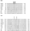
Fig. 1
Fig. 1
Location of mutations carried out in this study, illustrated through an alignment of the TM3 and TM7 of selected receptor subtypes. Bold, residues mutated in A2A receptors (present study) and in a chimeric bovine A1/rat A3 construct (18). Accession numbers are h A2a (human) P29274, rA2a (rat) P30543, cA2a (dog) P11617, gpA2a (guinea pig) U04201 , hA2b (human) P29275, hAl (human) P30542, hA3 (human) P33765, rA3 (rat) P28647, m3 (rat) P08483, β2 (hamster) P04274, and NK1 (human) P25103. In TM3, the canine and guinea pig A2A sequences are identical to the rat A2A sequence.
FIg. 2
FIg. 2
Structures of agonist (A) and antagonist (B) ligands used in this study.
Fig. 3
Fig. 3
Displacement of binding of the agonist radioligand [3H]CGS 21680 from HA-tagged A2A wildtype (WT) and Q89A mutant receptors expressed in COS-7 cells. Competitors used were IB-MECA (A) and ADAC (B). Competition binding studies were carried out using membrane homogenates prepared from transfected COS-7 cells, as described in Experimental Procedures. Data are from a representative experiment performed in duplicate.
Fig. 4
Fig. 4
Plot of affinity (Ki in nA2A) in competition binding experiments of three adenosine receptor ligands at Q89 mutant receptors as a function of the amino acid residue. The amino acids were arranged in order of increasing solvent-accessible surface area, using theoretical values for each isolated amino acid as calculated by Hubbard et al. (36). Ki values for the agonist (NECA and lB-MECA) and antagonist (XAC) ligands (structures in Fig. 2) were determined in [3H]CGS 21680 (15 nM) competition binding studies using membrane homogenates prepared from transiently transfected COS-7 cells, as described in Experimental Procedures. Ki values were calculated from IC50 values by using the KaleidaGraph program. All constructs contain the HA epitope tag sequence at the amino terminus (15). ***, p < 0.001; **, p < 0.01; *, p < 0.05; n.s., not significant.
Fig. 5
Fig. 5
Stimulation of adenylyl cyclase in COS-7 cells transiently expressing HA-tagged A2A wild-type (WT) or mutant A2A-adenosine receptors in the presence of 2 units/mI adenosine deaminase and 0.1 m rolipram. The following receptors were studied: wild-type, T88A, T88R, and T88S mutant receptors. Transfected COS-7 cells were incubated for 30 mm at 37° (for details, see Experimental Procedures) with increasing concentrations of CGS 21680. Data are presented as percentage of maximal increase in cAMP above basal levels in the absence of CGS 21680 for a representative experiment. For each curve, the maximal stimulation represents a 4–5-fold stimulation over basal levels. At agonist concentrations of ≥100 μm, the stimulation observed in nontransfected COS-7 cells (15) was subtracted. EC50 values (average of three independent experiments, each carried out in duplicate) were wild-type receptor, 0.915 ± 0.213 nm; T88A, 14.4 ± 0.423 μm; and T88S, 0.323 ± 0.048 μm.
Fig. 6
Fig. 6
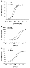
Stimulation of adenylyl cyclase in COS-7 cells transiently expressing HA-tagged A2A wild-type (WT) or mutant A2A-adenosine receptors in the presence of 2 units/mI adenosine deaminase and 0.1 m rolipram. The following receptors were studied: wild-type and Q89A mutant receptors with CGS 21680 (top), NECA (middle), or DPMA (bottom). Transfected COS-7 cells were incubated for 30 mm at 37° (for details, see Experimental Procedures) with increasing concentrations of agonist. Data are presented as percentage of maximal increase in cAMP above basal levels in the absence of agonist for a representative experiment. For each curve, the maximal stimulation represents a 4-5-fold stimulation over basal levels. EC50 values were calculated averaged over three independent experiments, each carried out in duplicate.

Similar articles

Cited by

References

    1. Jacobson KA, van Galen PJM, Williams M. Adenosine receptors: pharmacology, structure activity relationships, and therapeutic potential. J. Med. Chem. 1992;35:407–422. - PMC - PubMed
    1. Libert F, Parmentier M, Lefort A, Dinsart C, van Sande J, Maenhaut C, Simons MJ, Dumont JE, Vassart G. Selective amplification and cloning of four new members of the G protein-coupled receptor family. Science (Washington D. C.) 1989;244:569–572. - PubMed
    1. Jacobson M. Molecular biology of adenosine receptors. In: Belardinelli L, Pelleg A, editors. Ad.snosine and Adenine Nucleotides: From Molecular Biology to Integrative Physiology. Kluver; Norwell, MA: 1995. pp. 5–14.
    1. Barraco RA, Martens K, Parizon M, Normile HJ. Role of adenosine A2a receptors in the nucleus-accumbens. Prog. Neuropsychopharmacol. Biol. Psychiatry. 1994;18:545–553. - PubMed
    1. Olsson RA, Pearson JD. Cardiovascular purinoceptors. Pharmacol. Rev. 1990;3:761–845. - PubMed

MeSH terms

LinkOut - more resources