. 1996 Sep 15;16(18):5715-26.
doi: 10.1523/JNEUROSCI.16-18-05715.1996.

Laser ablation of Drosophila embryonic motoneurons causes ectopic innervation of target muscle fibers

Collaborators, Affiliations

Laser ablation of Drosophila embryonic motoneurons causes ectopic innervation of target muscle fibers

T N Chang et al. J Neurosci. .

Abstract

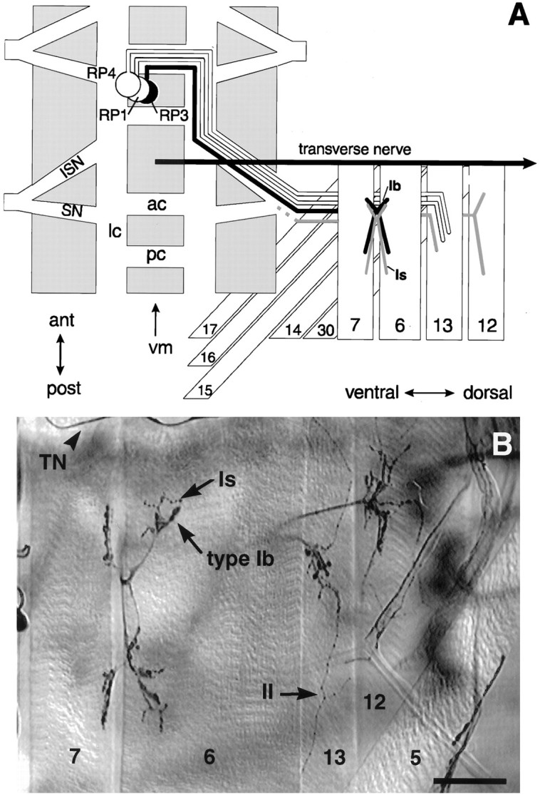
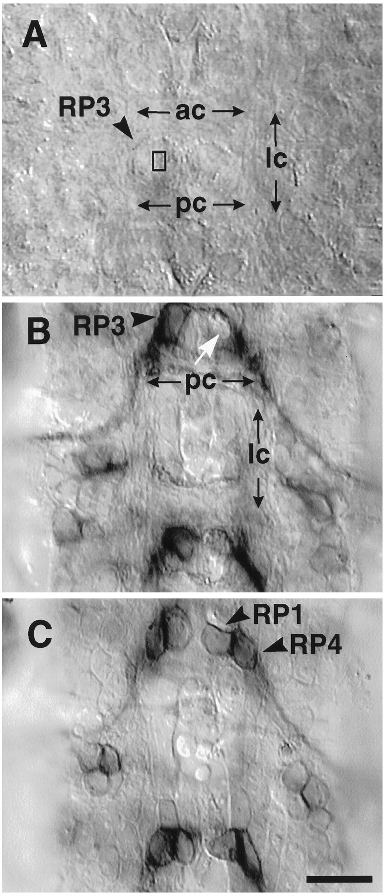
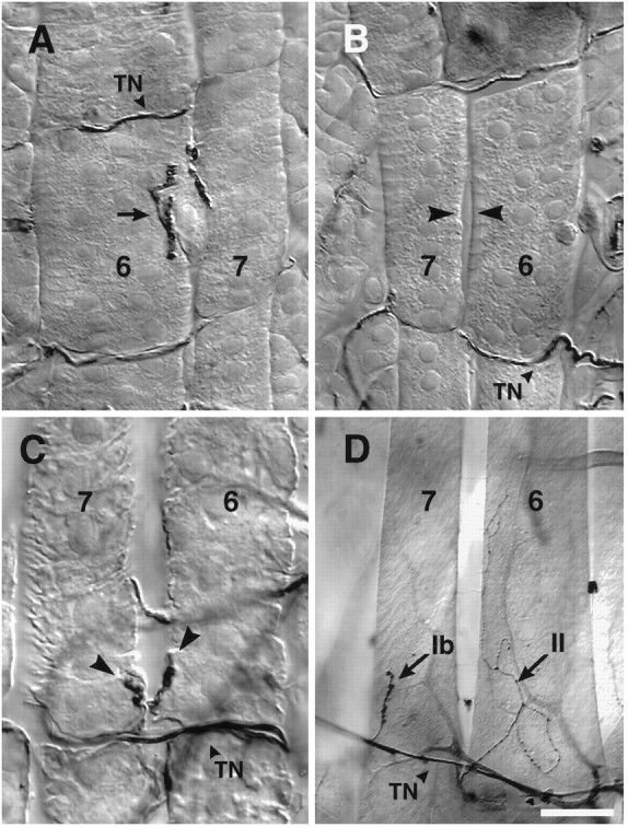
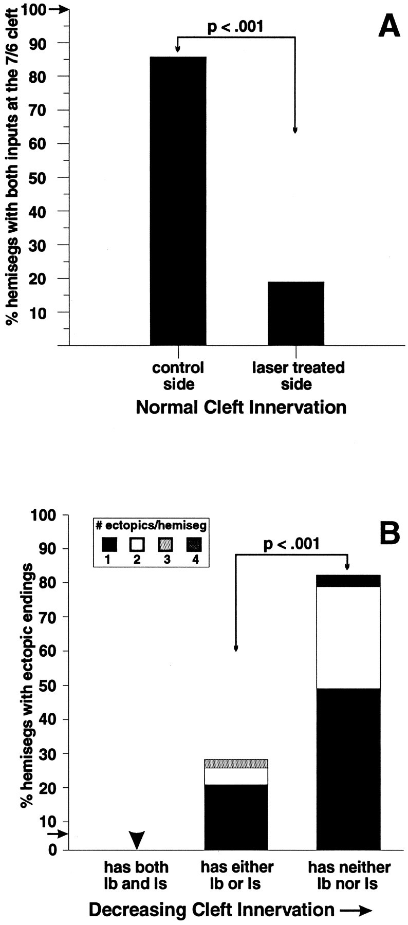
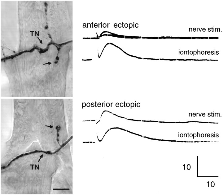
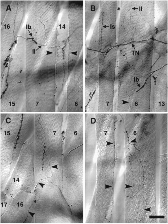
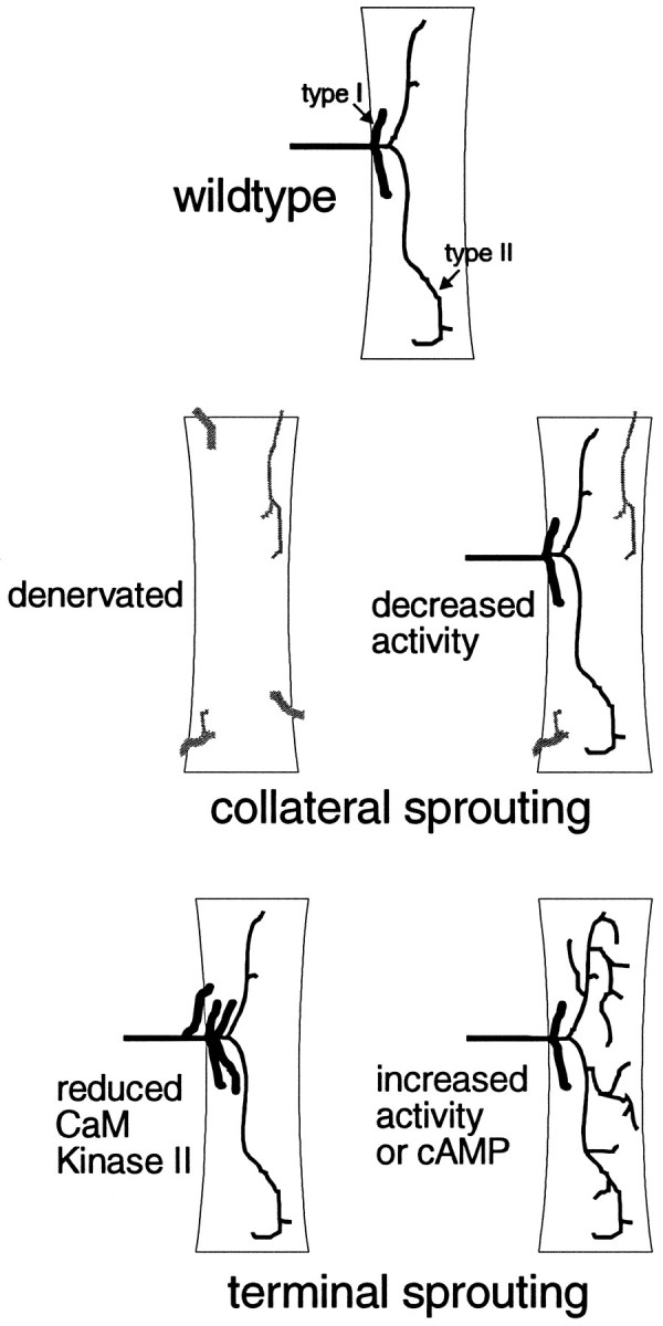
We have tested the effects of neuromuscular denervation in Drosophila by laser-ablating the RP motoneurons in intact embryos before synaptogenesis. We examined the consequences of this ablation on local synaptic connectivity in both 1st and 3rd instar larvae. We find that the partial or complete loss of native innervation correlates with the appearance of alternate inputs from neighboring motor endings and axons. These collateral inputs are found at ectopic sites on the denervated target muscle fibers. The foreign motor endings are electrophysiologically functional and are observed on the denervated muscle fibers by the 1st instar larval stage. Our data are consistent with the existence of a local signal from the target environment, which is regulated by innervation and influences synaptic connectivity. Our results show that, despite the stereotypy of Drosophila neuromuscular connections, denervation can induce local changes in connectivity in wild-type Drosophila, suggesting that mechanisms of synaptic plasticity may also be involved in normal Drosophila neuromuscular development.


Figures

Fig. 1.
Fig. 1.
The cell body locations, muscle targets, and axonal pathways of RP motoneurons discussed in this study.A, Schematic drawing of one abdominal hemisegment showing the pathway for RP3, and the neighboring motoneurons RP1 and RP4. The segmentally repeated RP3 motoneuron cell bodies are located on the dorsal aspect of the CNS (gray) on either side of the midline (vm). Each RP3 axon (black) crosses the midline in the anterior commissure (ac) and turns posteriorly in the longitudinal connective (lc) to exit the CNS via the intersegmental nerve tract (ISN). There it joins the segmental nerve (SN) to innervate muscle fibers7 and 6 in the next posterior segment. The axon projects within the cleft between the two fibers to establish a characteristic nerve branch and site of innervation at stage16 (Halpern et al., 1991; Sink and Whitington, 1991a,b;Broadie and Bate, 1993a) (for review, see Broadie et al., 1994). The second input to the 7/6 cleft (gray) also innervates other ventral muscle targets. Its cell body location is not established. RP1 and RP4 continue laterally to innervate muscle fiber13 (Halpern et al., 1991; Sink and Whitington, 1991a). The transverse nerve (TN) runs along the borders of each segment. B, The ventral musculature of a 3rd instar larval fillet stained for motoneurons with an antibody to horseradish peroxidase. There are at least three morphologically distinct ending types: type Ib (large boutons), type Is(intermediate-sized boutons), and type II (small boutons, more branched and extensive). These have been shown by bouton backfills to belong to distinct motoneurons. Note thatDrosophila muscle fibers can be polyinnervated. Scale bar, 50 μm.
Fig. 2.
Fig. 2.
Laser ablation of RP3 in live stage late 15/early 16 embryos. A, The cell body of RP3 is imaged through the ventral surface of the dechorionated embryo. RP3’s cell body is located near the dorsal (internal) surface of the ventral nerve cord. Two RP3 cell bodies (arrowhead) can clearly be seen flanking the midline and bordered by the lateral commissures (lc) on the left and right, and above and below by the anterior and posterior commissures (ac,pc). The rectangle indicates the ∼1 × 2 μm target site of the laser. Every visible RP3 on one side was targeted. The contralateral RP3 seconds were left intact as internal controls. B, C, To test the specificity of the laser ablation with respect to neighboring cells, operated embryos were acutely filleted and stained with anti-Fasciclin III, which labels a subset of neurons, including RP1,RP3, RP4. B, In this segment seen from the dorsal view, there is no staining in the location of RP3 (white arrow), whereas the contralateral RP3 is still immunopositive for FasIII. C, At the dorsal surface of the CNS, one cell layer above RP3, both RP1and RP4 can be seen on both sides. Scale bar, 10 μm.
Fig. 3.
Fig. 3.
Ablation of RP3 leads to denervation of target muscle fibers 7 and 6 and the appearance of ectopic inputs.AC, 1st instar larvae, (D) 3rd instar. A and Bshow, respectively, the left and right ventral hemisegment of a 1st instar larva after embryonic laser ablation. In B, the cleft is denervated on the side where RP3 normally innervates (arrowheads). On the contralateral untreated side of the same animal (A), the 7/6 cleft is innervated with well defined boutons characteristic of 1st instar control animals.C, Collateral inputs (arrowheads) from the transverse nerve onto the denervated 7 and6 muscle fibers of a 1st instar larva. D, Ectopic inputs on a cleft-denervated 3rd instar hemisegment. Indicated are a type Ib input from the transverse nerve onto the posterior end of muscle fiber 7, and a typeII input onto muscle fiber 6, traced back to the input on the next posterior muscle fibers 15/16 (source not shown). Scale bars: AC, 10 μm;D, 40 μm.
Fig. 4.
Fig. 4.
Reduction in innervation at muscle fibers 7 and 6 leads to the appearance of collateral inputs. A, In control animals, hemisegments A2–A3 always have both type Ib and type Is inputs at the cleft (frequency shown by arrow;n = 32 hemisegments, 8 larvae). In contrast, in laser-treated animals, on the experimental side (contralateral to laser ablation) a significantly smaller number of hemisegments had both cleft inputs present at the 7/6 cleft than on the control side (ipsilateral to laser ablation; n = 21 larvae, 74 hemisegments, A2–A3). B, Increased cleft denervation correlates with an increased probability and frequency of collateral innervation. Muscle fibers 7 and 6 are examined in segments A2–A3. The black bars indicate the percent of hemisegments with ectopic endings that have only one ectopic ending. Bar heights indicate total percent of hemisegments with ectopic endings in each group. Fromleft to right: hemisegments that have both Ib and Is at the cleft (“normal” cleft innervation;n = 6 hemisegments), hemisegments that have one or the other (partial innervation; n = 41 hemisegments), and hemisegments that have neither (complete denervation; n = 27 hemisegments, 21 laser-treated larvae examined in A2–A3). In control animals, ectopically placed inputs were seen at low frequency (shown by arrow;n = 89 hemisegments, 8 animals).
Fig. 5.
Fig. 5.
Ectopic inputs are physiologically functional. After embryonic laser ablation, 3rd instar larvae were filleted in insect saline and incubated with fluoresceinated anti-HRP to vitally image ectopic inputs on denervated muscle fibers 7 and 6. The pictured ectopic inputs from the transverse nerve (TN) are located at the anterior and posterior ends of the same muscle fiber 7. An intracellular electrode in the muscle fiber recorded a postsynaptic potential in response to both shocking the transverse nerve using a suction electrode (top trace) and iontophoresis of glutamate above the boutons (bottom trace; scale shown is 10 mV, 10 msec). The fillet was then fixed and relabeled with anti-HRP. The results indicate that both pre- and postsynaptic elements of these collateral inputs are functional. Scale bar, 10 μm.
Fig. 6.
Fig. 6.
Collateral inputs are formed on denervated muscle fibers from nearby motor axons and terminals. A, An example of collateral inputs from the muscle15/16 cleft innervating muscle fiber7 from the medial aspect. Both type Iband type II endings are present.Arrowheads indicate the denervated cleft between muscle fibers 7 and 6. The inputs visible between the arrowheads are on the underlying muscle fiber 14. B, The three different types of ending morphology are evident in this example: an Isending from the transverse nerve (TN) to the posterior end of the anterior muscle fiber 7, a Ibinput from SNb branching onto the lateral edge of muscle fiber6, and two type II inputs: one arising laterally from SNb onto the anterior muscle fiber 6(source not shown), and the other laterally from SNb onto muscle fiber6 (arrowhead). C, A type II input from the next posterior 15/16cleft (arrowhead). Also visible are type Ib boutons that appear to be from the same source (see Fig. 7A).D, An example of a large, branched type II input from the 7/6 cleft ramifying over both muscle fibers 7 and 6(arrowheads). In contrast, type II inputs at the cleft in control animals are rare and small (see Results). Scale bar:A, C, D, 30 μm; B, 50 μm.
Fig. 7.
Fig. 7.
Summary of motoneuronal plasticity inDrosophila. Wild-type muscle fibers are innervated by type I (Ib and/or Is) motoneuronal endings. Additionally, they may have type II endings, which are more branched with smaller boutons. Collateral sprouting can result from either denervation (Halfon et al., 1994) (this study) or decreased activity (Jarecki and Keshishian, 1995). Terminal sprouting of type I endings can be caused by reduction of CaM kinase II, whereas terminal sprouting of type II endings can result from increased activity or increased cAMP (Budnik et al., 1990;Zhong et al., 1992).

References

    1. Aigner L, Arber S, Kapfhammer JP, Laux T, Schneider C, Botteri F, Brenner H-R, Caroni P. Overexpression of the neural growth-associated protein GAP-43 induces nerve sprouting in the adult nervous system of transgenic mice. Cell. 1995;83:269–278. - PubMed
    1. Atwood HL, Govind CK, Wu C-F. Differential ultrastructure of synaptic terminals on ventral longitudinal abdominal muscles in Drosophila larvae. J Neurobiol. 1993;24:1008–1024. - PubMed
    1. Benoit P, Changeux J-P. Consequences of tenotomy on the evolution of multi-innervation in developing rate soleus muscle. Brain Res. 1975;99:345–358. - PubMed
    1. Broadie KS, Bate M. Development of the embryonic neuromuscular synapse of Drosophila melanogaster . J Neurosci. 1993a;13:144–66. - PMC - PubMed
    1. Broadie KS, Bate M. Innervation directs receptor synthesis and localization in Drosophila embryo synaptogenesis. Nature. 1993b;361:350–353. - PubMed

Publication types

LinkOut - more resources