Comparative Study
. 1996 Sep 15;16(18):5795-811.
doi: 10.1523/JNEUROSCI.16-18-05795.1996.

Comparison of neurodegenerative pathology in transgenic mice overexpressing V717F beta-amyloid precursor protein and Alzheimer's disease

Affiliations
Comparative Study

Comparison of neurodegenerative pathology in transgenic mice overexpressing V717F beta-amyloid precursor protein and Alzheimer's disease

E Masliah et al. J Neurosci. .

Abstract

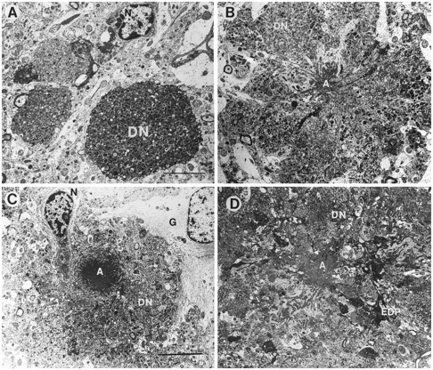
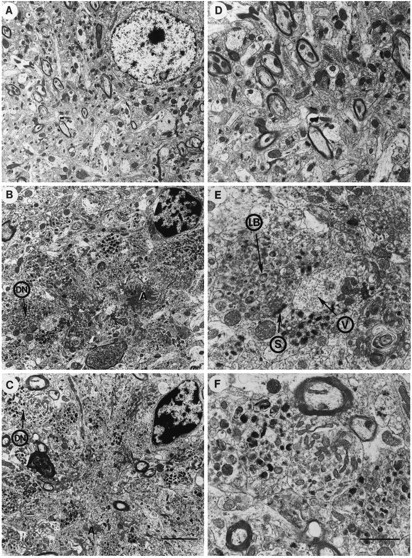
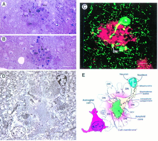
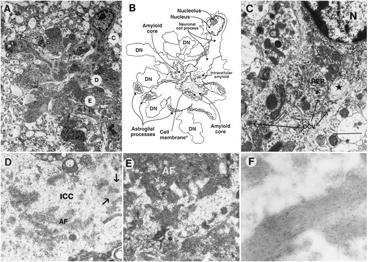
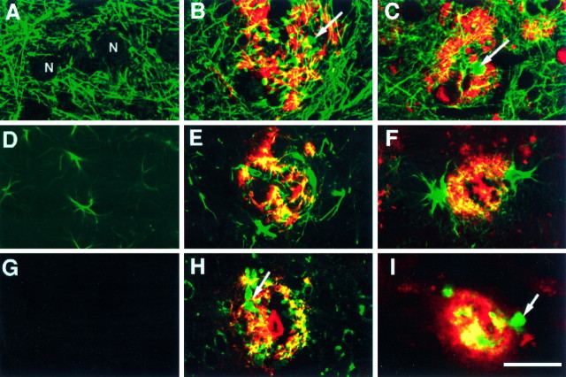
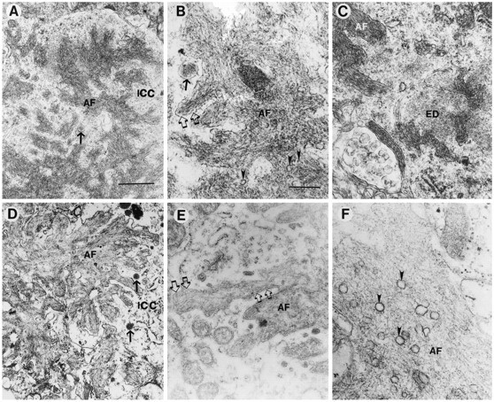
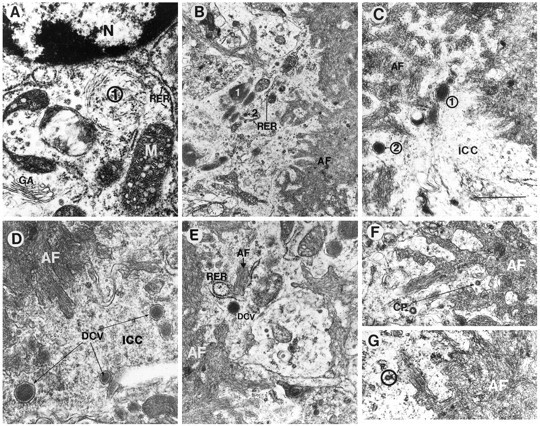
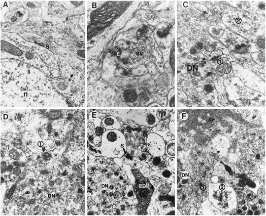
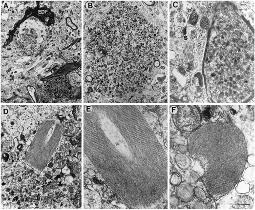
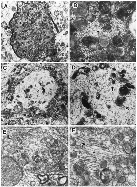
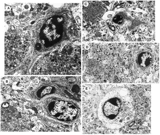
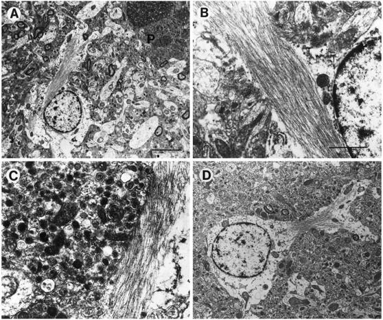
Overexpression of mutated human amyloid precursor protein (hAPP717V-->F) under control of platelet-derived growth factor promoter (PDAPP minigene) in transgenic (tg) mice results in neurodegenerative changes similar to Alzheimer's disease (AD). To clarify the pathology of these mice, we studied images derived from laser scanning confocal and electron microscopy and performed comparisons between PDAPP tg mice and AD. Similar to AD, neuritic plaques in PDAPP tg mouse contained a dense amyloid core surrounded by anti-hAPP- and antineurofilament-immunoreactive dystrophic neurites and astroglial cells. Neurons were found in close proximity to plaques in PDAPP tg mice and, to a lesser extent, in AD. In PDAPP tg mice, and occasionally in AD, neuronal processes contained fine intracellular amyloid fibrils in close proximity to the rough endoplasmic reticulum, coated vesicles, and electron-dense material. Extracellular amyloid fibrils (9-11 nm in diameter) were abundant in PDAPP tg and were strikingly similar to those observed in AD. Dystrophic neurites in plaques of PDAPP tg mouse and AD formed synapses and contained many dense multilaminar bodies and neurofilaments (10 nm). Apoptotic-like figures were present in the tg mice. No paired helical filaments have yet been observed in the heterozygote PDAPP tg mice. In summary, this study shows that PDAPP tg mice develop massive neuritic plaque formation and neuronal degeneration similar to AD. These findings show that overproduction of hAPP717V-->F in tg mice is sufficient to cause not only amyloid deposition, but also many of the complex subcellular degenerative changes associated with AD.


Figures

Fig. 1.
Fig. 1.
Types of neuritic plaques in the hippocampal CA1 pyramidal cell layer of PDAPP tg mice. A, Ultrastructural analysis revealed the presence of clusters of dystrophic neurites (DN) containing abundant electrodense multilaminar bodies. No amyloid was observed in this type of lesion. N indicates the nucleus of cells associated with the plaque. B, C, Classical neuritic plaques contained dystrophic neurites (DN), an amyloid core (A), astrocytic glial cells (G), and an associated neuronal cell (N). D, A third type of plaque presented degenerating and dystrophic neurites (DN) in the periphery, and abundant very dense amyloid cores (A) with prominent fusiform electrodense processes (EDP). Scale bar, 15 μm.
Fig. 2.
Fig. 2.
Comparison of neuritic plaques between PDAPP tg mice and AD. A, Low-power view of the frontal cortex in control non-tg mice shows preserved neuronal structure.B, Low-power view of a classical neuritic plaque in the frontal cortex of a PDAPP tg mouse shows abundant dystrophic neurites (DN), amyloid deposits (A), and an associated cell that displays chromatin clumps beneath the nuclear envelope. C, Low-power view of a classical neuritic plaque in the frontal cortex of an AD case that displayed abundant dystrophic neurites (DN), amyloid deposits (A), and an associated cell that also presents some nuclear chromatin aggregation. D, Higher-power view of the neuropil in the control non-tg mice shows preservation of the neuritic and synaptic structure. E, Higher-power view of the dystrophic neurites in the PDAPP tg mouse shows similar electrodense laminar bodies (LB) to the ones observed in AD. The dystrophic neurites made synaptic contacts (S) and contained abundant small vesicles (V).F, Higher-power view of a dystrophic neurite in AD that shows abundant laminar dense bodies and mitochondria. Scale bars:B, 10 μm; C, 1.5 μm.
Fig. 3.
Fig. 3.
Cellular alterations associated with the neuritic plaque in the PDAPP tg mice. A, B, Semithin section (250 nm) of a neuritic plaque in the hippocampus (pyramidal cell layer) stained with toluidine blue demonstrates dystrophic neurites (DN), amyloid (A), neuronal cell involvement (N), and glial cells (arrows).C, The dystrophic neurites (DN) and a neuronal cell body (N) embedded in the plaque were strongly immunoreactive with SMI312, a monoclonal antibody that identifies phosphorylated neurofilaments (green). The amyloid component was immunolabeled with the polyclonal antibody against Aβ R1280 (red). This neuritic plaque was identified in the hippocampus and imaged with the LSCM as described previously (Masliah et al., 1993b). D, Electron micrograph of a classical neuritic plaque in the hippocampus shows that these lesions were composed of a prominent neuronal element accompanied by extensive neuritic dystrophy, amyloid deposits, and astroglial cell reaction. E, Diagrammatic representation of neuritic plaque illustrated in D. Scale bars: A, 30 μm; C, 10 μm; D, 25 μm.
Fig. 6.
Fig. 6.
Subcellular neuronal alterations associated with the neuritic plaques in PDAPP tg mouse. Electron micrographs were taken from a lesion in the hippocampus. A, Low-power view of a neuritic plaque composed of a prominent neuronal element accompanied by extensive neuritic dystrophy, amyloid deposits, and astroglial cell reaction. The letters inside circles correspond to subsequent panels that identify various characteristics of the neuronal element in the plaque in greater detail. B, Diagrammatic representation of the neuritic plaque illustrated in A.C, Proximal to the nucleus (and distal to the amyloid core), the neuron displayed abundant RER, neurosecretory dense-core vesicles (long arrows), and peculiar elliptical bodies containing fibrilo-tubular material (*). Scale bar, 3 μm.D, Distal to the cell nucleus (and proximal to the amyloid core), the intracellular cytoplasmic compartments (ICC) of the neuronal process displayed amyloid fibrils (AF) and amorphous electrodense granular material (arrows) surrounded by a membrane-forming digitating processes (E). The neuronal cytoplasm surrounding the extracellular amyloid fibrils (AF) showed diffuse granular material and mitochondria. F, Immunogold labeling with a monoclonal antibody against β-amyloid (3D6) showed amyloid fibrils decorated with the 5 nm gold particles.
Fig. 4.
Fig. 4.
Comparison of the neuritic and glial components in the PDAPP tg model and AD. Sections were double-immunolabeled and imaged with the laser scanning confocal microscope. Images obtained in the Texas Red channel correspond to β-amyloid (3D6) and in the FITC channel (green) to phosphorylated neurofilament (SMI312) (AC), GFAP (DF), or hAPP (8E5) (GI). A, Non-tg control mice displayed a well organized and preserved neuritic structure. The neuronal cell bodies (N) were not labeled with the anti-neurofilament antibody. PDAPP tg (B) and AD (C) cases displayed significant disruption of the neuritic structure and positive anti-SMI312 immunoreactivity in the dystrophic neurites (arrow) associated with the plaque.D, Non-tg control mice displayed occasional GFAP-immunoreactive astroglial cells. PDAPP tg (E) and AD (F) cases displayed hypertrophic GFAP-immunoreactive astroglial cells in the periphery of the plaque.G, Non-tg control mice were negative with the antibody specific for hAPP. PDAPP tg (H) and AD (I) cases displayed positive anti-hAPP immunoreactivity in the dystrophic neurites (arrow) associated with the plaque. Scale bar, 20 μm.
Fig. 5.
Fig. 5.
Comparison of the amyloid deposits in the PDAPP tg model and AD. Electron micrographs were obtained from the hippocampal region in the PDAPP tg mouse and from the frontal cortex in AD. Low-power (A) and high-power (B) view of amyloid fibrils (AF) in the PDAPP tg mouse showing dense deposits surrounded by a membrane (open arrows) and the neuronal intracytoplasmic compartment (ICC) containing electrodense granular material and dense-core neurosecretory vesicles (arrow). Occasional poorly defined clear vesicles (arrowheads) were associated with the amyloid fibrils in PDAPP mouse plaques.C, In the PDAPP tg mouse, the cytoplasmic component of neurons close to the extracellular amyloid fibrils (AF) showed the presence of amorphous electrodense material (ED). Low-power (D) and high-power (E) view of the amyloid deposits in AD showed abundant dense fibrils (AF) surrounded by a membrane (open arrows) and a cytoplasmic compartment (ICC) containing dense-core neurosecretory vesicles (arrows) and electrodense granular material.F, Clear vesicles were prominently associated with the amyloid fibrils (AF) in AD plaques (arrowheads). Scale bars: A, 1 μm;B, 100 nm.
Fig. 7.
Fig. 7.
Additional neuronal subcellular characteristics associated with amyloid formation in the PDAPP tg mouse. Electron micrographs were obtained from the hippocampus and are derived from the neuritic plaque presented at the bottom of Figure 3D.A, Proximal to the nucleus (N) (and distal to the amyloid core), the neuron displayed, in addition to RER and elliptical inclusion bodies (1), a prominent Golgi apparatus (GA) and mitochondria (M). B, Distal to the cell nucleus (and proximal to the amyloid core), the cytoplasm of the neuronal process displayed the presence of compact electrodense organelles surrounded by a membrane (1) adjacent to RER, mitochondria, and neurosecretory vesicles (2).C, D, Dense-core neurosecretory vesicles (1, 2) were abundant in the intracytoplasmic compartment (ICC) of the neuron adjacent to the extracellular amyloid fibrils (AF). E, Intracellular amyloid fibrils (AF, black letterhead) adjacent to RER were present in the cytoplasmic compartment of the neurons near the extracellular amyloid fibrils (AF, white letterhead). Dense-core neurosecretory vesicles (DCV) were also present. F,G, Coated pits (CP) in the cytoplasmic compartment of the neurons were closely associated with the plasma membrane (circle) surrounding extracellular amyloid deposits (AF). Scale bar, 400 nm.
Fig. 8.
Fig. 8.
Synaptic alterations in PDAPP tg mice. Electron micrographs were obtained from the hippocampus. A, Neuronal cells associated with the plaques showed synaptic terminals (S) associated with the perikaryon (n, nucleus; arrow, neurosecretory vesicle).B, Presynaptic terminals in the periplaque region displayed amorphous electrodense material (E) in addition to synaptic vesicles (SV).C, D, Some synaptic terminals in the periplaque region showed decreased numbers of synaptic vesicles (1), whereas others were distended and contained abundant vesicles (2) (DN, dystrophic neurites). E, Axonal terminals in the midst of the plaque were distended and displayed accumulations of multilaminar, multivesicular, and electrodense bodies. Dystrophic neurites (DN) made occasional synaptic contacts (S) with neighboring neuritic processes. Other neuritic elements in the plaque were electrodense (EDP).F, Adjacent to the extracellular amyloid fibrils (AF), the axonal terminals were distended and contained abundant dense vesicles (1) or clear vesicles (2). A dystrophic neurite (DN) was also present. Scale bar, 1.5 μm.
Fig. 9.
Fig. 9.
Neuritic alterations in PDAPP tg mice. Electron micrographs were obtained from the hippocampus. In PDAPP tg mice, the dystrophic unmyelinated (A, B) and myelinated neurites (C) contained abundant electrodense laminar and multivesicular bodies. Some neurites contained fine filaments (10 nm; arrow) and were surrounded by electrodense processes (EDP). Other neurites contained characteristic crystals displaying an array of symmetrically organized tubules (DF). Scale bar, 2 μm.
Fig. 10.
Fig. 10.
Comparison of cytoskeletal alterations between the PDAPP tg mouse and AD. Low-power (A) and high-power (B) views of a myelinated dystrophic neurite in the PDAPP tg containing abundant laminated bodies and filamentous material (10 nm in diameter; arrows). Low-power (C) and high-power (D) views of a dystrophic neurite in AD containing multilaminar bodies and neurofilamentous material similar to the one observed in the PDAPP mouse (arrows). Low-power (E) and high-power (F) views of a dystrophic neurite in AD containing classical paired helical filaments (arrows). Scale bars: E, 5 μm;F, 200 nm.
Fig. 11.
Fig. 11.
Apoptotic-like changes in neocortical cells of PDAPP tg mice. Electron micrographs were obtained from layer 5 of the frontal cortex. AD, Neuronal nuclei contained chromatin segmentation and condensation. Some cells displayed dense intracytoplasmic or intranuclear inclusions surrounded by a membrane (*). These cells also contained RER and neurosecretory vesicles. E, Other cells displayed, in addition to the chromatin segmentation, cytoplasmic distention and vacuolization. Scale bar, 10 μm.
Fig. 12.
Fig. 12.
Glial cell alterations in PDAPP tg mice. Electron micrographs were obtained from the frontal cortex. A, Prominent astrocytic cells were observed associated with the neuritic plaques (P). These cells displayed some cytoplasmic swelling and contained abundant intermediate filaments.B, C, Higher-power view of the astrocytic processes in the plaque. D, View of an enlarged astrocyte distant from the plaque. Scale bars: A, 10 μm; B, 2 μm.

References

    1. Bonhomme V, Hans P, Collette J, Moonen G. Neuron-specific enolase as a marker of in vitro neuronal damage. II. Investigation of the astrocyte protective effect against kainate-induced neurotoxicity. J Neurosurg Anesthesiol. 1993;5:117–120. - PubMed
    1. Campbell G, Lieberman AR, Anderson PN, Turmaine M. Regeneration of adult rat CNS axons into peripheral nerve autografts: ultrastructural studies of the early stages of axonal sprouting and regenerative axonal growth. J Neurocytol. 1992;21:755–787. - PubMed
    1. Chartier-Harlin M-C, Crawford F, Houlden H, Warren A, Hughes D, Fidani L, Goate A, Rossor M, Roques P, Hardy J, Mullan M. Early-onset Alzheimer’s disease caused by mutations at codon 717 of the β-amyloid precursor protein gene. Nature. 1991;353:844–846. - PubMed
    1. Clark RF, Goate AM. Molecular genetics of Alzheimer’s disease. Arch Neurol. 1993;50:1164–1172. - PubMed
    1. Cork LC, Masters C, Beyreuther K, Price DL. Development of senile plaques: relationships of neuronal abnormalities and amyloid deposits. Am J Pathol. 1990;137:1383–1392. - PMC - PubMed

Publication types