Review
. 1996:25:163-95.
doi: 10.1146/annurev.bb.25.060196.001115.

Use of 19F NMR to probe protein structure and conformational changes

Affiliations
Review

Use of 19F NMR to probe protein structure and conformational changes

M A Danielson et al. Annu Rev Biophys Biomol Struct. 1996.

Abstract

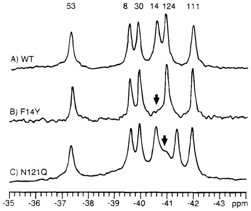
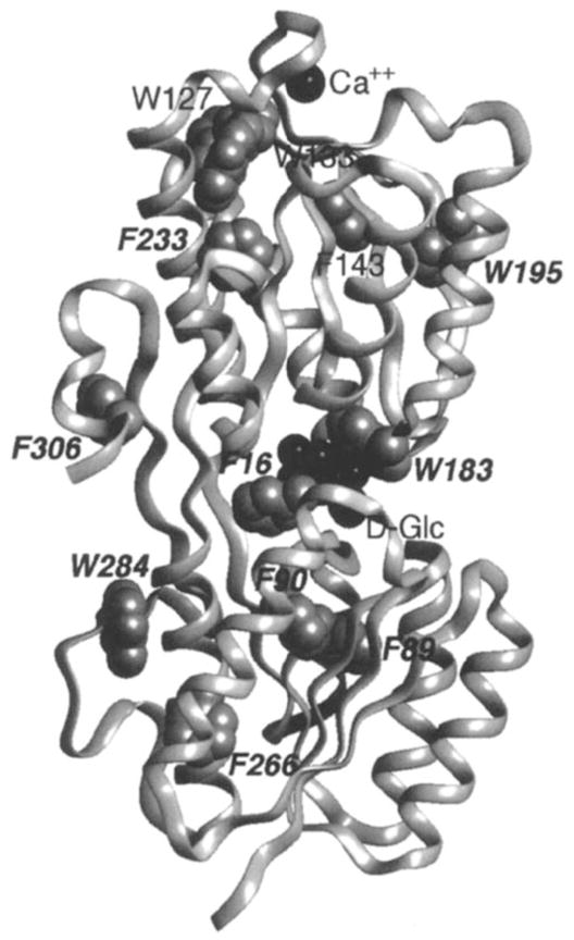
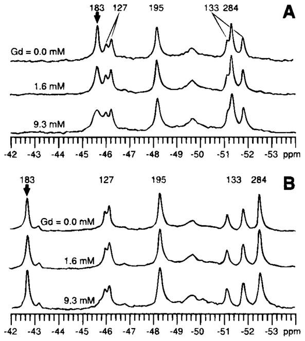
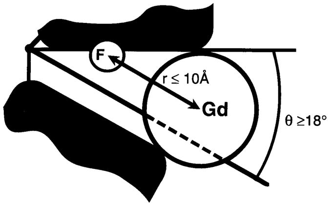
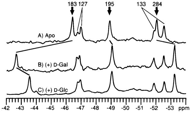
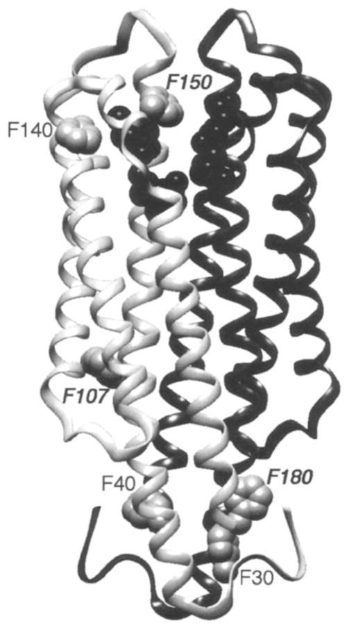
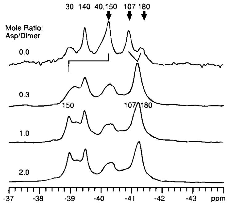
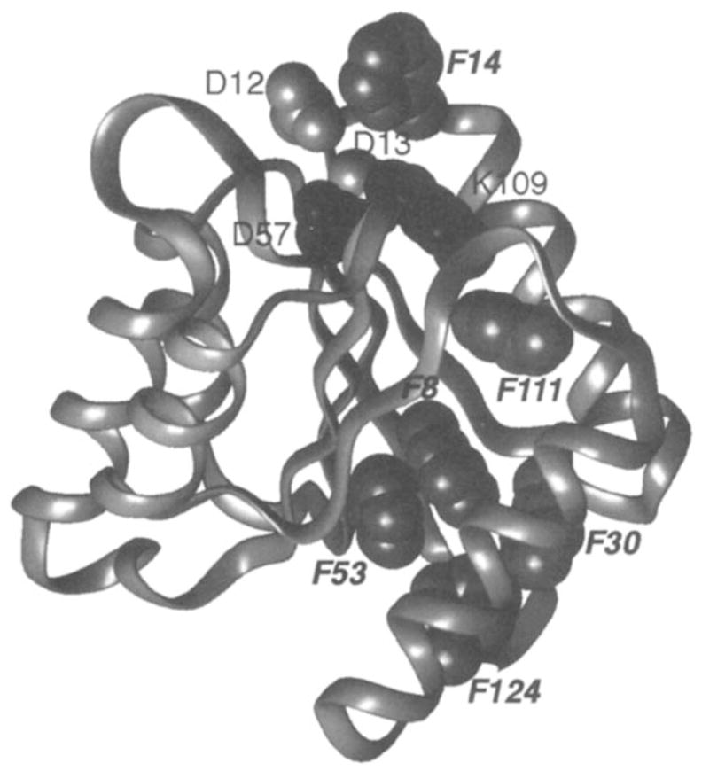
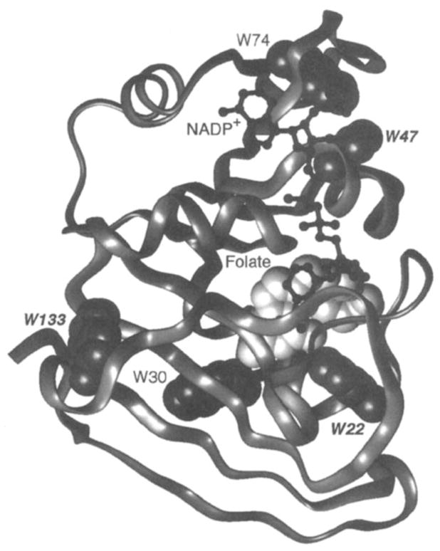
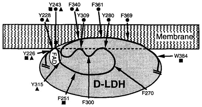
19F NMR has proven to be a powerful technique in the study of protein structure and dynamics because the 19F nucleus is easily incorporated at specific labeling sites, where it provides a relatively nonperturbing yet sensitive probe with no background signals. Recent applications of 19F NMR in mapping out structural and functional features of proteins, including the galactose-binding protein, the transmembrane aspartate receptor, the CheY protein, dihydrofolate reductase, elongation factor-Tu, and D-lactose dehydrogenase, illustrate the utility of 19F NMR in the analysis of protein conformational states even in molecules too large or unstable for full NMR structure determination. These studies rely on the fact that the chemical shift of 19F is extremely sensitive to changes in the local conformational environment, including van der Waals packing interactions and local electrostatic fields. Additional information is provided by solvent-induced isotope shifts or line broadening of the 19F resonance by aqueous and membrane-bound paramagnetic probes, which may reveal the proximity of a 19F label to bulk solvent or a biological membrane. Finally, the effect of exchanging conformations on the 19F resonance can directly determine the kinetic parameters of the conformational transition.


Figures

Figure 1
Figure 1
19F NMR spectra illustrating the two protein engineering approaches used to assign resonances of the phospho-signaling protein CheY (22). (A) The protein is labeled at its six phenylalanine positions with 4-F-Phe, thereby giving rise to a well-resolved 19F NMR spectrum at 470 MHz (B) The assignment of the Phe14 resonance by direct replacement with tyrosine. (C) The nudge assignment of Phe124 by replacement of the adjacent Asn121. The same two methods have also assigned the remaining resonances, as indicated.
Figure 2
Figure 2
Backbone ribbon structure of the galactose-binding protein (93), which illustrates bound ligands (Ca2+ and D-glucose, both in black) and the 12 fluorine probe positions. The protein has been labeled at its five tryptophan positions with 5-F-Trp, or its seven phenylalanine positions with 3-F-Phe (gray VDW surfaces), thereby enabling the use of 19F NMR to probe ligand-induced conformational changes (–56). The binding of D-galactose or D-glucose generates a long-range conformational change, thus yielding significant chemical shift changes for the nine probe positions indicated by italics (see Figure 5). In contrast, the three remaining probes, all in the vicinity of the Ca2+ binding site, are unperturbed. When Ca2+ binds, the converse pattern of frequency changes is observed, such that only the resonances of the latter three probes are observed to shift.
Figure 3
Figure 3
Effect of the aqueous paramagnetic probe Gd3+ EDTA on the 19F NMR spectrum of the galactose-binding protein (470 MHz) (56). The resonance of the 5-F-Trp probe at the Trp183 position within the sugar-binding cleft is broadened selectively by increasing concentrations of the paramagnet when the cleft is empty (A), whereas the same probe resonance in the closed cleft containing bound D-glucose is unaffected (B). The observed broadening of the Trp183 resonance in the empty cleft has been used to place an upper bound on the closest approach of the paramagnet (see Figure 4). The duplicity observed for the Trp127 and 133 resonances stems from two kinetically stable conformations of their environment.
Figure 4
Figure 4
Schematic model for the empty sugar cleft of the galactose-binding protein (56). Addition of the aqueous paramagnet Gd3+ EDTA (Gd) to the empty cleft is observed to broaden the 19F NMR resonance of the 5-F-Trp probe (F) located at the Trp183 position within the cleft (see Figure 3). The resulting broadening indicates that the paramagnet can approach the fluorine probe at a distance of 10 Å or less, which places the paramagnet within the cleft. To accommodate the paramagnet, the known structure of the closed cleft must open by an angle of at least 18 degrees.
Figure 5
Figure 5
Effect of D-galactose and D-glucose on the 19F NMR spectrum of the 5-F-Trp labeled galactose-binding protein (470 MHz) (54). (A) The spectrum of the sugar-empty protein. (B) The protein saturated with D-galactose. (C) The protein saturated with D-glucose. (bold arrows) Resonances for which significant ligand-induced chemical shift changes are observed. The greatest changes are observed for the resonance from the Trp183 position, which lies in van der Waals contact with the bound sugar molecule.
Figure 6
Figure 6
Ribbon backbone structure of the ligand-binding domain from the aspartate receptor (59), which shows the residues (black) in one of the two symmetric ligand-binding sites, as well as the six fluorine probe positions in one of the two symmetric subunits. The isolated domain has been labeled with 4-F-Phe at all six phenylalanines (gray VDW surfaces), thereby enabling the use of 19F NMR to detect ligand-induced chemical shift changes (see Figure 7) (19). Aspartate is observed to induce a long-range conformational change, which yields significant changes in chemical shift for the three probe positions indicated by italics. In the intact receptor, two transmembrane helices from each subunit would extend downward into the bilayer located below the figure, thus carrying the ligand-induced signal across the membrane.
Figure 7
Figure 7
The ligand-binding domain of the aspartate receptor: titration of the 19F NMR spectrum with aspartate (470 MHz) (19). (upper) The spectrum of the 4-F-Phe-labeled apo-domain to which is added increasing concentrations of ligand aspartate. (bold arrows) The resonances that undergo chemical shift changes on ligand binding. The final chemical shifts are observed at a mole ratio of one aspartate molecule per dimer, which indicates half-of-sites occupancy. In addition, the Phe 150 resonance is observed to disappear at an intermediate loading (mole ratio = 0.3), then reappears at a new frequency on saturation (mole ratio = 1.0), thus demonstrating that this resonance passes through the intermediate exchange limit.
Figure 8
Figure 8
Ribbon backbone structure of the phospho-signaling protein CheY (92). Shown are Asp57, the site of phosphorylation, the adjacent highly conserved Lys109 (black), two other conserved active site residues (Asp12,13) and the fluorine probe positions. The protein has been labeled with 4-F-Phe at all six phenylalanines (gray VDW surfaces), thereby enabling the use of 19F NMR to detect activation-induced chemical shift changes (7,22). Phosphorylation of Asp57 is observed to generate significant chemical shift changes at all six probe positions, indicated by italics. In contrast, constitutive activation of the protein by the D13K mutation gives rise to large chemical shift changes for only the Phe14 and Phe111 resonances, both of which arise from the vicinity of the active site.
Figure 9
Figure 9
Ribbon backbone structure of the biosynthetic enzyme dihydrofolate reductase (10). Shown are the bound ligands NADP+ (black ball and stick) and folate (light VDW surface), as well as the fluorine probe positions. The protein has been labeled with 6-F-Trp at its five tryptophan positions (gray VDW surfaces), thereby enabling 19F NMR studies of ligand-induced chemical shift changes (40). Binding of methotrexate to the site that contains folate triggers a long-range conformational change, thereby yielding chemical shift changes at the probe positions indicated by italics and increasing the structural homogeneity of the protein (see text). In contrast, the binding of NADPH generates a more localized conformational change, detected by chemical shifts changes of the Trp22 and Trp74 resonances that arise from the NADPH binding pocket itself.
Figure 10
Figure 10
Schematic low-resolution structural model for the membrane-binding domain of D-LDH, developed by 19F NMR studies (84). Shown are the predicted locations of the FAD binding site and the locations of fluorine probes within the proposed membrane-binding domain, spanning residues 226–384. The protein has been labeled with 5-F-Trp at its native tryptophan sidechains. In addition, engineered tryptophans have been substituted for other aromatic sidechains and labeled with 5-F-Trp as indicated. (filled squares) Probe positions judged to be exposed to aqueous solvent by their solvent isotope-induced shifts; (filled triangles) positions yielding FAD-induced chemical shift changes; and (filled circles) positions for which paramagnetic line broadening is observed on addition of a spin-labeled fatty acid, which implies close proximity to the membrane-binding surface.

References

    1. Abeles RH, Alston TA. Enzyme inhibition by fluoro compounds. J Biol Chem. 1990;265:16705–8. - PubMed
    1. Arseniev AS, Kuryatov AB, Tsetlin VI, Bystrov VF, Ivanov VT, Orchinnikov YA. 19F NMR study of 5-fluorotrytophan-labeled bacteriorhodopsin. FEBS Lett. 1987;213:283–88.
    1. Augspurger J, Pearson JG, Oldfield E, Dykstra CE, Park KD, Schwartz D. Chemical-shift ranges in proteins. J Magn Reson. 1992;100:342–57.
    1. Bellsolell L, Prieto J, Serrano L, Coll M. Magnesium binding to the bacterial chemotaxis protein CheY results in large conformational changes involving its functional surface. J Mol Biol. 1994;238:489–95. - PubMed
    1. Biemann H-P, Koshland DE., Jr Aspartate receptors of Escherichia coli and Salmonella typhimurium bind ligand with negative and half-of-sites cooperativity. Biochemistry. 1994;33:629–34. - PubMed

Publication types

MeSH terms

LinkOut - more resources