. 1996 Oct 1;16(19):6146-56.
doi: 10.1523/JNEUROSCI.16-19-06146.1996.

Dynamics of cell migration from the lateral ganglionic eminence in the rat

Affiliations

Dynamics of cell migration from the lateral ganglionic eminence in the rat

J A de Carlos et al. J Neurosci. .

Abstract

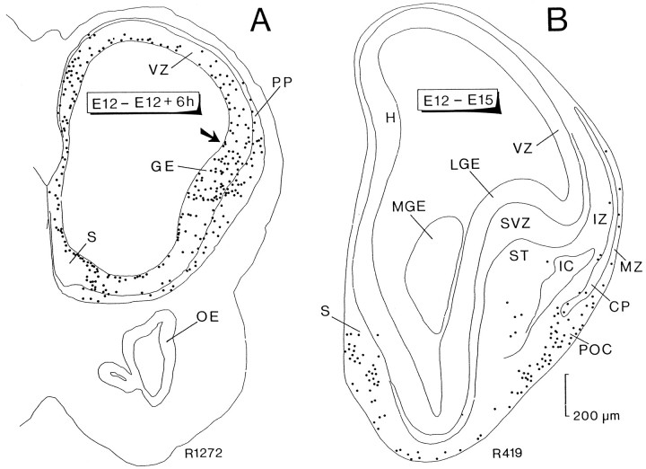
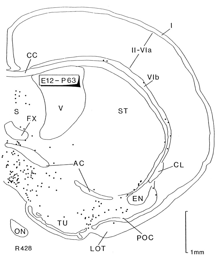
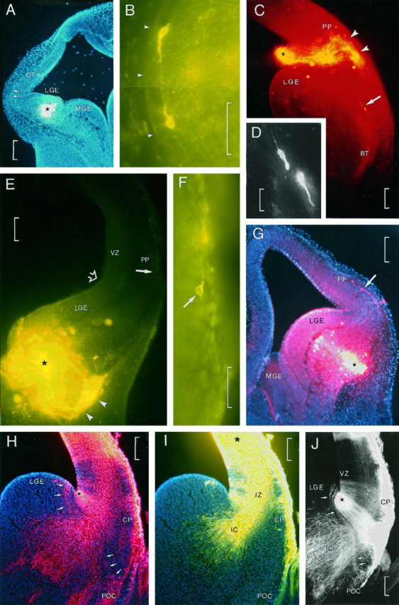
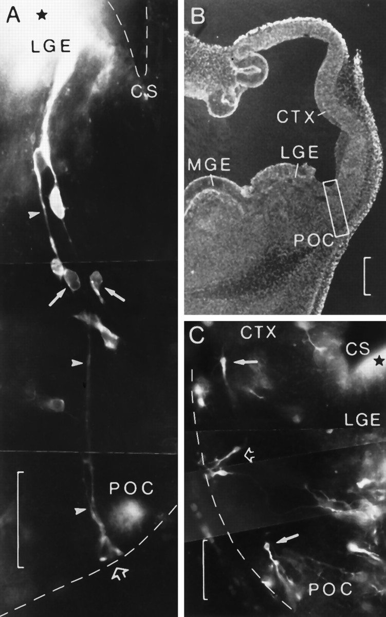
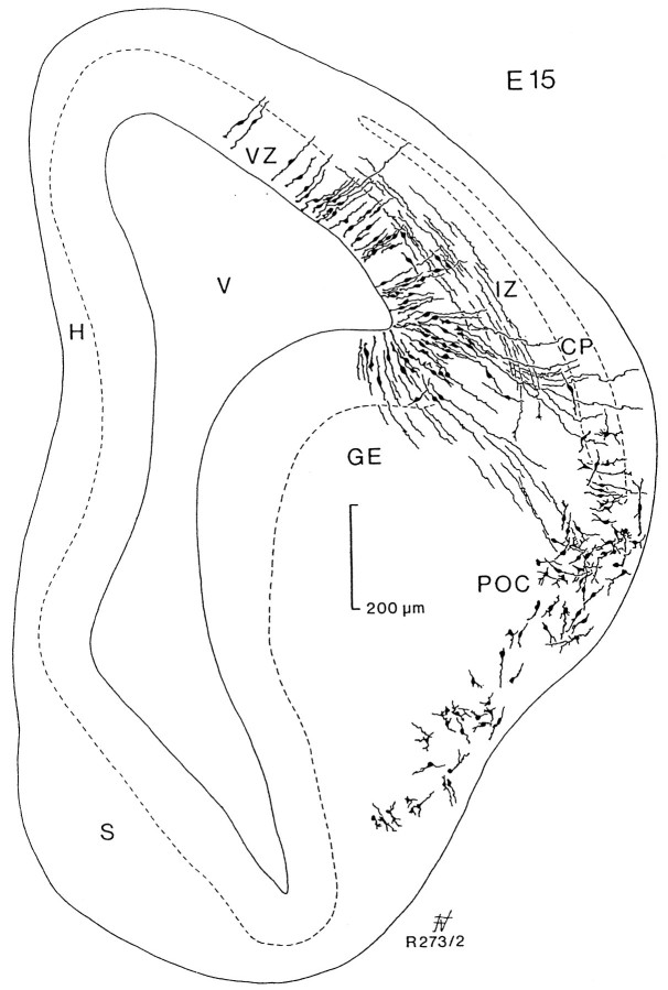
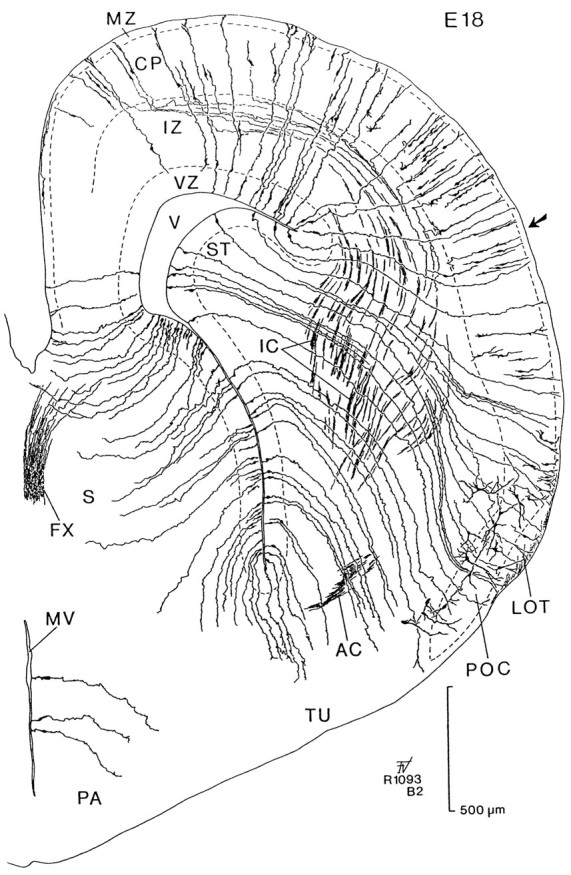
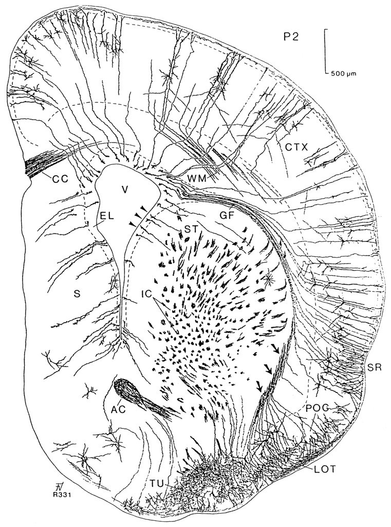
From previous developmental studies, it has been proposed that the neurons of the ventrolateral cortex, including the primary olfactory cortex, differentiate from progenitor cells in the lateral ganglionic eminence. The objective of the present study was to test this hypothesis. The cells first generated in the forebrain of the rat migrate to the surface of the telencephalic vesicle by embryonic day (E) 12. Using [3H]thymidine, we found that most of these cells contributed to the formation of the deep layer III of the primary olfactory cortex. To study the migratory routes of these cells, we made localized injections of the carbocyanine fluorescent tracers Dil and DiA into various parts of the lateral ganglionic eminence in living embryos at E12-E14 and subsequently maintained the embryos in a culture device for 17-48 hr. After fixation, most migrating cells were located at the surface of the telencephalic vesicle, whereas others were seen coursing tangentially into the preplate. Injections made at E13 and in fixed tissue at E15 showed that migrating cells follow radial glial fibers extending from the ventricular zone of the lateral ganglionic eminence to the ventrolateral surface of the telencephalic vesicle. The spatial distribution of radial glial fibers was studied in Golgi preparations, and these observations provided further evidence of the existence of long glial fibers extending from the ventricular zone of the lateral ganglionic eminence to the ventrolateral cortex. We conclude that cells of the primary olfactory cortex derive from the lateral ganglionic eminence and that some early generated cells migrating from the lateral ganglionic eminence transgress the cortico-striatal boundary entering the preplate of the neocortical primordium.


Figures

Fig. 1.
Fig. 1.
Computer printouts of the distribution of heavily labeled cells after [3H]thymidine injection at E12 and perfusion 6 hr later (A) and at E15 (B). Transverse sections through the anterior part of the forebrain. InA, most labeled cells are located in the ventricular zone (VZ), corresponding to progenitor cells in various phases of the mitotic cycle or to migrating cells after completing their last division. Heavily labeled cells outside the VZ appear to be located in the surface in the ventrolateral part of the TV and in the emerging preplate (PP). They correspond to the first-generated cells in the TV. The arrow points to the incipient cortico-striatal sulcus. In B, heavily labeled cells are distributed in several regions of the basal forebrain; numerous cells are concentrated in the septal area (S), prospective primary olfactory cortex (POC), and basal forebrain. Scattered labeled cells are dispersed in the base of the striatum (ST), the marginal zone (MZ) bordering the incipient cortical plate (CP), and below it in the intermediate zone (IZ). Each dot represents one heavily labeled cell. Both printouts are reproduced at the same magnification.GE, Ganglionic eminence; H, differentiating hippocampal field; IC, internal capsule;LGE, lateral ganglionic eminence; MGE, medial ganglionic eminence; OE, olfactory epithelium;ST, striatum; SVZ, subventricular zone.
Fig. 2.
Fig. 2.
Computer printout of the distribution of heavily labeled cells after [3H]thymidine injection at E12 and perfusion at P63. Transverse section through the level of the anterior commissure. Cells in the deep part (layer III) of the primary olfactory cortex (POC) are heavily labeled. On the medial side, numerous labeled cells appear to be located in the septal area (S) and below the anterior commissure (AC). Scattered cells, some of them forming clusters containing heavily labeled cells (not seen in the figure), can be observed in the base of the striatum (ST). Labeled cells are present in sublayer VIb (VIb), whereas no labeled cells were detected in the neocortical layer I (I). These groups of labeled cells, except those in layer I, are homologous to the labeled groups represented in Figure1B. Each dot represents one heavily labeled cell. CC, Corpus callosum;CL, claustrum; EN, endopiriform nucleus;FX, fornix; LOT, lateral olfactory tract;ON, optic nerve; TU, olfactory tubercle;V, lateral ventricle; II-VIa, neocortical layers.
Fig. 3.
Fig. 3.
A–G, Patterns of migrating cells after injections of carbocyanine dyes in living embryos.H–J, Patterns of radial glial fibers in fixed DiI-injected embryos. A, A small injection of carbocyanine type DiA in the lateral ganglionic eminence (LGE) of an embryo at E12 cultured for 24 hr. The injection site is marked by a star, and the section was counterstained with bisbenzimide. The two arrows in the lateral border of the cortex (CTX) point to two (Figure legend continues) migrating cells as shown in B.B, High-power view of the two cells marked inA. Arrowheads delimit the pial surface.C, Small injection of carbocyanine type DiI in the ventricular angle of the lateral ganglionic eminence (LGE) in an embryo at E13 cultured for 17 hr. The injection site is marked by a star.Arrowheads point to groups of migrating cells that have reached the surface at the level of the preplate (PP). Two cells, indicated by arrows, are seen migrating downward. D, High magnification of the two migrating cells indicated by the arrow inC. E, Large injection of carbocyanine type DiA in the lower part of the lateral ganglionic eminence (LGE) in an embryo at E12 cultured for 48 hr. Astar marks the center of the injection site.Arrowheads point to the migrating cells at the surface of the TV. Open arrow points to the cortico-striatal sulcus. An arrow at the lateral border points to one migrating cell as shown in F. F, High-magnification view of the cell indicated by anarrow in E. G, Injection of carbocyanine type DiI in the basal part of the lateral ganglionic eminence (LGE) in an embryo at E13 cultured for 24 hr. Astar marks the injection site. Arrowpoints to a migrating horizontal cell in the preplate (PP). H, I, Double injection of carbocyanine types DiI (H) and DiA (I) and counterstained with bisbenzimide in a fixed embryo at E15; H shows a small deposit of carbocyanine placed at the ventricular angle of the cortico-striatal sulcus (star); I shows a large infiltration of carbocyanine in the primordium of the neocortex (center of injection is marked by a star). H andI represent the same section photographed with different filters (H, DiI, rhodamine filter; I, DiA, fluorescein filter). With the rhodamine filter, DiA labeling is also visible. In H, arrows at the left of the injection site point to a group of radial glial fibers that can be traced from the VZ of the lateral ganglionic eminence (LGE) to the prospective primary olfactory cortex (POC, lower three arrows). InI, the radial glia, labeled with DiI, are not visible. The injection of DiA produced a large deposit, labeling projecting fibers of the internal capsule (IC). Cells of the incipient cortical plate (CP in H andI) and primary olfactory cortex (POC in H) appear intesely labeled. J, Double-exposure photograph of the section in H and I showing double labeling. From the DiI injection site, radial glial fibers are seen streaming toward the surface of the TV. Radial glial cells and fibers that can be traced from the lateral ganglionic eminence (LGE) to the primary olfactory cortex (POC) are marked by arrows. Fibers of the internal capsule (IC), labeled with DiA, are passing between radial glial fibers. All photomicrographs in this figure represent transverse sections made through the anterior part of the TV.BT, Basal telencephalon; MGE, medial ganglionic eminence; IZ, intermediate zone;VZ, ventricular zone. Scale bars: 200 μm inA, G, and J; 50 μm inB and F; 100 μm in C,E, H, and I; and 20 μm in D.
Fig. 4.
Fig. 4.
Small injections of carbocyanine type DiI in two embryos at E12 cultured for 48 hr. A, Transverse section showing the injection site (star) in the lateral ganglionic eminence (LGE) close to the cortico-striatal sulcus (CS). Radial glial fibers (arrowheads) can be followed to where they form their endfeet at the pial surface (open arrow) in a region corresponding to the prospective primary olfactory cortex (POC). Several cells appear to migrate along glial fibers. Note the bright endocytic vesicles in the two migrating cells (arrows). B, Low-power view of the adjacent section. Boxed area delimits approximately the zone reproduced in A, medial being to theleft. C, Transverse section through the right TV in another embryo showing a small injection site (star) in the ventricular surface of the lateral ganglionic eminence (LGE) close to the cortico-striatal sulcus (CS). After culturing for 48 hr, migrating cells appear to have reached the surface at the ventrolateral side of the TV, adopting tangential (arrows) and vertical (open arrow) directions. Medial is to the right.CTX, Neocortical primordium; MGE, medial ganglionic eminence. Scale bars: 50 μm in A andC; 200 μm in B.
Fig. 5.
Fig. 5.
Camera lucida drawing of a transverse section passing through the anterior part of the forebrain vesicle stained by the Golgi method in an embryo of 15 d. Staining is almost absent on the medial side. The lateral part shows radial glial cells of the ventricular zone (VZ) coursing perpendicular to the surface. Migrating horizontal cells can be seen in the intermediate zone (IZ) below the cortical plate (CP). From the ganglionic eminence (GE), glial fibers extend to the prospective primary olfactory cortex (POC), where numerous cells were stained. H, Differentiating hippocampal field; S, septal area; V, lateral ventricle.
Fig. 6.
Fig. 6.
Camera lucida drawing of a transverse section stained by the Golgi method in an embryo of 18 d. This brain was sectioned at an angle to the transverse plane so that the lateral part (right side) is anterior with respect to the medial part (left side, see Fig. 7C). Radial glial fibers extend from the ventricular zone (VZ) to the surface of the brain. Cells of the emerging cortical plate (CP) send axons through the intermediate zone (IZ) that enter the internal capsule (IC). The tilt of these sections allows us to follow radial glial fibers from the VZ of the striatal primordium (ST) to the primary olfactory cortex (POC). Note that radial glia from the lateral ventricular angle can be traced to a level in the cortex (indicated by an arrow). AC, Anterior commissure;FX, fornix; LOT, lateral olfactory tract;MV, third ventricle; MZ, marginal zone;PA, preoptic area; S, septum;TU, olfactory tubercle; V, lateral ventricle.
Fig. 7.
Fig. 7.
Camera lucida drawing of a transverse section stained using the Golgi method at postnatal day 2 at the level of the AC. This section shows young cortical cells with axons projecting to the white matter (WM). Radial glial cells extend from the surface of the lateral ventricle (V) to the cortex (CTX) following diverse trajectories; glial fibers (GF) from the lateral ventricular angle can be traced to the cortex above the sulcus rhinalis (SR). A compact bundle of radial glial fibers (arrows) is located on the lateral side of the striatum (ST). These fibers spread in a fantail manner when they traverse through the primary olfactory cortex (POC). Unlike the remaining glial fibers, they are not in the same plane; the continuity of the cut ends (indicated byarrows) was followed to the bodies of the glial cells (arrowheads) at the lateral border of the lateral ventricle after reconstruction in the two subsequent anterior sections.AC, Anterior commissure; CC, corpus callosum; EL, ependymal layer; IC, internal capsule; LOT, lateral olfactory tract;TU, olfactory tubercle.

References

    1. Altman JB. Proliferation and migration of undifferentiated precursor cells in the rat during postnatal gliogenesis. Exp Neurol. 1966;16:263–278. - PubMed
    1. Altman J, Das GD. Autoradiographic and histological studies of postnatal neurogenesis. I. A longitudinal investigation of the kinetics, migration and transformation of cells incorporating tritiated thymidine in neonate rats, with special reference to postnatal neurogenesis in some brain regions. J Comp Neurol. 1966;127:337–390. - PubMed
    1. Alvarez-Bolado G, Rosenfeld MG, Swanson LW. Model of forebrain regionalization based on spatiotemporal patterns of POU-III homeobox gene expression, birthdates, and morphological features. J Comp Neurol. 1995;355:237–295. - PubMed
    1. Angevine JB, McConnell JA. Time of origin of striatal neurons in the mouse. Anat Rec. 1974;178:300.
    1. Austin CP, Cepko CL. Cellular migration patterns in the developing mouse cerebral cortex. Development. 1990;110:713–732. - PubMed

Publication types

LinkOut - more resources