Comparative Study
. 1996 Oct 1;16(19):6219-35.
doi: 10.1523/JNEUROSCI.16-19-06219.1996.

Functional anatomic studies of memory retrieval for auditory words and visual pictures

Affiliations
Comparative Study

Functional anatomic studies of memory retrieval for auditory words and visual pictures

R L Buckner et al. J Neurosci. .

Abstract

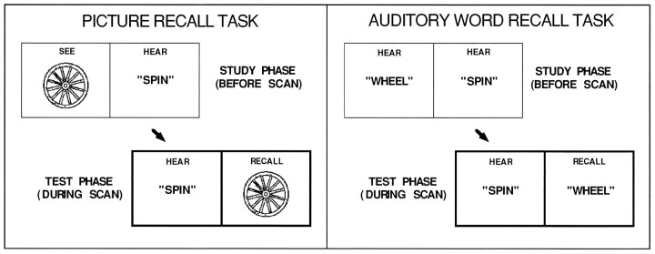
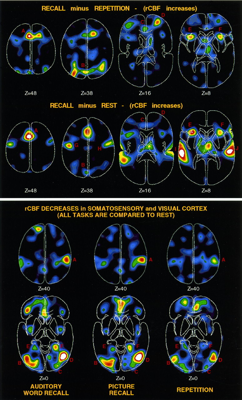
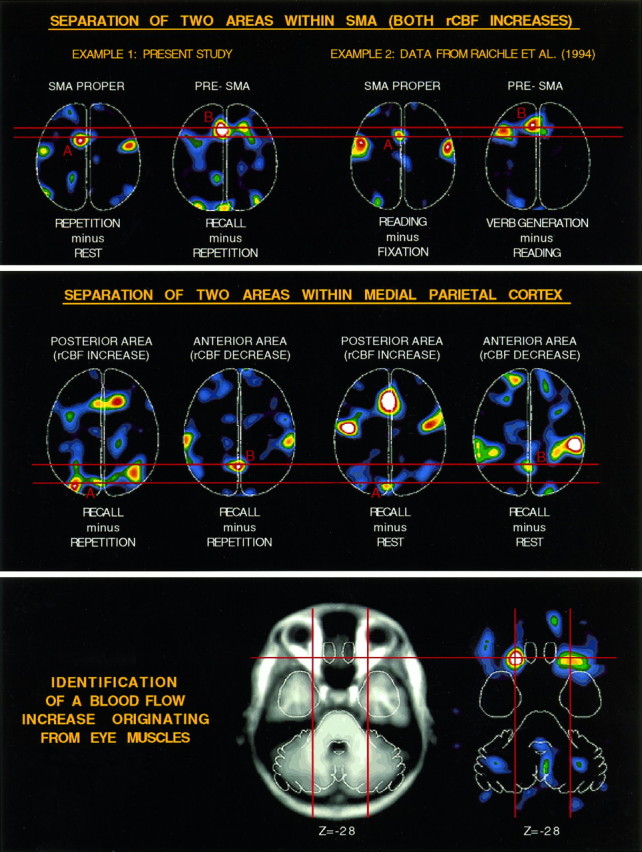
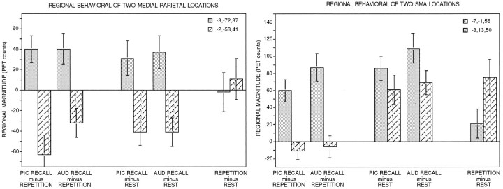
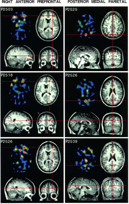
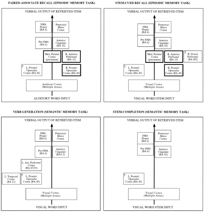
Functional neuroimaging with positron emission tomography was used to study brain areas activated during memory retrieval. Subjects (n = 15) recalled items from a recent study episode (episodic memory) during two paired-associate recall tasks. The tasks differed in that PICTURE RECALL required pictorial retrieval, whereas AUDITORY WORD RECALL required word retrieval. Word REPETITION and REST served as two reference tasks. Comparing recall with repetition revealed the following observations. (1) Right anterior prefrontal activation (similar to that seen in several previous experiments), in addition to bilateral frontal-opercular and anterior cingulate activations. (2) An anterior subdivision of medial frontal cortex [pre-supplementary motor area (SMA)] was activated, which could be dissociated from a more posterior area (SMA proper). (3) Parietal areas were activated, including a posterior medial area near precuneus, that could be dissociated from an anterior parietal area that was deactivated. (4) Multiple medial and lateral cerebellar areas were activated. Comparing recall with rest revealed similar activations, except right prefrontal activation was minimal and activations related to motor and auditory demands became apparent (e.g., bilateral motor and temporal cortex). Directly comparing picture recall with auditory word recall revealed few notable activations. Taken together, these findings suggest a pathway that is commonly used during the episodic retrieval of picture and word stimuli under these conditions. Many areas in this pathway overlap with areas previously activated by a different set of retrieval tasks using stem-cued recall, demonstrating their generality. Examination of activations within individual subjects in relation to structural magnetic resonance images provided an-atomic information about the location of these activations. Such data, when combined with the dissociations between functional areas, provide an increasingly detailed picture of the brain pathways involved in episodic retrieval tasks.


Figures

Fig. 1.
Fig. 1.
Diagrams illustrate the two different episodic memory retrieval tasks that were studied: PICTURE RECALL and AUDITORY WORD RECALL.
Fig. 2.
Fig. 2.
An example of the stimuli used for the after-scan picture recognition task.
Fig. 3.
Fig. 3.
Horizontal sections show PET data from several of the subtraction images analyzed. All data are raw subtraction data displayed without threshold so that the quality of the image data can be observed. Brighter colors represent larger rCBF changes (peak = white). Activations in the cerebellum are not shown but are listed in Tables 2, 4, and 6. Top, rCBF increases are shown (scaled to 70 PET counts). Several areas, only some of which are labeled, were found to be activated in RECALL minus REPETITION including SMA (A), posterior medial parietal cortex (B), anterior cingulate (C), right anterior prefrontal cortex (D), and bilateral frontal-opercular cortex (E, F). RECALL minus REST (scaled to 100 PET counts) revealed many of the same activations with only minimal activation of right anterior prefrontal cortex (D) and robust activation of motor cortex (G, H) and auditory cortex (I, J). Bottom, rCBF decreases are shown for all of the tasks involving auditory stimulation (AUDITORY WORD RECALL, PICTURE RECALL, and REPETITION) compared with REST. Robust rCBF decreases in somatosensory cortex (A) (right > left) and visual cortex (B–F) are seen across all comparisons (scaled to −100 PET counts).
Fig. 4.
Fig. 4.
Horizontal sections show PET data as in Figure 3. Top, rCBF change for two distinct areas along medial frontal cortex are shown separately for two different sets of tasks. REPETITION minus REST reveals a posterior medial frontal activation in SMA (SMA PROPER). In addition to this activation, RECALL minus REPETITION reveals a second more anterior activation (PRE-SMA). The same separation between posterior and anterior divisions of medial frontal cortex is shown for READING minus FIXATION (SMA PROPER) and VERB GENERATION minus READING(PRE-SMA). Middle, Two separate medial parietal areas show rCBF change. A more posterior area shows an rCBF increase in RECALL compared with either REPETITION or REST, whereas a second more anterior area shows a reliable rCBF decrease in the same subtraction images. Bottom, A bilateral rCBF increase (right) is shown that appears to originate from somewhere within the eye muscles as demonstrated by comparison to an averaged MRI image (left).
Fig. 5.
Fig. 5.
Regional activation magnitudes are displayed with SE bars. Each region was tracked across multiple subtraction pairs as listed on the x-axis. Left panel, Regional rCBF change for two separate parietal regions. The two regions, which show completely opposite behavior, change rCBF in relation to the RECALL task compared with either of the reference tasks, whereas both show no rCBF change when the two reference tasks are compared directly. Right panel, Regional rCBF change for two separate divisions of medial frontal cortex. One area (SMA proper), shown with lighter shading, increased rCBF for both RECALL and REPETITION compared with REST, whereas the second area (pre-SMA) showed an rCBF increase only during RECALL.
Fig. 6.
Fig. 6.
Within-subject PET activation data are shown coregistered with structural MRI data. Three single subject activations for each of two areas are shown (see Results). The color sections are transverse PET images with raw subtracted PET data from each subject. Images are scaled near the slice maximum. Three separate MRI views of each subject are aligned to center on the peak activation identified in the PET image (see Materials and Methods).Red lines and square markings show corresponding locations across the different modality types and views.
Fig. 7.
Fig. 7.
Examples of slow drift and erratic movement as measured by the horizontal EOG for subject p2537. Vertical displacement on the record reflects horizontal eye movements. Time bar indicates scale of horizontal axis.
Fig. 8.
Fig. 8.
Heuristic diagrams illustrate current functional anatomic characterizations of four verbal tasks relying on long-term memory retrieval (see Discussion). These diagrams are tentative. The areas activated by each task are indicated by boxes, with similarly localized areas occurring in consistently positioned boxes across diagrams. In each diagram, theleft-most boxes reflect left-lateralized brain areas and the right-most boxes reflect right-lateralized brain areas. Question marks are included in several of the boxes to indicate that there is some uncertainty over whether one or multiple areas are included. A set of areas (shown in thick outlined boxes) demonstrates activation during episodic memory retrieval and may reflect areas being recruited to guide processes selectively demanded by episodic retrieval tasks. SMA proper and pre-SMA are joined in stem-cued recall and stem completion to reflect the fact that the activations could not be separated in those studies. Subcortical structures, including consistently activated cerebellar areas, are not included in these diagrams for simplicity.

Similar articles

Cited by

References

    1. Andreasen NC, O’Leary DS, Arndt S, Cizadlo T, Hurtig R, Rezai K, Watkins GL, Boles L, Ponto B, Hichwa R. Short-term and long-term verbal memory: a positron emission tomography study. Proc Natl Acad Sci USA. 1995a;92:5111–5115. - PMC - PubMed
    1. Andreasen NC, O’Leary DS, Cizadlo T, Arndt S, Rezai K, Watkins GL, Boles Ponto LL, Hichwa RD. Remembering the past: two facets of episodic memory explored with positron emission tomography. Am J Psychiatry. 1995b;152:1576–1585. - PubMed
    1. Buckner RL. Beyond HERA: contributions of specific prefrontal brain areas to long-term memory retrieval. Psych Bull Rev. 1996;3:149–158. - PubMed
    1. Buckner RL, Petersen SE. What has neuroimaging told us about prefrontal cortex involvement in long-term memory retrieval? Semin Neurosci. 1996;8:47–55.
    1. Buckner RL, Petersen SE, Ojemann JG, Miezin FM, Squire LR, Raichle ME. Functional anatomical studies of explicit and implicit memory retrieval tasks. J Neurosci. 1995a;15:12–29. - PMC - PubMed

Publication types

MeSH terms