

Spinal cord terminations of the medial wall motor areas in macaque monkeys
- PMID: 8815929
- PMCID: PMC6578918
- DOI: 10.1523/JNEUROSCI.16-20-06513.1996
Spinal cord terminations of the medial wall motor areas in macaque monkeys
Abstract
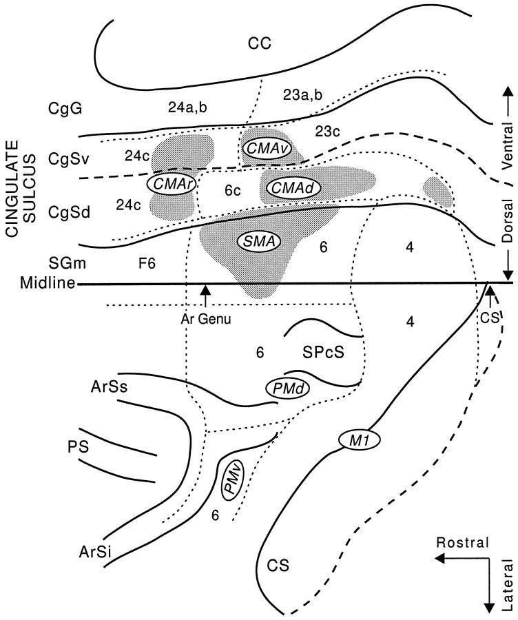
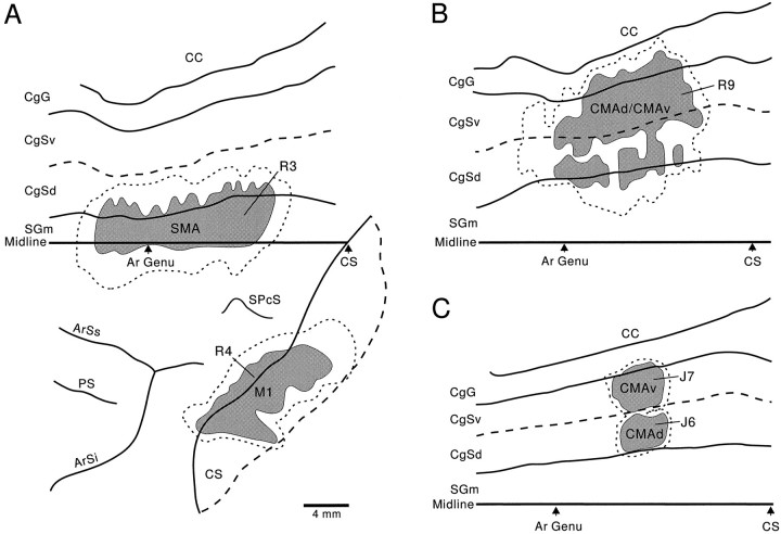
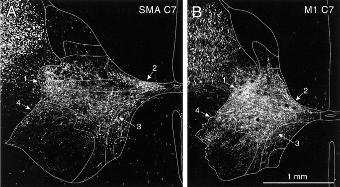
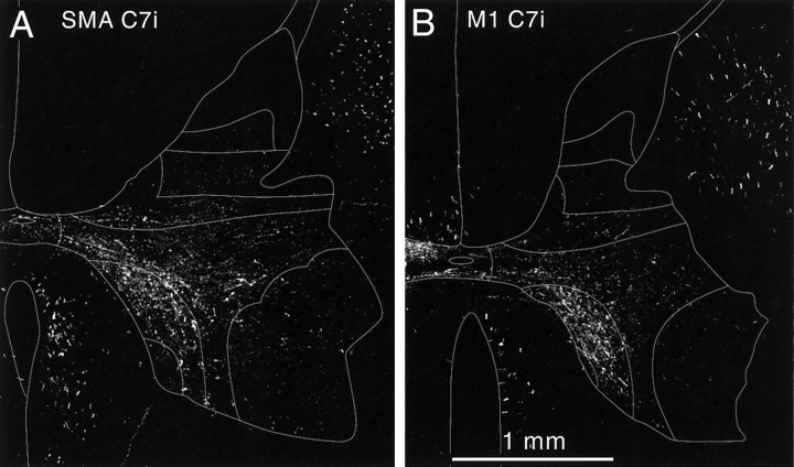
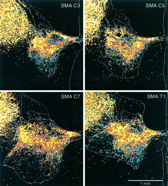
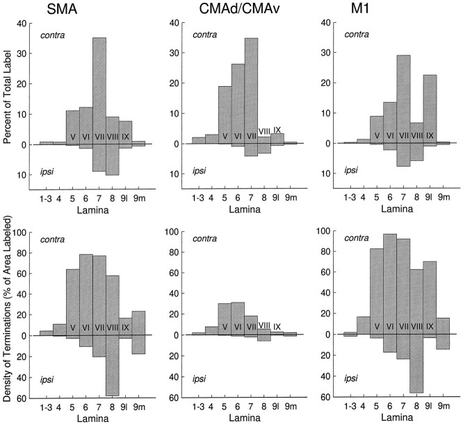
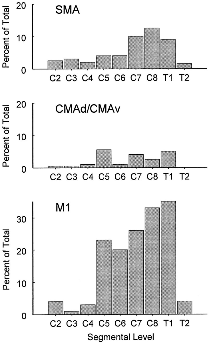
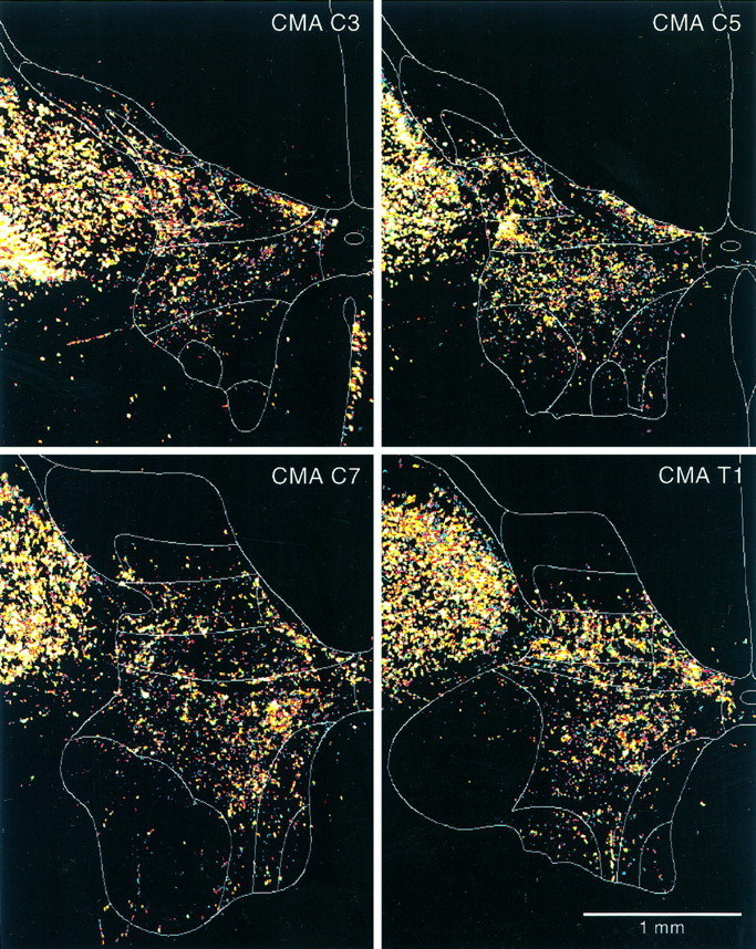
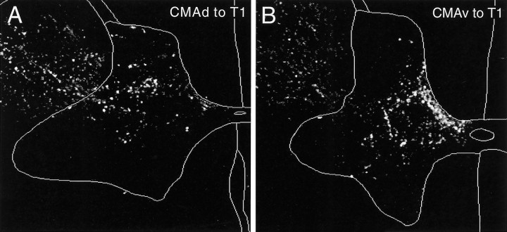
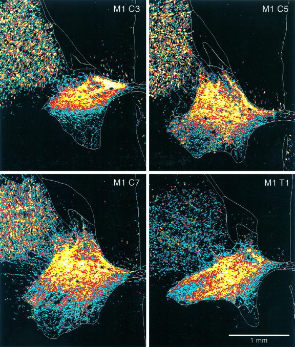
We used anterograde transport of wheat germ agglutinin-horseradish peroxidase to examine the pattern of spinal termination of efferents from the supplementary motor area (SMA) and the two caudal cingulate motor areas (CMAd and CMAv). Our analysis was limited to cervical segments of the macaque. For comparison, we also examined the pattern of termination of efferents from the primary motor cortex (M1). The SMA, CMAd, CMAv, and M1 all terminate in the ventral horn (lamina IX). Thus, all of these motor areas appear to have direct connections with spinal motoneurons, particularly those innervating muscles of the fingers and wrist. All of the motor areas also terminate in the intermediate zone of the spinal cord (laminae V-VIII). Terminations from the SMA and M1 were densest in three regions: (1) dorsolaterally within laminae V-VII; (2) dorsomedially within lamina VI; and (3) ventromedially within lamina VII and adjacent lamina VIII. In contrast, efferents from the CMAd terminate most densely in the dorsolateral portion of the intermediate zone, whereas those from the CMAv were concentrated in the dorsomedial region. Thus, the CMAd and CMAv may innervate distinct sets of interneurons that project directly to motoneurons, and thereby influence specific aspects of segmental motor control. These results suggest that corticospinal projections from the SMA, CMAd, and CMAv are in many respects similar to those of efferents from M1. Consequently, each of the motor areas on the medial wall has the potential to generate and control movement at the level of the spinal cord and may provide an anatomical substrate for the recovery of motor function that follows damage to M1.
Figures

References
-
- Aizawa H, Inase M, Mushiake H, Shima K, Tanji J. Reorganization of activity in the supplementary motor area associated with motor learning and functional recovery. Exp Brain Res. 1991;84:668–671. - PubMed
-
- Apkarian AV, Hodge CJ. Primate spinothalamic pathways. I. A quantitative study of the cells of origin of the spinothalamic pathwayi. J Comp Neurol. 1989;288:447–473. - PubMed
-
- Biber MP, Kneisley LW, LaVail JH. Cortical neurons projecting to the cervical and lumbar enlargements of the spinal cord in young and adult rhesus monkeys. Exp Neurol. 1978;59:492–508. - PubMed
-
- Brinkman C. Supplementary motor area (SMA) and premotor area (PM) of the monkey’s brain: distribution of degeneration in the spinal cord after unilateral lesions. Neurosci Lett. 1982;8:36.
Publication types
MeSH terms
Grants and funding
LinkOut - more resources
Full Text Sources